. 2015 Jun 12:6:7199.
doi: 10.1038/ncomms8199.

De novo mutations in PLXND1 and REV3L cause Möbius syndrome

Affiliations

De novo mutations in PLXND1 and REV3L cause Möbius syndrome

Laura Tomas-Roca et al. Nat Commun. .

Abstract

Möbius syndrome (MBS) is a neurological disorder that is characterized by paralysis of the facial nerves and variable other congenital anomalies. The aetiology of this syndrome has been enigmatic since the initial descriptions by von Graefe in 1880 and by Möbius in 1888, and it has been debated for decades whether MBS has a genetic or a non-genetic aetiology. Here, we report de novo mutations affecting two genes, PLXND1 and REV3L in MBS patients. PLXND1 and REV3L represent totally unrelated pathways involved in hindbrain development: neural migration and DNA translesion synthesis, essential for the replication of endogenously damaged DNA, respectively. Interestingly, analysis of Plxnd1 and Rev3l mutant mice shows that disruption of these separate pathways converge at the facial branchiomotor nucleus, affecting either motoneuron migration or proliferation. The finding that PLXND1 and REV3L mutations are responsible for a proportion of MBS patients suggests that de novo mutations in other genes might account for other MBS patients.


Figures

Figure 1
Figure 1. Clinical features of Möbius syndrome patients and de novo mutations in PLXND1 and REV3L.
(a) Clinical features of the patients carrying the mutations in PLXND1 (P1, P9 and P10). Patient P1 shows an impairment of ocular abduction in a relaxed facial position; she is not able to smile and she cannot close her eyes completely. Patient P9 in a relaxed facial position presents left upper facial paralysis. Patient P10 showing bilateral facial paralysis, inability to close his mouth and lagophthalmos. (b) Genomic structure of the PLXND1 gene and the PLXND1 protein structure and the position of de novo mutations. The different protein domains are depicted. GAP, GTPase-activating protein; IPT/TIG, immunoglobulin-like fold, plexins, transcription factors/transcription factor immunoglobin; D1-PBM, PDZ-binding motif; MET, mesenchymal–epithelial transition. (c) Clinical features of the patients carrying the mutations in REV3L (P5, P11 and P12). Patient P5 in a relaxed position, showing the characteristic inability of MBS patients to smile in the second photograph. Both eyes show incomplete closure. There is agenesis of the left pectoralis muscle (Poland variant) and absence of digits from the left hand. Patient P11 has both eyes fixed in straight position and a complete deficiency of both abduction and adduction. There is a complete inability to follow objects laterally. Furthermore, a bilateral facial nerve palsy is present producing a lagophthalmos on eye closure in both eyes, and oral rim asymmetry with an inability to smile. Patient P12 in a relaxed facial position. The ability to smile of P12 has been improved following plastic surgery at age of 13 years. Inappropriate closure of both eyes is still present. (d) Schematic structure of human REV3L gene (left) and REV3L protein (right). MBS-associated mutations detected in three patients (P5, P11 and P12) are indicated. The coloured bands represent the known domains of the protein. Rev7-binding domain, site of binding of the heterodimeric Rev3l partner Rev7; ZF, zinc finger.
Figure 2
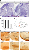
Figure 2. Plxnd1 mouse brain characterization.
(a) Paraffin sagittal sections of embryonic mouse brains processed with Nissl staining at E16.5. These two pictures are medial sections of a view of the corpus callosum (CC), the anterior commisure (ac) and the fasciculus retroflexus (rf). These three structures appear hypoplastic in Plxnd1−/− mice. Scale bars, 500 μm. (b) Graphic representation of the number of motoneurons in facial motor nucleus in wt, heterozygous and homozygous Plxnd1 knockout (mean±s.d.). Unpaired t-test was used to calculate the P value=0.0049 (N=5). (c) Schematic representation of the facial nerve migratory process along the hindbrain. The motoneuron migratory pathway is indicated with a red arrow. The rhombomeres (r3–r11) boundaries are marked with a line. Cb, cerebellum; FBM, facial motor nucleus; SpC, spinal cord. The upper part in (d) shows the immunohistochemical staining of facial FBM motoneurons with anti-Islet-1 antibody (dark brown). The rhombomere units (r3–r7) are marked with a dashed line. Black arrows point the motoneurons migration across the rhombomeres in the heterozygous and the homozygous Plxnd1 knockout mouse. The area of the facial motor nucleus is marked by the square. Scale bar, 300 μm. A detailed view of the facial motor nucleus and motoneurons migration is shown in the lower part of d. Motoneurons appear outside the facial nucleus along the migratory path of the facial nerve in both the heterozygous and the homozygous genotypes. Scale bar, 100 μm. All reported P values tested were calculated by the unpaired t-test using Graphpad software.
Figure 3
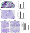
Figure 3. Morphological analysis of the brains of Rev3l embryonic and newborn mice.
(a) Example of a sagittal section of the embryonic mouse head at E16.5 stage used for subarachnoid volume rendering, processed with Nissl staining. The subarachnoid space is shown in green. Scale bar, 1 mm. Right: subarachnoid volume rendering measures of both genotypes. In all Rev3l heterozygous embryos the subarachnoid space is significantly increased as compared with wt embryos (Unpaired t-test P value=0.0047, N=5). Scale bar, 500 μm (b) Example of one of the hindbrain sagittal sections at P0 used for hindbrain volume rendering. We measured the volume of five wt and five heterozygous mice. The hindbrain boundaries are indicated by dashed lines. Scale bar, 500 μm. Right: Rev3l heterozygous mice show a significant decrease of hindbrain volume (Unpaired t-test P value=0.015, N=5). (c) Lateral hindbrain sections at the P0 stage of a view of the facial motor nucleus, inside the square. Scale bar, 300 μm. (d) Higher magnification of the facial motor nucleus showing paucity of Rev3l heterozygous motoneurons. Scale bar, 100 μm. Right: quantification of motoneurons in the facial motor nucleus, in each side of the hindbrain, in five wt and five heterozygous P0 mice (mean±s.d.). The difference is statistically highly significant (Unpaired t-test P value<0.0001, N=10). We do not show both genotypes because there are no visible differences between them. 5N, trigeminal nucleus; 7N, facial nucleus; Cb, cerebellum; HB, hindbrain; Is, isthmus; r1–r11, rhombomeres r1 to r11; Sb, subarachnoid space; Sk, skull; SpC, spinal cord. A P value smaller than 0.05 was considered to be statistically significant (*). P values smaller than 0.01 (**) or 0.001 (***) were considered highly significant.
Figure 4
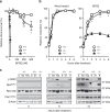
Figure 4. Survival, replication of bulky DNA lesions and DNA damage signaling in Rev3l-mutated MEF lines.
(a) Survival of wt (+/+), Rev3l heterozygous (+/−) and Rev3l-deficient (−/−) MEF lines after mock exposure, or after exposure to Benzo(a)pyrene diolepoxyde (BPDE). Rev3l heterozygous MEFs are slightly sensitive to BPDE indicating haploinsufficiency. Experiments were performed in triplicate. Error bars, s.e.m. The asterisks indicate a significant difference between the wt and the heterozygous lines (unpaired t-test, P<0.05). (b) Representation of replication fork progression after exposure to BPDE, or mock exposure. Rev3l heterozygous MEFs display normal fork progression at undamaged DNA (mock treated, left panel), but a marginal defect in replication of bulky lesion-containing DNA (BPDE, right panel). Error bars, s.e.m. (c) Immunoblots to detect the phosphorylation of DNA damage-signalling markers upon treatment with BPDE. γ-H2AX: phosphorylated histone H2AX. RpaS4/8-P: 32 kDa subunit of single-stranded DNA-binding protein Rpa, phosphorylated at Ser 4 and/or Ser 8. Total levels of Rpa are somewhat variable consequent to slight differences in proliferation rate between the lines. Chk1S354-P Checkpoint kinase-1, phosphorylated at Ser 354. β-actin: loading control. Rev3l-heterozygous MEFs display DNA damage signalling that is almost as strong as Rev3l-deficient MEFs.

Similar articles

Cited by

References

    1. von Graefe A. In: Handbuch der Gesammten Augenheilkunde eds von Graefe A., Saemisch T. Vol. 6, 148W Engelmann (1880).
    1. Möbius P. J. Ueber angeborene doppelseitige Abducens-Facialis-Lähmung. Münchener Medicinische Wochenschrift. About Congenital 35, 91–94 (1888).
    1. Verzijl H. T., van der Zwaag B., Lammens M., ten Donkelaar H. J. & Padberg G. W. The neuropathology of hereditary congenital facial palsy vs Möbius syndrome. Neurology 64, 649–653 (2005). - PubMed
    1. Assaf A. A. Congenital innervation dysgenesis syndrome (CID)/congenital cranial dysinnervation disorders (CCDDs). Eye (Lond) 25, 1251–1261 (2011). - PMC - PubMed
    1. Rucker J. C. et al.. Characterization of ocular motor deficits in congenital facial weakness: Moebius and related syndromes. Brain 137, 1068–1079 (2014). - PMC - PubMed

Publication types

Substances