. 2010 Oct 18;191(2):415-28.
doi: 10.1083/jcb.201004108.

A mechanism for vertebrate Hedgehog signaling: recruitment to cilia and dissociation of SuFu-Gli protein complexes

Affiliations

A mechanism for vertebrate Hedgehog signaling: recruitment to cilia and dissociation of SuFu-Gli protein complexes

Hanna Tukachinsky et al. J Cell Biol. .

Abstract

In vertebrates, Hedgehog (Hh) signaling initiated in primary cilia activates the membrane protein Smoothened (Smo) and leads to activation of Gli proteins, the transcriptional effectors of the pathway. In the absence of signaling, Gli proteins are inhibited by the cytoplasmic protein Suppressor of Fused (SuFu). It is unclear how Hh activates Gli and whether it directly regulates SuFu. We find that Hh stimulation quickly recruits endogenous SuFu-Gli complexes to cilia, suggesting a model in which Smo activates Gli by relieving inhibition by SuFu. In support of this model, we find that Hh causes rapid dissociation of the SuFu-Gli complex, thus allowing Gli to enter the nucleus and activate transcription. Activation of protein kinase A (PKA), an inhibitor of Hh signaling, blocks ciliary localization of SuFu-Gli complexes, which in turn prevents their dissociation by signaling. Our results support a simple mechanism in which Hh signals at vertebrate cilia cause dissociation of inactive SuFu-Gli complexes, a process inhibited by PKA.


Figures
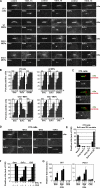
Figure 1.
Figure 1.
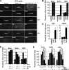
Endogenous SuFu is rapidly recruited to primary cilia by Hh signaling, paralleling recruitment of endogenous Smo, Gli2, and Gli3-FL. (A) Fluorescence micrographs of cilia from untreated cells or cells treated with Shh. Cilia were detected by staining against acetylated tubulin. Because the anti-GliC antibody detects both Gli2 and Gli3-FL, Gli2−/− and Gli3−/− MEFs are shown to demonstrate ciliary recruitment of Gli2 and Gli3-FL separately. The tip of the cilium points to the left. (B) Cells were treated with Shh for varying amounts of time, and ciliary recruitment of SuFu, Smo, Gli2, and Gli3-FL was determined. Asterisks indicate p-values for ciliary recruitment at 1 h compared with t = 0 (*, P < 0.05; **, P < 0.01; ***, P < 0.001). P < 0.05 for all later time points. (C) In NIH-3T3 cells stimulated with Shh for 1 h, SuFu and Gli proteins localize at the tip, whereas Smo localizes along the length of cilia. Cilia were stained as in A, and centrioles were stained with anti–γ-tubulin. (D) Endogenous SuFu and Gli proteins colocalize at the tips of primary cilia in SAG-treated NIH-3T3 cells. (left) Cilia costained for endogenous SuFu (rabbit antibody) and Gli (goat antibody). (right) Cilia costained for Smo (rabbit antibody) and Gli (goat antibody). (E) Cilia counts for the experiment in D (left). Endogenous SuFu and Gli colocalize both in the resting and stimulated states of the Hh pathway. (F) Recruitment of SuFu, Smo, and Gli to cilia in response to Hh stimulation does not require new protein synthesis. Ciliary localization was determined in NIH-3T3 cells treated or not with Shh in the presence or absence of CHX. (G) Inhibition of protein synthesis does not block the transcriptional output of the Hh pathway. Transcription of the direct transcriptional targets Gli1 and Ptch1 was assayed by Q-PCR after 3 and 6 h of stimulation with Shh in the presence or absence of CHX. wt, wild type. Error bars indicate mean ± SD for three independent counts. Bars, 2 µm.
Figure 2.
Figure 2.
Hh-dependent recruitment of SuFu and Gli proteins to cilia requires active Smo. (A) NIH-3T3 cells were treated with the Smo agonist SAG or with the antagonist Cyc. SuFu and Gli are recruited to cilia by SAG but not by Cyc, although both SAG and Cyc recruit Smo to cilia. The tips of cilia point to the left. Bar, 2 µm. (B) Cilia counts for the experiment in A. (C) Q-PCR assay of Hh pathway target genes for the experiment in A. (D) Maintaining increased levels of SuFu and Gli at cilia is continuously dependent on active Smo. Cyc was added in the presence of Shh to NIH-3T3 cells prestimulated with Shh for 3 h. Ciliary localization was determined before and after 3 h of Shh stimulation and 1 and 3 h after Cyc addition. (E) NIH-3T3 cells were stimulated with Shh for 3 h followed by incubation with the Smo antagonist SANT-1 for 3 h. Ciliary localization of SuFu, Gli, and Smo was measured at the indicated times. P < 0.002 for the recruitment of Smo, SuFu, and Gli by Shh stimulation. P-values for exit from the cilium were calculated relative to ciliary localization after 3 h of Hh stimulation. Asterisks indicate the p-values for ciliary exit (*, P < 0.05; **, P < 0.01; ***, P < 0.001). Error bars indicate mean ± SD.
Figure 3.
Figure 3.
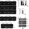
Localization of endogenous SuFu and Gli to cilia is antagonized by PKA. (A) Activation of PKA by FSK blocks localization of endogenous SuFu and Gli proteins to cilia. NIH-3T3 cells were treated with or without Shh and FSK. Shh, FSK, or Shh and FSK recruit Smo to the cilium; in contrast, endogenous SuFu and Gli are removed from cilia by FSK, both in the presence and absence of Shh stimulation. (B) Cilia counts for the experiment in A. (C) Q-PCR analysis of the experiment in A. Inhibition of SuFu and Gli ciliary localization by FSK correlates with complete inhibition of the transcriptional output of the Hh pathway. Error bar indicates mean ± SD. (D) FSK inhibits localization of SuFu and Gli to primary cilia in Smo−/− MEFs. Percentages shown indicate corresponding ciliary counts. (E) NIH-3T3 cells were incubated with or without Shh (left) or with or without FSK (right) followed by immunoblotting for SuFu, Gli3-FL, GSK3, and α-tubulin. (top) Numbers shown indicate the levels of Gli3-FL in each lane relative to α-tubulin. FSK treatment causes only a slight reduction in Gli3-FL, which is much smaller than the decrease caused by Shh. (F) NIH-3T3 cells stably expressing a Gli1–SuFu fusion were incubated with control media, SAG, or FSK. The Gli1–SuFu fusion localizes to cilia in unstimulated cells, and its localization is increased by SAG. FSK treatment completely blocks ciliary localization of the Gli1–SuFu fusion. Percentages shown indicate ciliary localization of the fusion. Bars, 2 µm.
Figure 4.
Figure 4.
Gli proteins are required to localize SuFu to cilia, but Gli proteins can localize to cilia in the absence of SuFu. (A) Wild-type and Gli2−/− Gli3−/− MEFs were incubated with or without Shh. SuFu does not localize to cilia with or without Shh stimulation in Gli2−/− Gli3−/− MEFs, whereas Smo recruitment is normal. (B) Cilia counts for a time course of ciliary recruitment of Smo, SuFu, and Gli in Gli2−/− Gli3−/− MEFs stimulated with Shh. (C) SuFu+/− and SuFu−/− MEFs were stimulated or not with Shh. Endogenous Gli proteins do not localize to cilia with or without Shh stimulation in the absence of SuFu. Recruitment of Smo is normal. (D) Immunoblot of SuFu−/− and SuFu+/− MEFs stably expressing Gli1HA and treated with the proteasome inhibitor bortezomib. Proteasome inhibition allows SuFu−/− cells to accumulate Gli1HA to levels similar to those in the control SuFu+/− cells. (E) Stably expressed Gli1HA localizes to ciliary tips in SuFu−/− MEFs stimulated with SAG in the presence of bortezomib. Percentages shown indicate corresponding ciliary counts. Bars, 2 µm.
Figure 5.
Figure 5.
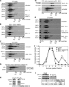
Biochemical evidence that Hh pathway activation causes rapid dissociation of endogenous SuFu–Gli complexes. (A–I) Endogenous SuFu–Gli complexes were analyzed by sucrose gradient centrifugation (A–G) and immunoprecipitation (H and I). (A) In untreated NIH-3T3 cells, the majority of endogenous SuFu (54 kD) exists as a monomer of similar size as the kinase GSK3-β (47 kD). A small fraction of SuFu from untreated cells forms a higher molecular mass complex (top, black lines), the level of which quickly drops in cells treated with Shh for 1 h (middle), an effect completely blocked if Smo is inhibited with 200 nM SANT-1 (bottom). The position in the gradient of two size markers run in parallel is shown below the Western blots (aldolase: molecular mass, 158 kD; Stokes radius, 48.1 Å; catalase: molecular mass, 232 kD; Stokes radius, 52.2 Å). (B) In Gli2−/− Gli3−/− MEFs, only the monomeric SuFu peak is seen by sucrose gradient centrifugation. Hh stimulation of Gli2−/− Gli3−/− MEFs does not change the size of the SuFu peak, although Smo is recruited to the cilia normally in these cells. (C) As in A, but cells were stimulated or not with SAG, and sucrose gradient fractions were immunoblotted for endogenous SuFu, GSK3, and Gli3-FL. The higher molecular mass SuFu peak overlaps with endogenous Gli3-FL in unstimulated cells. Acute Hh pathway stimulation causes the simultaneous disappearance of the overlapping, higher molecular mass SuFu and Gli3-FL peaks. (D) To prevent dissociation of SuFu from Gli, a direct fusion of Gli1 to SuFu was generated. NIH-3T3 cells stably expressing this Gli1–SuFu fusion were stimulated or not with SAG. The apparent size of the Gli1–SuFu fusion peak does not change upon Hh pathway activation. (E) Treatment of NIH-3T3 cells with SAG causes complete disappearance of the SuFu–Gli complex, which is not reversed by inhibition of the proteasome with bortezomib. In contrast, activation of PKA with FSK completely blocks SuFu–Gli dissociation induced by SAG stimulation. (F) Quantification of the experiment in E. The amount of SuFu in each fraction was measured relative to the amount of SuFu in the input lane. The first fraction represents the top of the sucrose gradient. (G) Mouse SuFu expressed in Xenopus embryos shows the same size distribution as endogenous SuFu in mammalian cultured cells, suggesting that SuFu forms a similar complex with endogenous Gli proteins in Xenopus embryos. (H) NIH-3T3 cells were incubated with or without SAG followed by immunoprecipitation (IP) with anti-SuFu antibodies. The level of Gli3-FL is similar in SAG-treated and untreated cells (left). Gli3-FL coimmunoprecipitates with SuFu only in untreated cells but not in SAG-stimulated cells (right), indicating that acute Hh pathway activation dissociates endogenous Gli3-FL from SuFu. (I) NIH-3T3 cells were incubated with control media, SAG, SAG and bortezomib, and SAG and FSK followed by immunoprecipitation with anti–Gli3-FL antibodies. Gli2−/− Gli3−/− MEFs were used as negative control (lanes 1 and 6). Endogenous SuFu does not coimmunoprecipitate with Gli3-FL in cells stimulated with SAG, although levels of Gli3-FL decrease only slightly. Proteasome inhibition by bortezomib (sufficient to abolish any decrease in the level of Gli3-FL) does not block dissociation of endogenous SuFu from Gli3-FL. In contrast, SAG-induced dissociation of SuFu from Gli3-FL is completely blocked by FSK.
Figure 6.
Figure 6.
A model for activation of Gli proteins during vertebrate Hh signaling. In the resting state of the Hh pathway (left), SuFu forms inactive complexes with Gli2 and Gli3-FL, which are sequestered in the cytoplasm. Without Hh stimulation, SuFu–Gli complexes traffic to the primary cilium at a low level independently of Smo; this basal ciliary trafficking is antagonized by PKA. Hh pathway stimulation (right) leads to the translocation of active Smo to the cilium, which, in turn, recruits SuFu–Gli complexes. Active Smo at cilia causes the dissociation of SuFu from Gli. Monomeric SuFu and Gli leave the cilium followed by Gli nuclear translocation and activation of the transcriptional program of the Hh pathway. PKA antagonizes Hh signaling by blocking ciliary localization of SuFu–Gli complexes, thus preventing coupling between active Smo and dissociation of SuFu–Gli complexes.

Similar articles

Cited by

References

    1. Alcedo J., Ayzenzon M., Von Ohlen T., Noll M., Hooper J.E. 1996. The Drosophila smoothened gene encodes a seven-pass membrane protein, a putative receptor for the hedgehog signal. Cell. 86:221–232 10.1016/S0092-8674(00)80094-X - DOI - PubMed
    1. Alexandre C., Jacinto A., Ingham P.W. 1996. Transcriptional activation of hedgehog target genes in Drosophila is mediated directly by the cubitus interruptus protein, a member of the GLI family of zinc finger DNA-binding proteins. Genes Dev. 10:2003–2013 10.1101/gad.10.16.2003 - DOI - PubMed
    1. Aza-Blanc P., Ramírez-Weber F.A., Laget M.P., Schwartz C., Kornberg T.B. 1997. Proteolysis that is inhibited by hedgehog targets cubitus interruptus protein to the nucleus and converts it to a repressor. Cell. 89:1043–1053 10.1016/S0092-8674(00)80292-5 - DOI - PubMed
    1. Barnfield P.C., Zhang X., Thanabalasingham V., Yoshida M., Hui C.C. 2005. Negative regulation of Gli1 and Gli2 activator function by Suppressor of fused through multiple mechanisms. Differentiation. 73:397–405 10.1111/j.1432-0436.2005.00042.x - DOI - PubMed
    1. Barzi M., Berenguer J., Menendez A., Alvarez-Rodriguez R., Pons S. 2010. Sonic-hedgehog-mediated proliferation requires the localization of PKA to the cilium base. J. Cell Sci. 123:62–69 10.1242/jcs.060020 - DOI - PubMed

Publication types

MeSH terms