. 2024 Apr 17;13(8):697.
doi: 10.3390/cells13080697.

Quantitative Phase Imaging as Sensitive Screening Method for Nanoparticle-Induced Cytotoxicity Assessment

Affiliations

Quantitative Phase Imaging as Sensitive Screening Method for Nanoparticle-Induced Cytotoxicity Assessment

Anne Marzi et al. Cells. .

Abstract

The assessment of nanoparticle cytotoxicity is challenging due to the lack of customized and standardized guidelines for nanoparticle testing. Nanoparticles, with their unique properties, can interfere with biochemical test methods, so multiple tests are required to fully assess their cellular effects. For a more reliable and comprehensive assessment, it is therefore imperative to include methods in nanoparticle testing routines that are not affected by particles and allow for the efficient integration of additional molecular techniques into the workflow. Digital holographic microscopy (DHM), an interferometric variant of quantitative phase imaging (QPI), has been demonstrated as a promising method for the label-free assessment of the cytotoxic potential of nanoparticles. Due to minimal interactions with the sample, DHM allows for further downstream analyses. In this study, we investigated the capabilities of DHM in a multimodal approach to assess cytotoxicity by directly comparing DHM-detected effects on the same cell population with two downstream biochemical assays. Therefore, the dry mass increase in RAW 264.7 macrophages and NIH-3T3 fibroblast populations measured by quantitative DHM phase contrast after incubation with poly(alkyl cyanoacrylate) nanoparticles for 24 h was compared to the cytotoxic control digitonin, and cell culture medium control. Viability was then determined using a metabolic activity assay (WST-8). Moreover, to determine cell death, supernatants were analyzed for the release of the enzyme lactate dehydrogenase (LDH assay). In a comparative analysis, in which the average half-maximal effective concentration (EC50) of the nanocarriers on the cells was determined, DHM was more sensitive to the effect of the nanoparticles on the used cell lines compared to the biochemical assays.

Keywords: digital holographic microscopy; in vitro; label-free cytotoxicity testing; nanoparticles; quantitative phase imaging.


Conflict of interest statement

The authors declare no conflicts of interest.

Figures
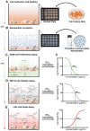
Figure 1
Figure 1
Experimental design and workflow for comparison of PACA nanoparticle in vitro cytotoxicity assessment by DHM with downstream WST-8 and LDH assays. (A) Seeding of NIH-3T3 and RAW 264.7 cells into 96-well plates. (B) Incubation of cells with PACA, cbz-loaded PACA nanoparticles and controls. (C) Label-free DHM QPI proliferation assay. (D) WST-8 cell viability assay. (E) LDH cell death assay. (F) Determination of EC50 values.
Figure 2
Figure 2
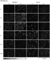
DHM QPI images of RAW 264.7 macrophages and NIH-3T3 fibroblasts incubated with unloaded PACA nanoparticles in five representatively selected concentrations (0.2, 2, 8, 32 and 256 µg/mL) vs. cell culture medium controls (0 µg/mL) at time points t = 0 and t = 24 h. For both cell lines, viable proliferated cells were observed after incubation with cell culture medium control and 0.2 and 2 µg/mL of unloaded PACA nanoparticles. RAW 264.7 cells with 8 µg/mL showed cell debris at t = 0, and after 24 h; NIH-3T3 cells showed cell detachment at t = 0 and proliferated cells after 24 h. For 32 and 256 µg/mL of unloaded PACA nanoparticles, cell debris was observed for RAW 264.7 macrophages after 24 h, and proliferated cells, detached cells and cell debris were observed for NIH-3T3 with 32 µg/mL. Corresponding bright-field images (Figure S1) and enlarged areas of DHM QPI and bright-field images (Figure S2), which allow for a more detailed investigation of the cellular morphology alterations, are provided in the Supplementary Materials.
Figure 3
Figure 3
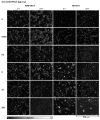
DHM QPI images of RAW 264.7 macrophages and NIH-3T3 fibroblasts after incubation with cbz-loaded PACA nanoparticles in five representatively selected concentrations (0.002, 0.2, 8, 32 and 256 µg/mL) vs. cell culture medium controls (0 µg/mL) at time points t = 0 and t = 24 h. For both cell lines, after incubation with cell culture medium control and 0.002 µg/mL of cbz-loaded PACA nanoparticles, viable cells were detected after 24 h. For 0.2 µg/mL, cell debris could be observed for RAW 264.7, and detached and swollen cells could be observed for NIH-3T3. Macrophages incubated with 8 µg/mL of cbz-loaded PACA showed a swollen but viable cell morphology after 24 h, and for NIH-3T3, detached cells and cell debris were visible at t = 0 24 h, but after 24 h, proliferated cells were visible. Cell debris was observed in both cell lines with 32 and 256 µg/mL of cbz-loaded nanoparticles after 24 h. Corresponding bright-field images (Figure S3) and enlarged areas of DHM QPI and bright-field images (Figure S4), which allow for a more detailed investigation of the cellular morphology alterations, are provided in the Supplementary Materials.
Figure 4
Figure 4
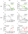
Dose–response relationship for unloaded PACA and cbz-loaded PACA nanoparticles on cell proliferation (DHM, green, (A) unloaded (D) cbz-loaded PACA), viability (WST-8, gray, (B) unloaded (E) cbz loaded PACA) and death (LDH, red, (C) unloaded (F) cbz loaded PACA) of RAW 264.7 macrophages and NIH-3T3 fibroblasts. Mouse RAW 264.7 macrophages and NIH-3T3 fibroblasts were seeded in 96-well plates incubated with unloaded and cbz-loaded PACA, and dry mass increments of cell populations were analyzed with DHM. Subsequently, the viability of the same cell populations was determined with a WST-8 metabolic activity assay, and the supernatants were analyzed in parallel for the release of LDH to detect cell death. The mean values ± SD from three independent experiments are shown (n = 3). Significance levels were given as p < 0.001 (***), p < 0.01 (**) and p < 0.05 (*).

Similar articles

  • Defining the optimum strategy for identifying adults and children with coeliac disease: systematic review and economic modelling.
    Elwenspoek MM, Thom H, Sheppard AL, Keeney E, O'Donnell R, Jackson J, Roadevin C, Dawson S, Lane D, Stubbs J, Everitt H, Watson JC, Hay AD, Gillett P, Robins G, Jones HE, Mallett S, Whiting PF. Elwenspoek MM, et al. Health Technol Assess. 2022 Oct;26(44):1-310. doi: 10.3310/ZUCE8371. Health Technol Assess. 2022. PMID: 36321689 Free PMC article.
  • Gallium Scan.
    Dittrich RP, De Jesus O. Dittrich RP, et al. 2022 Dec 26. In: StatPearls [Internet]. Treasure Island (FL): StatPearls Publishing; 2025 Jan–. 2022 Dec 26. In: StatPearls [Internet]. Treasure Island (FL): StatPearls Publishing; 2025 Jan–. PMID: 33620825 Free Books & Documents.
  • Diffusion Tensor Imaging.
    Ranzenberger LR, Das JM, Snyder T. Ranzenberger LR, et al. 2023 Nov 12. In: StatPearls [Internet]. Treasure Island (FL): StatPearls Publishing; 2025 Jan–. 2023 Nov 12. In: StatPearls [Internet]. Treasure Island (FL): StatPearls Publishing; 2025 Jan–. PMID: 30726046 Free Books & Documents.
  • Depressing time: Waiting, melancholia, and the psychoanalytic practice of care.
    Salisbury L, Baraitser L. Salisbury L, et al. In: Kirtsoglou E, Simpson B, editors. The Time of Anthropology: Studies of Contemporary Chronopolitics. Abingdon: Routledge; 2020. Chapter 5. In: Kirtsoglou E, Simpson B, editors. The Time of Anthropology: Studies of Contemporary Chronopolitics. Abingdon: Routledge; 2020. Chapter 5. PMID: 36137063 Free Books & Documents. Review.
  • Interventions to reduce harm from continued tobacco use.
    Lindson-Hawley N, Hartmann-Boyce J, Fanshawe TR, Begh R, Farley A, Lancaster T. Lindson-Hawley N, et al. Cochrane Database Syst Rev. 2016 Oct 13;10(10):CD005231. doi: 10.1002/14651858.CD005231.pub3. Cochrane Database Syst Rev. 2016. PMID: 27734465 Free PMC article. Review.

References

    1. Sun L., Liu H., Ye Y., Lei Y., Islam R., Tan S., Tong R., Miao Y.B., Cai L. Smart nanoparticles for cancer therapy. Signal Transduct. Target. Ther. 2023;8:418. doi: 10.1038/s41392-023-01642-x. - DOI - PMC - PubMed
    1. Mazzotta E., De Santo M., Lombardo D., Leggio A., Pasqua L. Mesoporous silicas in materials engineering: Nanodevices for bionanotechnologies. Mater. Today Bio. 2022;17:100472. doi: 10.1016/j.mtbio.2022.100472. - DOI - PMC - PubMed
    1. Domingues C., Santos A., Alvarez-Lorenzo C., Concheiro A., Jarak I., Veiga F., Figueiras A. Where is nano today and where is it headed? A review of nanomedicine and the dilemma of nanotoxicology. ACS Nano. 2022;16:9994–10041. doi: 10.1021/acsnano.2c00128. - DOI - PubMed
    1. Huang H.J., Lee Y.H., Hsu Y.H., Liao C.T., Lin Y.F., Chiu H.W. Current strategies in assessment of nanotoxicity: Alternatives to in vivo animal testing. Int. J. Mol. Sci. 2021;22:4216. doi: 10.3390/ijms22084216. - DOI - PMC - PubMed
    1. OECD . OECD Guidelines for the Testing of Chemicals. OECD; Paris, France: 1994.

Publication types

Grants and funding

LinkOut - more resources