Review
. 2023 Jul 5;47(4):fuad034.
doi: 10.1093/femsre/fuad034.

Gas and light: triggers of c-di-GMP-mediated regulation

Affiliations
Review

Gas and light: triggers of c-di-GMP-mediated regulation

Zhaoqing Yu et al. FEMS Microbiol Rev. .

Abstract

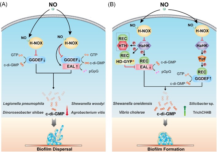
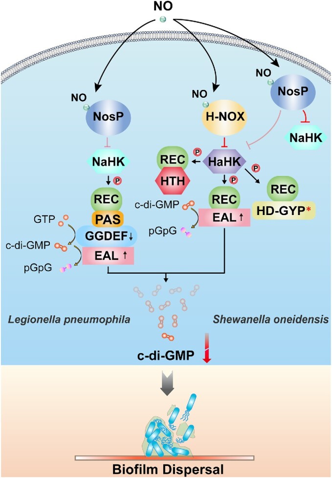
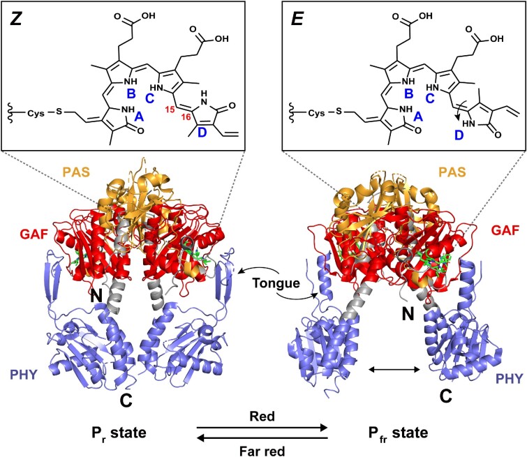
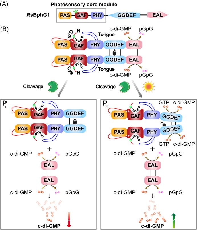
The widespread bacterial second messenger c-di-GMP is responsible for regulating many important physiological functions such as biofilm formation, motility, cell differentiation, and virulence. The synthesis and degradation of c-di-GMP in bacterial cells depend, respectively, on diguanylate cyclases and c-di-GMP-specific phosphodiesterases. Since c-di-GMP metabolic enzymes (CMEs) are often fused to sensory domains, their activities are likely controlled by environmental signals, thereby altering cellular c-di-GMP levels and regulating bacterial adaptive behaviors. Previous studies on c-di-GMP-mediated regulation mainly focused on downstream signaling pathways, including the identification of CMEs, cellular c-di-GMP receptors, and c-di-GMP-regulated processes. The mechanisms of CME regulation by upstream signaling modules received less attention, resulting in a limited understanding of the c-di-GMP regulatory networks. We review here the diversity of sensory domains related to bacterial CME regulation. We specifically discuss those domains that are capable of sensing gaseous or light signals and the mechanisms they use for regulating cellular c-di-GMP levels. It is hoped that this review would help refine the complete c-di-GMP regulatory networks and improve our understanding of bacterial behaviors in changing environments. In practical terms, this may eventually provide a way to control c-di-GMP-mediated bacterial biofilm formation and pathogenesis in general.

Keywords: NO sensors; c-di-GMP; c-di-GMP-specific phosphodiesterase; diguanylate cyclase; photoreceptors; sensory domains.


Conflict of interest statement

None declared.

Figures

Figure 1.
Figure 1.
Crystal structures of two types of heme-based O2 sensors. (A) Crystal structure of the heme-containing PAS domain from Escherichia coli EcDosP (PDB entry: 1V9Z) (Kurokawa et al. 2004). Its five β-strands (Aβ, Bβ, Gβ, Hβ, and Iβ) and four flanking α-helices (Cα, Dα, Eα, and Fα) are labeled as indicated. (B) Crystal structure of the heme-containing GCS domain from Escherichia coli EcDosC (PDB entry: 4ZVB) (Tarnawski et al. 2015). Each monomer contains eight α-helices, which are named Zα, Aα, Bα, Cα, Eα, Fα, Gα, and Hα according to the classical globin nomenclature. The heme ligand in each domain is indicated by arrows.
Figure 2.
Figure 2.
Redox-induced changes at the distal heme site of EcDosP. EcDosP is O2-dependent and its activity is regulated by the transition between the ferric form (PDB entry:1V9Y) (Kurokawa et al. 2004) and ferrous form (PDB entry:1V9Z) (Kurokawa et al. 2004) of the heme-PAS domain, a process accompanied by the change of distal axial ligands. When the heme-PAS domain is in the ferric form, the distal axial ligand of its complex is a water molecule W1, stabilized by another water molecule W2 (marked by the magenta dashed box); when it is reduced to the ferrous form, the distal axial ligand of its complex is changed to Met95 (marked by the blue dashed box) of the FG-loop (shown in yellow). Also indicated is the iron-binding His77.
Figure 3.
Figure 3.
O2 sensors EcDosC and EcDosP mediate c-di-GMP-dependent RNA processing in Escherichia coli. (A) Domain organization of EcDosC and EcDosP. EcDosP contains a degenerate GGDEF domain with the EGTQF motif at its active site. (B) Possible scheme for O2-dependent RNA degradation in the EcDosCP complex (based on Tuckerman et al. 2011). Under anaerobic conditions (upper panel), EcDosP and EcDosC are unliganded (deoxygenated), and the DGC activity of EcDosC is activated (marked with an asterisk), producing c-di-GMP to activate the receptor PNPase in the RNA degradation complex. Under aerobic conditions (lower panel), EcDosP and EcDosC are liganded (oxygenated) and the PDE activity of EcDosP is induced (marked with an asterisk). EcDosP hydrolyzes c-di-GMP to pGpG, which drastically decreases PNPase activity. mRNAs that depend on O2 for preservation and degradation may be selected by a mechanism involving sRNAs and Hfq, where sRNAs serve as mediators to recognize target mRNAs, and the RNA chaperone Hfq catalyzes this hybridization.
Figure 4.
Figure 4.
Crystal structures of two classes of heme-based NO sensors. (A) Crystal structure of the H-NOX domain from Caldanaerobacter subterraneus (PDB entry: 5JRU) (Hespen et al. 2016). The H-NOX fold consists of seven α-helices (Aα–Fα, and Kα) and a four-stranded antiparallel β-sheet (Gβ, Hβ, Iβ, and Jβ). Located on the α-helix Fα, His102 is the proximal axial ligand for heme iron and is highly conserved across all H-NOX domains. Tyr131, Ser133, and Arg135 are strictly conserved residues in the YXSXR motif. The heme ligand is also indicated by arrows. (B) Predicted structure of Pseudomonas aeruginosa NosP (PA1975), obtained from the AlphaFold website (https://alphafold.ebi.ac.uk/entry/Q9I2D0). This model contains 10 α-helices and 21 β-sheets.
Figure 5.
Figure 5.
Mechanisms of NO-induced biofilm dispersal or formation via H-NOX domain. CMEs in bacteria function normally in the absence of NO. However, when bacteria are exposed to a certain concentration of NO, it would affect the activities of some CMEs, changing the cellular c-di-GMP concentrations and affecting the formation of bacterial biofilm. (A) NO may directly affect the protein–protein interaction between H-NOX proteins and HaCMEs, thereby altering their catalytic activities. When such HaCME has only a separate GGDEF domain (in some bacteria, it may have an additional degenerated EAL domain), the binding of NO to H-NOX inhibits the DGC activity of such HaCME. When the HaCME contains both GGDEF and EAL domains, binding of NO to H-NOX will maintain or even down-regulate the DGC activity of this HaCME, or activate its PDE activity. These signal events would reduce the cellular c-di-GMP concentrations in bacteria, ultimately leading to biofilm dispersal. (B) NO may also affect the interaction of H-NOX protein with HaHK, thereby affecting the transfer of the phosphoryl group to indirectly regulate the activity of response regulator HaCME. Such HaCME proteins usually fuse the phospho-signaling receptor REC domain and the GGDEF/EAL domain. Binding of NO to H-NOX protein inhibits the autophosphorylation of HaHK, hindering the downstream transmission of the phosphoryl group, and changes the phosphorylation state of HaCME, thereby inhibiting the PDE activity or activating the DGC, resulting in an elevated c-di-GMP level, which ultimately promotes the formation of bacterial biofilms. * indicates domain degeneration and a lack of catalytic activity. The arrows on the c-di-GMP metabolic domains represent an increase or decrease in activities of the corresponding enzymes. The protein shown in the dashed box in Fig. 5(B) is HnoC, which may not be present in the signaling networks of some bacteria.
Figure 6.
Figure 6.
Mechanisms of NO-induced biofilm dispersal via the NosP domain. Possible NosP signaling pathway in Legionella pneumophila (left, based on Fischer et al. 2019). Binding of NO to LpNosP weakens the interaction between LpNosP and LpNahK and diminishes the inhibitory effect of LpNosP on the autophosphorylation of LpNaHK. LpNaHK can thus transfer the phosphoryl group to the downstream bifunctional LpNaCME, which exhibits reduced DGC activity and increased PDE activity, causing a decrease in the cellular c-di-GMP concentrations, and ultimately leading to biofilm dispersal. SoNosP is a master regulator of the multicomponent No/c-di-GMP signaling network in Shewanella oneidensis (right, based on Nisbett et al. 2019). When the bacteria are not exposed to NO, iron-free SoNosP strongly inhibits the autophosphorylation activity of SoNaHK and SoHaHK, thereby preventing downstream components of the phosphate transport chain from being phosphorylated. However, when NO is present, SoNosP attenuates the inhibitory effect on SoHaHK, enabling the transfer of the phosphoryl group to SoHaCME and enhancing the PDE activity of SoHaCME to induce biofilm dispersion. * indicates that the domain is degraded and lacks catalytic activity. The arrows on the c-di-GMP metabolic domains represent an increase or decrease in activities of the corresponding enzymes.
Figure 7.
Figure 7.
Light-driven changes in the structure and spectral properties of Deinococcus radiodurans Bph. The biliverdin chromophore contains four pyrrole rings, named A, B, C, and D rings. Under the irradiation by red and far-red light, biliverdin undergoes reversible Z/E isomerization around the C15/C16 double bond in the methine bridge between the C ring and the D ring, resulting in the rotation of the D ring and causing atomic rearrangements in the chromophore-binding pocket, which in turn leads to repositioning of the PHY domain and refolding of the tongue-like structure therein. In the Pr state, the tongue-like region appears as a β-sheet (PDB entry:4O0P) (Takala et al. 2014), while in the Pfr state, it transforms into an α-helix (PDB entry:4O01) (Kurokawa et al. 2004).
Figure 8.
Figure 8.
The photoreceptor protein RsBphG1 from Rhodobacter sphaeroides is involved in regulating c-di-GMP levels in bacteria. (A) Schematic representation of the domain composition of RsBphG1 that includes the classic PAS-GAF-PHY photosensory module and the GGDEF–EAL functional modules. Green stars represent the biliverdin chromophore. (B) An “EAL lock” model can be applied to explain how RsBphG1 modulates its catalytic activity in response to light signals (based on Tarutina et al. 2006). The EAL domain and the linker between GGDEF and EAL of RsBphG1 tend to homodimerize, although dimerization of the EAL domains is not required for PDE activity. Protein–protein interaction between the EAL domains can form an “EAL lock” to restrict the mobility of the upstream GGDEF domains. In this process, homodimer formation of GGDEF domains is necessary to receive signals from the sensory domain to undergo a conformational change and complete the transition from a nonproductive state with low or no enzymatic activity to a productive state with high activity. When “EAL lock” is present, the GGDEF domain cannot obtain enough mobility to change conformation, so DGC activity is inhibited; in other words, RsBphG1 gets locked in PDE mode. However, bacteria may use a cleavage mechanism to unlock this “EAL lock,” eventually splitting full-length RsBphG1 into two proteins. The smaller species was identified as the EAL domain and the linker with PDE activity, while the larger species was the rest of RsBphG1. When the unlocked RsBphG1 is not activated by light, its GGDEF domain is in a nonproductive state; when activated by light, the conformation of its GGDEF domain will shift to a productive mode, increasing the cellular c-di-GMP concentrations. RsBphG1 usually exists as a tetramer or a higher-order oligomer, but was drawn as a dimer in this figure for ease of presentation.
Figure 9.
Figure 9.
c-di-GMP is involved in regulating Synechocystis motility. (A) Motility behaviors of Synechocystis sp. Its phototaxis is spectrally dependent, and the phototaxis behaviors have a transition point at a wavelength of about 470 nm. When the wavelength is higher than 470 nm, the cells show positive phototaxis, while when the wavelength is lower than 470 nm, the cells either do not move (e.g. under blue light) or exhibit negative phototaxis (e.g. from ultraviolet light) (Fiedler et al. , Chau et al. 2017). This process may involve c-di-GMP, since artificial degradation of c-di-GMP in Synechocystis activates bacterial motility, while synthetic c-di-GMP inhibits phototaxis (Savakis et al. , Wallner et al. 2020). (B) Schematic illustration of the domain composition of SyCph2 and its interaction partners that regulate phototaxis in Synechocystis. The PCB chromophore (purple star) and tetrapyrrole chromophore (cyan star) are covalently bound to the GAF1 and GAF3 domains of SyCph2, respectively, conferring SyCph2 light-sensing capabilities. (C) SyCph2-dependent model of Synechocystis blue light avoidance behaviors (based on Wallner et al. 2020). SyCph2, SySlr1143, and SySlr1142 may be able to form a protein complex that can flexibly adjust cellular c-di-GMP concentrations in response to different light signals. Under blue light irradiation, the C-terminal GGEDF domain of SyCph2 is activated to synthesize c-di-GMP. Elevated levels of c-di-GMP affect the expression of minor pilin operon (pilA5-pilA6 and pilA9-slr2019) and chemotaxis gene operon (tax2), in particular, up-regulated expression of pilA9-slr2019 significantly induces flocculation of Synechocystis, and this operon has also been shown to play an important role in the phototaxis motility of Synechocystis. Besides, the c-di-GMP receptor CdgR has been recently identified in cyanobacteria and demonstrated to control the cell size (Zeng et al. 2023). The possible existence of yet unknown c-di-GMP receptors that could regulate cyanobacterial motility remains to be investigated. Furthermore, this c-di-GMP-dependent signaling network also appears to crosstalk with the cAMP signal transduction system and the Hfq regulatory system due to the involvement of two CRP-like transcription factors, SyCRP1 (a cAMP receptor protein) and SyCRP2 (a potential c-di-GMP-dependent transcription factor lacking the key amino acids for binding cAMP) (Fu et al. 2021). The dotted lines represent mechanisms that have yet to be studied.
Figure 10.
Figure 10.
Structures of blue light photoreceptors in bacteria. (A) Crystal structure of the LOV domain of Bacillus subtilis YvtA (PDB entry: 2PR5) (Möglich and Moffat 2007). The YvtA core domain consists of a five-stranded antiparallel β-sheet (Aβ, Bβ, Gβ, Hβ, and Iβ) and four α-helices (Cα, Dα, Eα, and Fα), with a short linker helix at the C-terminus of the core domain. Critical Cys62 are highlighted and drawn in ball-and-stick. (B) Crystal structure of the BLUF domain of Rhodobacter sphaeroides AppA with FMN replacing the FAD cofactor of the native AppA (PDB entry: 1YRX) (Anderson et al. 2005). It contains two α-helices (Bα and Eα) and a five-stranded mixed β-sheet (Aβ, Cβ, Dβ, Gβ, and Fβ). (C) Crystal structure of Ectothiorhodospira halophila PYP (PDB entry: 2PHY) (Borgstahl et al. 1995). PYP consists of a central six-stranded β-sheet (Cβ, Dβ, Gβ, Jβ, Kβ, and Lβ) and six α-helices (Aα, Bα, Eα, Fα, Hα, and Iα). Key Cys69 covalently binding the chromophore are highlighted and drawn in ball-and-stick. (D) Protein fold of the Synechocystis sp. PCC6803 cryptochrome DASH (PDB entry: 1NP7) (Brudler et al. 2003). It contains several 310 helices (magenta) in addition to α-helices (red) and β-sheets (cyan). The chromophores in each domain are also indicated by arrows.

Similar articles

Cited by

References

    1. Abreu IA, Lourenço AI, Xavier AV et al. A novel iron centre in the split-soret cytochrome c from Desulfovibrio desulfuricans ATCC 27774. J Biol Inorg Chem. 2003;8:360–70. - PubMed
    1. Agari Y, Kashihara A, Yokoyama S et al. Global gene expression mediated by Thermus thermophilus SdrP, a CRP/FNR family transcriptional regulator. Mol Microbiol. 2008;70:60–75. - PubMed
    1. Ali-Ahmad A, Fadel F, Sebban-Kreuzer C et al. Structural and functional insights into the periplasmic detector domain of the GacS histidine kinase controlling biofilm formation in Pseudomonas aeruginosa. Sci Rep. 2017;7:11262. - PMC - PubMed
    1. Almblad H, Randall TE, Liu F et al. Bacterial cyclic diguanylate signaling networks sense temperature. Nat Commun. 2021;12:1986. - PMC - PubMed
    1. Anderson S, Dragnea V, Masuda S et al. Structure of a novel photoreceptor, the BLUF domain of AppA from Rhodobacter sphaeroides. Biochemistry. 2005;44:7998–8005. - PMC - PubMed

Publication types