Nanomaterials and Coatings for Managing Antibiotic-Resistant Biofilms
- PMID: 36830221
- PMCID: PMC9952333
- DOI: 10.3390/antibiotics12020310
Nanomaterials and Coatings for Managing Antibiotic-Resistant Biofilms
Abstract
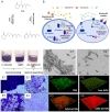
Biofilms are a global health concern responsible for 65 to 80% of the total number of acute and persistent nosocomial infections, which lead to prolonged hospitalization and a huge economic burden to the healthcare systems. Biofilms are organized assemblages of surface-bound cells, which are enclosed in a self-produced extracellular polymer matrix (EPM) of polysaccharides, nucleic acids, lipids, and proteins. The EPM holds the pathogens together and provides a functional environment, enabling adhesion to living and non-living surfaces, mechanical stability, next to enhanced tolerance to host immune responses and conventional antibiotics compared to free-floating cells. Furthermore, the close proximity of cells in biofilms facilitates the horizontal transfer of genes, which is responsible for the development of antibiotic resistance. Given the growing number and impact of resistant bacteria, there is an urgent need to design novel strategies in order to outsmart bacterial evolutionary mechanisms. Antibiotic-free approaches that attenuate virulence through interruption of quorum sensing, prevent adhesion via EPM degradation, or kill pathogens by novel mechanisms that are less likely to cause resistance have gained considerable attention in the war against biofilm infections. Thereby, nanoformulation offers significant advantages due to the enhanced antibacterial efficacy and better penetration into the biofilm compared to bulk therapeutics of the same composition. This review highlights the latest developments in the field of nanoformulated quorum-quenching actives, antiadhesives, and bactericides, and their use as colloid suspensions and coatings on medical devices to reduce the incidence of biofilm-related infections.
Keywords: antiadhesion; antimicrobial; biofilm inhibition; nanoparticles; quorum quenching; quorum sensing.
Conflict of interest statement
The authors declare no conflict of interest.
Figures


Similar articles
-
Bacterial extracellular vesicles: Modulation of biofilm and virulence properties.Acta Biomater. 2024 Apr 1;178:13-23. doi: 10.1016/j.actbio.2024.02.029. Epub 2024 Feb 27. Acta Biomater. 2024. PMID: 38417645 Review.
-
Biofilm in implant infections: its production and regulation.Int J Artif Organs. 2005 Nov;28(11):1062-8. doi: 10.1177/039139880502801103. Int J Artif Organs. 2005. PMID: 16353112 Review.
-
Novel approaches to control biofilm infections.Curr Med Chem. 2009;16(12):1512-30. doi: 10.2174/092986709787909640. Curr Med Chem. 2009. PMID: 19355904 Review.
-
Biofilms as Promoters of Bacterial Antibiotic Resistance and Tolerance.Antibiotics (Basel). 2020 Dec 23;10(1):3. doi: 10.3390/antibiotics10010003. Antibiotics (Basel). 2020. PMID: 33374551 Free PMC article. Review.
-
The clinical impact of bacterial biofilms.Int J Oral Sci. 2011 Apr;3(2):55-65. doi: 10.4248/IJOS11026. Int J Oral Sci. 2011. PMID: 21485309 Free PMC article. Review.
Cited by
-
Surfactants' Interplay with Biofilm Development in Staphylococcus and Candida.Pharmaceutics. 2024 May 15;16(5):657. doi: 10.3390/pharmaceutics16050657. Pharmaceutics. 2024. PMID: 38794319 Free PMC article.
-
Anti-Biofilm Strategies: A Focused Review on Innovative Approaches.Microorganisms. 2024 Mar 22;12(4):639. doi: 10.3390/microorganisms12040639. Microorganisms. 2024. PMID: 38674584 Free PMC article. Review.
-
Biofilm Prevention and Removal in Non-Target Pseudomonas Strain by Siphovirus-like Coliphage.Biomedicines. 2024 Oct 9;12(10):2291. doi: 10.3390/biomedicines12102291. Biomedicines. 2024. PMID: 39457603 Free PMC article.
-
Cytotoxicity and Antibiofilm Activity of Silver-Polypropylene Nanocomposites.Antibiotics (Basel). 2023 May 17;12(5):924. doi: 10.3390/antibiotics12050924. Antibiotics (Basel). 2023. PMID: 37237827 Free PMC article.
-
Light-Based Anti-Biofilm and Antibacterial Strategies.Pharmaceutics. 2023 Aug 9;15(8):2106. doi: 10.3390/pharmaceutics15082106. Pharmaceutics. 2023. PMID: 37631320 Free PMC article. Review.
References
Publication types
Grants and funding
LinkOut - more resources
Full Text Sources

