Clinical neurogenetics: autosomal dominant spinocerebellar ataxia
- PMID: 24176420
- PMCID: PMC3818725
- DOI: 10.1016/j.ncl.2013.04.006
Clinical neurogenetics: autosomal dominant spinocerebellar ataxia
Abstract
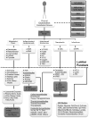
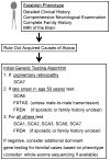
The autosomal dominant spinocerebellar ataxias are a diverse and clinically heterogeneous group of disorders characterized by degeneration and dysfunction of the cerebellum and its associated pathways. Clinical and diagnostic evaluation can be challenging because of phenotypic overlap among causes, and a stratified and systematic approach is essential. Recent advances include the identification of additional genes causing dominant genetic ataxia, a better understanding of cellular pathogenesis in several disorders, the generation of new disease models that may stimulate development of new therapies, and the use of new DNA sequencing technologies, including whole-exome sequencing, to improve diagnosis.
Keywords: Ataxia; Autosomal dominant; Cerebellum; SCA; Spinocerebellar.
Copyright © 2013 Elsevier Inc. All rights reserved.
Figures


Similar articles
-
[Clinical features and diagnosis of spinocerebellar ataxia].Ideggyogy Sz. 2004 Jan 20;57(1-2):11-22. Ideggyogy Sz. 2004. PMID: 15042864 Review. Hungarian.
-
Spinocerebellar ataxia: an update.J Neurol. 2019 Feb;266(2):533-544. doi: 10.1007/s00415-018-9076-4. Epub 2018 Oct 3. J Neurol. 2019. PMID: 30284037 Free PMC article. Review.
-
Spinocerebellar ataxia types 2 and 3 segregating simultaneously in a single family.Mov Disord. 2006 Jul;21(7):1051-3. doi: 10.1002/mds.20893. Mov Disord. 2006. PMID: 16628604
-
Spinocerebellar ataxias: genotype-phenotype correlations in 104 Brazilian families.Clinics (Sao Paulo). 2012;67(5):443-9. doi: 10.6061/clinics/2012(05)07. Clinics (Sao Paulo). 2012. PMID: 22666787 Free PMC article.
-
Spinocerebellar ataxia type 4 is not detected in a cohort from Hokkaido, the northernmost island of Japan.J Neurol Sci. 2024 May 15;460:122974. doi: 10.1016/j.jns.2024.122974. Epub 2024 Mar 20. J Neurol Sci. 2024. PMID: 38523039 No abstract available.
Cited by
-
Spinocerebellar Ataxia 12 Patients have better Quality of Life than Spinocerebellar Ataxia 1 and 2.Ann Indian Acad Neurol. 2022 Jul-Aug;25(4):647-653. doi: 10.4103/aian.aian_611_21. Epub 2022 Apr 6. Ann Indian Acad Neurol. 2022. PMID: 36211176 Free PMC article.
-
Developing a smartphone application, triaxial accelerometer-based, to quantify static and dynamic balance deficits in patients with cerebellar ataxias.J Neurol. 2020 Mar;267(3):625-639. doi: 10.1007/s00415-019-09570-z. Epub 2019 Nov 11. J Neurol. 2020. PMID: 31713101 Clinical Trial.
-
Microstructural Alterations in Asymptomatic and Symptomatic Patients with Spinocerebellar Ataxia Type 3: A Tract-Based Spatial Statistics Study.Front Neurol. 2017 Dec 22;8:714. doi: 10.3389/fneur.2017.00714. eCollection 2017. Front Neurol. 2017. PMID: 29312133 Free PMC article.
-
The impact of histone post-translational modifications in neurodegenerative diseases.Biochim Biophys Acta Mol Basis Dis. 2019 Aug 1;1865(8):1982-1991. doi: 10.1016/j.bbadis.2018.10.019. Epub 2018 Oct 20. Biochim Biophys Acta Mol Basis Dis. 2019. PMID: 30352259 Free PMC article. Review.
-
Abnormal Findings in Polysomnographic Recordings of Patients with Spinocerebellar Ataxia Type 2 (SCA2).Cerebellum. 2019 Apr;18(2):196-202. doi: 10.1007/s12311-018-0982-x. Cerebellum. 2019. PMID: 30264264
References
-
- Durr A. Autosomal dominant cerebellar ataxias: polyglutamine expansions and beyond. Lancet Neurol. 2010;9(9):885–94. - PubMed
-
- Klockgether T, et al. The natural history of degenerative ataxia: a retrospective study in 466 patients. Brain. 1998;121(Pt 4):589–600. - PubMed
-
- Fogel BL, Perlman S. An approach to the patient with late-onset cerebellar ataxia. Nat Clin Pract Neurol. 2006;2(11):629–35. quiz 1 p following 635. - PubMed
-
- Fogel BL, Perlman S. Cerebellar disorders: Balancing the approach to cerebellar ataxia. In: Gálvez-Jiménez N, Tuite PJ, editors. Uncommon Causes of Movement Disorders. Cambridge ; New York: Cambridge University Press; 2011. pp. 198–216.
Publication types
MeSH terms
Substances
Grants and funding
LinkOut - more resources
Full Text Sources
Other Literature Sources
Medical

