. 2012 Mar 4;44(4):381-9, S1-2.
doi: 10.1038/ng.1106.

Mutations in axonemal dynein assembly factor DNAAF3 cause primary ciliary dyskinesia

Affiliations

Mutations in axonemal dynein assembly factor DNAAF3 cause primary ciliary dyskinesia

Hannah M Mitchison et al. Nat Genet. .

Abstract

Primary ciliary dyskinesia most often arises from loss of the dynein motors that power ciliary beating. Here we show that DNAAF3 (also known as PF22), a previously uncharacterized protein, is essential for the preassembly of dyneins into complexes before their transport into cilia. We identified loss-of-function mutations in the human DNAAF3 gene in individuals from families with situs inversus and defects in the assembly of inner and outer dynein arms. Knockdown of dnaaf3 in zebrafish likewise disrupts dynein arm assembly and ciliary motility, causing primary ciliary dyskinesia phenotypes that include hydrocephalus and laterality malformations. Chlamydomonas reinhardtii PF22 is exclusively cytoplasmic, and a PF22-null mutant cannot assemble any outer and some inner dynein arms. Altered abundance of dynein subunits in mutant cytoplasm suggests that DNAAF3 (PF22) acts at a similar stage as other preassembly proteins, for example, DNAAF2 (also known as PF13 or KTU) and DNAAF1 (also known as ODA7 or LRRC50), in the dynein preassembly pathway. These results support the existence of a conserved, multistep pathway for the cytoplasmic formation of assembly competent ciliary dynein complexes.


Figures
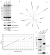
Figure 1
Figure 1. The Chlamydomonas PF22 locus encodes a conserved cytoplasmic protein important for axonemal dynein assembly
(a) Demembranated flagellar axonemes from wild type, pf22, and the pf22 strain transformed with untagged (R22) or cMyc-tagged (Myc22) wild type PF22 genes probed for the presence of assembled dynein subunits. Upper panel, Coomassie-stained gel of total axonemal proteins, showing an overall reduction of high molecular weight dynein heavy chain bands in pf22 axonemes (arrowhead). Lower panels, Western blots (WB) probed for ODA subunits (IC2) and subunits of three IDAs, showing ODAs and IDA “b” and “c” missing from pf22 axonemes, whereas IDA “f” is retained at normal levels. Assembly of all three missing dyneins is rescued by transformation with untagged or Myc-tagged gene copies. (b) Dendrogram of sequence relationships among PF22 eukaryotic orthologs shows the presence of a single orthologous sequence in each genome. (c) Dot matrix representation of sequence similarity in a pair-wise comparison of human and Chlamydomonas PF22 protein sequences. Similarity extends throughout both sequences except for two insertions specific to the algal protein. (d) Blots of cell fractions from Chlamydomonas transformed with Myc-tagged PF22 probed using anti-Myc antibody to show the relative abundance of PF22 in cytoplasmic and flagellar fractions. Upper panel: extracts from untagged (WT) and Myc-tagged (Myc22) strains show a single 60 kDa band in the transformed strain, as well as several non-specific bands. Flagellar axoneme protein loaded at a 1:1 or 50:1 stoichiometric ratio to the extract lanes does not have any detectable 60 kDa band. Lower panel: identical samples probed with anti-IC2 as a control to show the relative abundance of axonemal dynein subunits in the cytoplasmic and flagellar fractions. Numbers are size markers (kDa). Protein sequences used in the alignments (b, c) are AEC04845 (Chlamydomonas reinhardtii); XP_003054829 (Micromonas pusilla, microalga); XP_814338 (Trypanosoma cruzi, trypanosome); CAK83719 (Paramecium tetraurelia, ciliate); ACI64850 (Thalassiosira pseudonana, diatom); EGF76589 (Batrachochytrium dendrobatidis, chytrid fungus); EGD81026 (Salpingoeca sp., choanozoan); NP_849159 (Homo sapiens, human); XP_002130641 (Ciona intestinalis, sea squirt); AAI08526 (Xenopus laevis, frog); XP_785142 (Strongylocentrotus purpuratus, sea urchin).
Figure 2
Figure 2. Identification of DNAAF3 mutations in PCD patients with dynein assembly defects
(a) Pedigrees of three families found to carry DNAAF3 mutations are shown. The individuals in generations I and II in each family are inferred to indicate consanguineous unions. Filled symbols indicate individuals with PCD, those with situs inversus denoted by an asterisk, and a double horizontal line represents a consanguineous marriage. UCL89 has two affected monozygous twins. (b) Sequence chromatograms for control individuals (top panels) and representative affected individuals from the families (bottom panels), show mutations in the DNA (arrows) and consequences for the protein, with the location of each mutation shown within the gene (bottom). (c) An extract of a multiple species alignment of DNAAF3/PF22 proteins is shown with the position of the highly conserved p.Leu108 residue indicated by a red arrow. Protein sequences used for the multispecies alignment are NP_849159 (Homo sapiens); NP_001028720 (Mus musculus); XP_541416 (Canis familiaris, dog); XP_001921018 (Danio rerio, zebrafish); XP_002938320 (Xenopus tropicalis); CAK83719 (Paramecium tetrauralia) and XP_001025714 (Tetrahymena thermophila). The Chlamydomonas reinhardtii protein sequence is newly reported here.
Figure 3
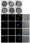
Figure 3. Absence of outer dynein arms in respiratory epithelial cell cilia of PCD patients carrying DNAAF3 mutations
(a) Transmission electron microscopy of representative cilia cross sections in family UCL89 showing loss of both outer and inner dynein arms in affected individuals. Top row, cross sections of cilia from a control and two unaffected individuals from UCL89: III.1 (unaffected father) and IV.6 (unaffected sibling). Bottom row, cross sections of cilia from three affected siblings: IV.2, IV.3 and IV.5. The outer and inner dynein arms are indicated by red arrows. Scale bar, 0.2 um. (b) Cells double-labeled for anti-alpha/beta-tubulin to label the cilia axoneme (red) and DNAH5 (green) show that both proteins colocalize along the entire cilia in cells from the unaffected control, but DNAH5 does not appear in the cilia of any of the three affected individuals IV.2, IV.3 or IV.5, showing that DNAAF3 is necessary for outer row dynein assembly. Reduced amounts of DNAH5 label appear in the apical cytoplasm in some DNAAF3 patient cells (IV.5), suggesting that some outer row dynein proteins are present in a form that cannot assemble into cilia. Nuclei are stained with Hoechst 33342. Scale bar, 5 um.
Figure 4
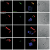
Figure 4. Absence of inner row dynein subunit DNALI1 in respiratory epithelial cell cilia of PCD patients carrying DNAAF3 mutations
Cells double-labeled for anti-alpha/beta tubulin to label the cilia axoneme (red) and DNALI1 (green) show that both proteins colocalize along the axoneme of cilia in cells from an unaffected control, but DNALI1 is absent from the cilia of the three affected individuals IV.2, IV.3 and IV.5, demonstrating the disruption of inner dynein arm assembly in airway cilia of these patients. Nuclei are stained with Hoechst 33342. Scale bar, 5 um.
Figure 5
Figure 5. Morpholino knockdown of dnaaf3 in zebrafish embryos results in axis curvature defects, kidney cysts, hydrocephalus, perturbed otolith development and laterality defects
(a-e) Representative images of phenotypes of dnaaf3 MO-injected zebrafish embryos at 72 hours post fertilization (hpf). Morphant fish display a curly-tail phenotype compared to an unaffected sibling (a, b), develop pronephric cysts (a-d, black arrows) and hydrocephalus (c-e, white asterisks). (e) An example of a dnaaf3 MO-injected zebrafish embryo displaying hydrocephalus (white asterisk) and an abnormal number (three) of inner-ear otoliths (white arrow). (f) Graph showing the percentage of embryos injected with dnaaf3 MO ex3 or dnaaf3 MO ex8 displaying the defects shown in a-e, compared to wildtype (302-315 embryos per group). Axis curvature was scored visually as normal (0-5% curl of the tail), mild with a slight curved or bent tail (5-50% curling of the tail) or severe (over 50% curling of the tail, sometimes curled over itself). The occurrence of pronephric cysts (KID), hydrocephalus (HYD) and abnormal otolith numbers (OTO) is also shown. The dnaaf3 morphant embryos had left-right axis determination defects visualized via whole-mount in situ hybridization for cmlc2 at 48 hpf (g, upper panel). (g, lower panel) Graph of the proportion of these phenotypes, comparing 48 hpf cmlc2 in situ results (N=144) with visual scoring at 72 hpf of heart looping (N=315). SS, situs solitus; SI, situs inversus; ML, bilateral expression with the heart positioned along the midline; Unclear, heart position undeterminable. (h, i) Transmission electron micrographs of olfactory placode cilia cross-sections show reduction and loss of dynein arms (red arrows) in morphants compared to wildtype siblings. Scale bars = 500 μm (a, b), 200 μm (c-e, g), or 100 nm (h, i).
Figure 6
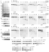
Figure 6. Altered ODA subunit abundance in the Chlamydomonas pf22 mutant cytoplasm
(a) Cytoplasmic abundance of ODA subunits that fail to assemble in pf22 flagella. Upper panel, stained gel shows equal loading of cytoplasmic extracts from wild type and assembly mutant strains. Lower panels, blots probed with antibodies to four ODA subunits. IC2 has an abnormally increased abundance in all three mutant strains. Dynein heavy chains appear at near-normal levels in pf22, but HCα is reduced in oda7, and both HCβ and HCγ are reduced in oda7 and in pf13. (b) Comparison of HCα and IC2 cytoplasmic abundance in single mutants pf22 and oda7 and in a double mutant pf22oda7 strain. HCα abundance is intermediate in the double mutant. CB, portion of Coomassie blue stained gel showing equal protein loads. (c-e) Proteolytic sensitivity of outer dynein arm heavy chains in the cytoplasm of Chlamydomonas assembly mutants. Extracts treated for 5 min with the indicated concentrations of trypsin were blotted for HCα (c), HCβ (d) or HCγ (e). Extracts from oda9, which does not alter heavy chain abundance, were used as controls. (c) Altered sensitivity of HCα is only seen in the oda7 cytoplasm, as evidenced by rapid loss of a high molecular weight band (arrowhead) and the appearance of a new band (arrow). One non-specific band appears only on the oda7 blot due to the 10-fold longer exposure time required to visualize HCα in this strain. (d-e) HCβ and HCγ show altered proteolytic patterns in both oda7 and pf13 cytoplasm, but not in pf22 cytoplasm. Arrows indicate bands that only appear in strains with increased heavy chain protease sensitivity. (f) Cytoplasmic heavy chain turnover tested by treating cells with cycloheximide (CHX) for the indicated number of hours. Whole cell samples were probed for outer dynein arm heavy chains HCα and HCβ. Control strains oda1 and oda16 have normal cytoplasmic assembly of dynein complexes (see text for details). The reductions in HCβ abundance in pf22 and pf13 (a,b) correlate with reduced protein half-lives (c-e). The half life of HCα is also shorter in the pf13 cytoplasm, but unaffected in pf22. The greatly reduced abundance of HCα in oda7 (a,b) correlates with a half-life reduced to less than 3 hr (f). The size of standards (kDa) is shown next to the stained gel in (a) and to the right of each oda7 panel in (c-e).
Figure 7
Figure 7. The Chlamydomonas pf22 mutant fails to correctly assemble outer dynein arms in the cytoplasm
Immunoprecipitation was used to test for formation of dynein complexes in the pf22 cytoplasm. (a) Immunoprecipitates produced with anti-HCβ monoclonal antibody were probed with antibodies to three outer row dynein subunits. All three co-precipitate from wild type cytoplasm, but the antibody failed to bind its antigen in the pf22 extract. (b) Immunoprecipitates produced with anti-HA epitope monoclonal antibody from extracts of cells expressing HA-IC2 were probed with anti-HA and chain-specific antibodies. All three heavy chains co-precipitate with IC2 from wild type extract, and reduced amounts co-precipitate from pf22 extract.
Figure 8
Figure 8. Hypothesized pathway of cytoplasmic assembly of axonemal outer row dynein
The three heavy chains present in Chlamydomonas outer dynein arms appear to require the action of ODA7 and PF13 for proper folding of their head domains, and all three assembly factors for their stability. These assembly factors may also be important for later steps in which heavy chains associate with intermediate chains. We hypothesize that these assembly factors work with a chaperone complex (labeled with “?”). In the absence of PF22, an epitope on the tail of HCβ is inaccessible; therefore PF22 may be required for dissociation of the chaperone complex at the completion of assembly. Order of addition of the three heavy chains (only two of which have orthologs in vertebrates) is speculative, but consistent with assembly defects observed previously in the absence of individual heavy chains.

Similar articles

Cited by

References

    1. Coren ME, Meeks M, Morrison I, Buchdahl RM, Bush A. Primary ciliary dyskinesia: age at diagnosis and symptom history. Acta Paediatr. 2002;91:667–669. - PubMed
    1. Bush A, Hogg C, Mitchison HM, Nisbet M, Wilson R. Update in primary ciliary dyskinesia. Clin. Pulm. Med. 2009;16:219–225.
    1. Bush A. Congenital heart disease in primary ciliary dyskinesia. Pediatr. Cardiol. 1998;19:191. - PubMed
    1. Kennedy MP, et al. Congenital heart disease and other heterotaxic defects in a large cohort of patients with primary ciliary dyskinesia. Circulation. 2007;115:2814–2821. - PubMed
    1. Tan SY, et al. Heterotaxy and complex structural heart defects in a mutant mouse model of primary ciliary dyskinesia. J Clin. Invest. 2007;117:3742–3752. - PMC - PubMed

Publication types

MeSH terms

Associated data