. 2010 Apr 30;285(18):13990-4004.
doi: 10.1074/jbc.M109.061309. Epub 2010 Feb 16.

AMSH interacts with ESCRT-0 to regulate the stability and trafficking of CXCR4

Affiliations

AMSH interacts with ESCRT-0 to regulate the stability and trafficking of CXCR4

Maria I Sierra et al. J Biol Chem. .

Abstract

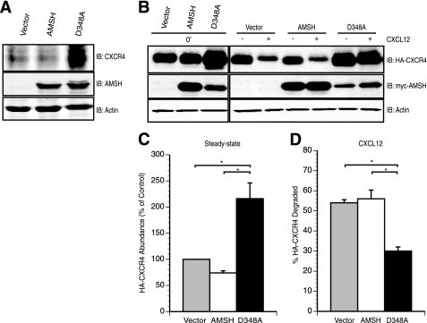
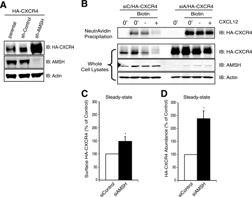
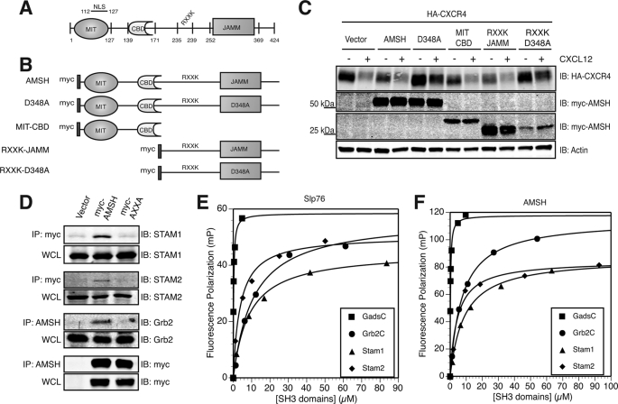
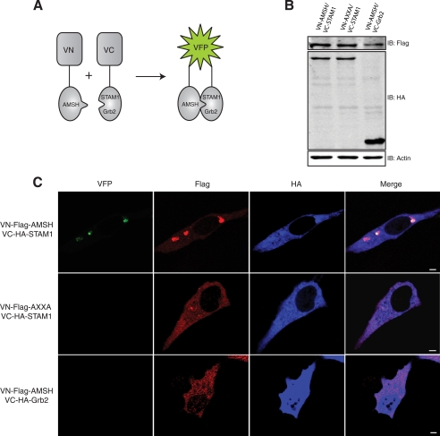
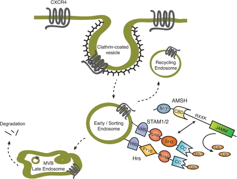
Reversible ubiquitination is essential for the endocytic sorting and down-regulation of G protein-coupled receptors, such as the chemokine receptor CXCR4. The deubiquitinating enzyme AMSH has been implicated in the endocytic sorting of both G protein-coupled receptors and receptor-tyrosine kinases. Herein, we examine the role of AMSH in the regulation of CXCR4 stability and trafficking and characterize protein-protein interactions critical for this function. Loss of AMSH catalytic activity or depletion by RNAi results in increased steady-state levels of CXCR4 under basal conditions. Analysis of truncation and point mutation of AMSH reveal the importance of an RXXK motif for CXCR4 degradation. The RXXK motif of AMSH interacts with the SH3 domains of the STAM and Grb2 families of adaptor proteins with high affinity. Cells expressing a catalytically inactive mutant of AMSH show basal hyperubiquitination, but not increased degradation, of the ESCRT-0 components STAM1 and Hrs. This is dependent on the RXXK motif of AMSH. Ubiquitination of endocytic machinery modulates their activity, suggesting that AMSH may directly regulate endocytic adaptor protein function. This is reflected in CXCR4 trafficking and provides a mechanism by which AMSH specifies the fate of endocytosed receptors. Taken together, these studies implicate AMSH as a key modulator of receptor fate determination through its action on components of the endocytic machinery.


Figures

FIGURE 1.
FIGURE 1.
AMSH activity modulates CXCR4 turnover. A, overexpression of a catalytically inactive AMSH led to accumulation of endogenous CXCR4 under steady-state conditions. Whole cell lysates derived from HeLa cells transiently expressing pcDNA3.1 vector (vector), wild type AMSH, or D348A, a catalytic point mutant of AMSH, were immunoblotted (IB) against endogenous CXCR4, AMSH, and β-actin proteins as indicated. B–D, AMSH regulates both basal turnover and ligand-mediated degradation of CXCR4. B, HEK293 cells transiently co-transfected with HA-tagged CXCR4 (HA-CXCR4) and either vector, wild type AMSH, or D348A were treated with vehicle alone (0′) or in the presence of cycloheximide with (+) or without (−) CXCL12 for 3 h. C, shown is a graphical representation of HA-CXCR4 protein levels relative to the control cells. D, percent HA-CXCR4 degraded after a 3-h stimulation with CXCL12 relative to treatment with cycloheximide alone. Quantification of HA-CXCR4 levels by immunoblot was performed using LiCOR Odyssey infrared imager and software. Significance was assessed using Student's t test with the asterisks corresponding to p < 0.05, n = 3. All graphs were generated using Delta graph 5.7.5 software.
FIGURE 2.
FIGURE 2.
AMSH depletion results in increased abundance and intracellular accumulation of CXCR4. A, stable ablation of AMSH led to HA-CXCR4 accumulation under steady-state conditions. Whole cell lysates derived from HeLa cells stably expressing HA-CXCR4 and a lentiviral shRNA targeting either AMSH (sh-AMSH) or a non-targeting control shRNA (sh-Control) were immunoblotted (IB) against HA-CXCR4, AMSH, and β-actin proteins as indicated. B, AMSH regulated CXCR4 trafficking to the cell surface. Surface biotinylation of HeLa cells transiently express HA-CXCR4 and oligo-induced RNAi against AMSH (siAMSH/siA) or non-targeting control (siControl/siC). Cells were treated with vehicle alone (0′) or in the presence of cycloheximide with (+) or without (−) CXCL12 for 3 h. Biotinylated material was precipitated with NeutrAvidin-agarose resin, and HA-CXCR4 was followed by an immunoblot. Graphical representations of surface (C) and total cellular HA-CXCR4 abundance (D) are shown; asterisks correspond to p < 0.05, n = 3.
FIGURE 3.
FIGURE 3.
The RXXK motif of AMSH interacts with a subset of SH3 domains and modulates CXCR4 stability. A, a schematic of domain organization of murine AMSH (GenBankTM accession no. BC025111). Amino acid boundaries of individual domains and motifs are as indicated; RXXK, conserved RXXK peptide sequence which interacts with SH3 domain. NLS, nuclear localization signal. B, shown are constructs of AMSH and mutants used in this study; D348A, catalytic domain mutant, MIT-CBD (amino acids 1- 187), RXXK-JAMM (amino acids 201–424), RXXK-D348A (amino acids 201–424). C, the RXXK motif of AMSH is required for AMSH-mediated regulation of CXCR4 stability. HEK293 cells co-transfected with HA-tagged CXCR4 and vector, wild type AMSH, D348A, MIT-CBD, RXXK-JAMM, or RXXK-D348A were treated with cycloheximide and vehicle (−) or CXCL12 (+) for 3 h. IB, immunoblot. D, the RXXK motif is essential for AMSH interaction with the adaptor proteins STAM1, STAM2, and Grb2. HeLa cells transiently expressing vector, wild type AMSH, or AXXA mutant were immunoprecipitated (IP) from Myc or AMSH (as indicated) to determine whether RXXK mutation to AXXA would alter the ability of AMSH to interact with endogenous binding partners. Corresponding input whole cell lysates (WCL) are shown. E and F, the RXXK motif of AMSH binds to Gads, Grb2, STAM1, and STAM2 SH3 domains with high affinity. Dissociation constants for the interactions between fluorescein-tagged peptides Slp76 (fluorescein-tagged APSIDRSTKPA) bound with affinities as shown to Gads-C (210 nm), STAM1 (8.6 μm), STAM2 (3.8 μm), Grb2-C (11 μm) (E), and AMSH (fluorescein-tagged PPVVDRSLKPG) bound with affinities as shown to Gads-C (240 nm), STAM1 (11 μm), STAM2 (4.8 μm), and Grb2-C (7 μm) (F). GST-SH3 domains were measured using fluorescence polarization (FP). Measurements (millipolarization units (mP)) were taken using Beacon 2000, and curve fitting was performed using DeltaGraph 5.7.5.
FIGURE 4.
FIGURE 4.
Subcellular localization of AMSH with STAM1 requires the RXXK/SH3 interaction. A, shown is a schematic representation of the BiFC assay. VN and VC represent the N-terminal and C-terminal fragments of the VFP, respectively, and are not singularly fluorescent. When the two fragments colocalize (green), fluorescence is observed. B and C, AMSH interacts directly with STAM1 via the RXXK motif but not with Grb2. B, HeLa cells transiently co-transfected with VN-AMSH/VC-STAM1 or VN-AXXA/VC-STAM1 or VN-AMSH/VC-Grb2 cells were lysed, and protein levels were detected by immunoblot (IB). C, cells were fixed and immunostained for FLAG (red) and HA (blue) to detect expression of VN-AMSH or VC-STAM1 and of VC-Grb2, respectively. Representative images are shown with scale bars corresponding to 5 μm.
FIGURE 5.
FIGURE 5.
The AMSH·STAM1 interaction occurs on sorting endosomes. Direct interactions between AMSH and STAM1 occur at the Hrs-positive/sorting endosome and not at the early (EEA1-positive) or late (CD63-positive) endosome. A, HeLa cells co-transfected with VN-AMSH and VC-STAM1 were fixed and immunostained for Hrs, EEA1, or CD63 (red). Scale bars correspond to 5 μm. B, shown is a graphic representation of colocalization between VFP and endosomal proteins, where 0 indicates no overlap, and 1 indicates complete overlap; ***, p < 0.001 n = 3.
FIGURE 6.
FIGURE 6.
AMSH regulates ESCRT-0 ubiquitination in a manner dependent upon the RXXK motif. The RXXK motif of AMSH is required in the spatial organization of AMSH to endosomes to deubiquitinate target protein cargo. A, HeLa cells co-transfected with HA-ubiquitin and vector, AMSH, D348A, or AXXA-D348A were fixed and immunostained with anti-STAM1 (red) and anti-HA (green). Representative images are shown with scale bars corresponding to 5 μm. B, quantification of colocalization between HA-ubiquitin and STAM1 from experiments in A; ***, p < 0.001, n = 2. C and D, the RXXK motif of AMSH is required for regulation of ubiquitin status of ESCRT-0 components. HeLa cells co-transfected with HA-ubiquitin and vector, AMSH, D348A, or AXXA-D348A were immunoprecipitated (IP) for endogenous proteins. STAM1 (C) and Hrs (D) and their ubiquitination status was assessed by immunoblot (IB). E, AMSH does not affect the stability of Hrs or STAM1. Corresponding whole cell lysates were analyzed by immunoblot against endogenous Hrs, STAM1, AMSH, and β-actin proteins.
FIGURE 7.
FIGURE 7.
AMSH regulates ubiquitin dynamics and trafficking of CXCR4 on STAM-positive endosomes. AMSH ablation results in accumulation of ubiquitinated cargo and CXCR4 on endosomes marked by endogenous STAM1 protein. HeLa cells transfected with HA-ubiquitin (A) or HA-CXCR4 (C) in combination with either siControl or siAMSH were fixed and immunostained for HA (green) and STAM1 (red). Representative images are shown with scale bars corresponding to 5 μm. Graphic representations of colocalization between STAM1 and HA-ubiquitin (B) or HA-CXCR4 (D)are shown; *, p < 0.05, ***, p < 0.001, n = 2. E, whole cell lysates corresponding to C were immunoblotted (IB) against HA-CXCR4, AMSH, and β-actin as indicated.
FIGURE 8.
FIGURE 8.
AMSH recruitment via the RXXK motif mediates steady-state CXCR4 trafficking. AMSH mediated trafficking of CXCR4 through Stam1-positive endosomes requires the RXXK motif of AMSH and the direct interaction of that motif with STAM. A, HEK293 cells co-transfected with HA-CXCR4 and vector, AMSH, D348A, or AXXA-D348A and immunoblotted (IB) against HA-CXCR4, AMSH, and β-actin as indicated. B, shown is quantification of steady-state HA-CXCR4 protein abundance relative to the vector control for the corresponding immunoblot shown in panel A; *, p < 0.05, n = 3. C, HeLa cells transiently coexpressing HA-CXCR4 (green) and vector, AMSH, D348A, or AXXA-D348A (blue) were fixed and immunostained for endogenous STAM1 (red). Scale bars correspond to 5 μm. D, shown is a graphic representation of colocalization between CXCR4 and STAM1; ***, p < 0.001. **, p < 0.01, n = 2.
FIGURE 9.
FIGURE 9.
Proposed model of AMSH function in the regulation of basal CXCR4 turnover. Constitutive endocytosis of CXCR4 via clathrin-coated pits results in trafficking of the receptor to the sorting endosome. At the sorting endosome, the ESCRT-0 complex, composed of Hrs and STAM, sort ubiquitinated (Ub) cargo toward either recycling or degradation. AMSH is recruited to the ESCRT-0 complex by direct interaction with STAM, mediated by the SH3 domain of STAM and the RXXK motif of AMSH. The RXXK/SH3 interaction is critical for AMSH-mediated deubiquitination of ESCRT-0 machinery. That ubiquitination of ESCRT machinery modulates the activity of individual components implicates AMSH as a regulator of endocytic adaptor protein function. As reflected in CXCR4 trafficking, this function of AMSH affects the fate of endocytosed receptors. UIM, ubiquitin-interacting motif; CC, coiled-coil; MVB, multivesicular body.

Similar articles

Cited by

References

    1. von Zastrow M. (2003) Life Sci 74, 217–224 - PubMed
    1. Sorkin A., Von Zastrow M. (2002) Nat. Rev. Mol. Cell Biol. 3, 600–614 - PubMed
    1. Lefkowitz R. J. (1998) J. Biol. Chem. 273, 18677–18680 - PubMed
    1. Tsao P., Cao T., von Zastrow M. (2001) Trends Pharmacol Sci 22, 91–96 - PubMed
    1. Di Fiore P. P., Polo S., Hofmann K. (2003) Nat. Rev. Mol. Cell Biol. 4, 491–497 - PubMed

Publication types

MeSH terms

Substances