. 2009 Oct;20(19):4216-24.
doi: 10.1091/mbc.e09-03-0218. Epub 2009 Aug 5.

Autocatalytic processing of m-AAA protease subunits in mitochondria

Affiliations

Autocatalytic processing of m-AAA protease subunits in mitochondria

Mirko Koppen et al. Mol Biol Cell. 2009 Oct.

Abstract

m-AAA proteases are ATP-dependent proteolytic machines in the inner membrane of mitochondria which are crucial for the maintenance of mitochondrial activities. Conserved nuclear-encoded subunits, termed paraplegin, Afg3l1, and Afg3l2, form various isoenzymes differing in their subunit composition in mammalian mitochondria. Mutations in different m-AAA protease subunits are associated with distinct neuronal disorders in human. However, the biogenesis of m-AAA protease complexes or of individual subunits is only poorly understood. Here, we have examined the processing of nuclear-encoded m-AAA protease subunits upon import into mitochondria and demonstrate autocatalytic processing of Afg3l1 and Afg3l2. The mitochondrial processing peptidase MPP generates an intermediate form of Afg3l2 that is matured autocatalytically. Afg3l1 or Afg3l2 are also required for maturation of newly imported paraplegin subunits after their cleavage by MPP. Our results establish that mammalian m-AAA proteases can act as processing enzymes in vivo and reveal overlapping activities of Afg3l1 and Afg3l2. These findings might be of relevance for the pathogenesis of neurodegenerative disorders associated with mutations in different m-AAA protease subunits.


Figures

Figure 1.
Figure 1.
Paraplegin is processed by MPP and Afg3l1/Afg3l2 upon import into mitochondria. (A) Impaired processing of paraplegin upon down-regulation of Afg3l1 and Afg3l2. MEFs were transfected twice with scrambled siRNA or siRNAs directed against Afg3l1, Afg3l2, or both. After 48 h, cells were lysed and analyzed by SDS-PAGE and immunoblotting using paraplegin-, Afg3l1-, and Afg3l2-specific antibodies and, as a gel loading control, with antibodies directed against the 70-kDa subunit of succinate dehydrogenase (Sdha). A larger paraplegin variant, which accumulates upon down-regulation of Afg3l1 and Afg3l2, is marked with an asterisk. (B) Two-step processing of newly imported paraplegin. 35S-labeled paraplegin was imported into isolated mouse liver mitochondria at 30°C in the absence (−ΔΨ) or presence (+ΔΨ) of a mitochondrial membrane potential. Import was halted at various time points by the addition of valinomycin (0.5 μM). Samples were incubated further at 30°C for 60 min when indicated (Chase). Nonimported precursor proteins were removed by treatment with trypsin (Tryp, 50 μg/ml). Samples were analyzed by SDS-PAGE and autoradiography. (C) The mitochondrial processing peptidase (MPP) cleaves paraplegin in vitro. 35S-labeled precursor proteins were incubated at 30°C for 5 or 20 min in the absence or the presence of purified MPP. 35S-labeled Su9 (1–69)-DHFR served as a positive control. Samples were analyzed by SDS-PAGE and autoradiography. (D) Accumulation of the intermediate form of paraplegin upon down-regulation of Afg3l1 and Afg3l2. The electrophoretic mobility of different paraplegin forms accumulating after incubation of 35S-labeled paraplegin with MPP in vitro, after import of 35S-labeled paraplegin into isolated liver mitochondria, and in MEFs after transfection with control siRNA or siRNA directed against Afg3l1 and Afg3l2 (L1/L2) were compared using SDS-PAGE. Paraplegin was detected either by autoradiography or immunoblotting (Western) using paraplegin-specific antibodies. p, precursor; i, intermediate form; m, mature form.
Figure 2.
Figure 2.
Assembly of newly imported paraplegin with Afg3l1 and Afg3l2. (A) Coimmunoprecipitation of newly imported 35S-labeled paraplegin with subunits of the m-AAA protease. Murine liver mitochondria (200 μg) containing 35S-labeled, newly imported paraplegin were lysed in digitonin. After removal of an input control (10%), extracts were subjected to coimmunoprecipitation using affinity-purified polyclonal antibodies directed against Afg3l1 or Afg3l2. Preimmune serum was used as a negative control. Precipitates were analyzed by SDS-PAGE and autoradiography. (B) Assembly of 35S-labeled paraplegin variants into high-molecular-weight complexes in mitochondria. After import of 35S-labeled paraplegin, a paraplegin variant lacking amino acid residues 48–105 (Δ48–105) or an unrelated precursor protein (control) into mouse liver mitochondria (100 μg), membranes were solubilized with digitonin and extracts were analyzed by BN-PAGE and autoradiography (left) or immunoblotting (right) using Afg3l2-specific antiserum (αAfg3l2). m-AAA protease complexes are marked with an asterisk. Thyroglobulin (669 kDa), apoferritin (440 kDa), and alcohol dehydrogenase (150 kDa) were used for calibration. (C) Paraplegin assembly does not require its processing to the mature form. m-AAA protease complexes containing newly imported paraplegin were isolated from BN-PAGE and fractionated by SDS-PAGE in a second dimension. Paraplegin species were detected by autoradiography. i, intermediate form of paraplegin; m, mature form. (D) Mitochondrial targeting of paraplegin depends on amino acid residues 1–48. 35S-labeled paraplegin or variants thereof (Δ1–48; Δ1–105; Δ48–105) were incubated with isolated mouse liver mitochondria at 30°C for 30 min in the absence (−ΔΨ) or presence (+ΔΨ) of a mitochondrial membrane potential. Import was halted by the addition of valinomycin (0.5 μM) and samples were incubated further at 30°C for 60 min when indicated (Chase). Nonimported precursor proteins were removed by treatment with trypsin (50 μg/ml). Samples were analyzed by SDS-PAGE and autoradiography.
Figure 3.
Figure 3.
Determination of processing sites in mouse m-AAA protease subunits. (A–C) Affinity purification of paraplegin (A), Afg3l1 (B), and Afg3l2 (C) expressed in yeast. Mitochondria harboring a C-terminal hexahistidine tagged murine m-AAA protease subunits were isolated (40 mg) and solubilized in NP-40 and extracts were fractionated by metal chelating chromatography. Different fractions were analyzed by SDS-PAGE and Coomassie staining. S, supernatant after solubilization; F, flow through; W, washing fraction (0.5% of each fraction); E, elution fraction (6.25%); i, intermediate form; m, mature form. (D) Alignment of the N-terminal amino acid sequence of mouse paraplegin, Afg3l1, and Afg3l2. Amino acid residues determined by Edman sequencing of intermediate and mature forms are underlined. The arrows indicate the processing sites.
Figure 4.
Figure 4.
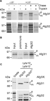
Mitochondrial import and processing of Afg3l1 and Afg3l2. (A) Afg3l1 and Afg3l2 are processed differently upon import into mitochondria. 35S-labeled Afg3l1 and Afg3l2 were imported for 30 min at 30°C into mouse liver mitochondria in the absence (−ΔΨ) or the presence (+ΔΨ) of a mitochondrial membrane potential. Import was halted by the addition of valinomycin (0.5 μM), and mitochondria were incubated further for 60 min at 30°C when indicated (Chase). Samples were further analyzed as described in Figure 1B. (B) Assembly of newly imported Afg3l1 with Afg3l2. 35S-labeled Afg3l1 was imported into mouse liver mitochondria (200 μg), which were lysed subsequently with digitonin. Ten percent of the sample was removed for control (Input). Coimmunoprecipitations were carried out using either preimmune serum or affinity-purified polyclonal Afg3l2-specific antibodies. Immunoprecipitates were analyzed as described in Figure 2A. (C) Processing of Afg3l1 and Afg3l2 in yeast. Afg3l1, Afg3l2 (Afg3lX), or their proteolytically inactive variants Afg3l1E567Q and Afg3lE574Q (Afg3lXEQ) were expressed individually in Δyta10Δyta12 yeast cells. Mitochondria isolated from these yeast strains were analyzed by SDS-PAGE and immunoblotting with Afg3l1- and Afg3l2-specific antibodies. 35S-labeled precursor forms of Afg3l1 and Afg3l2 were examined in parallel by SDS-PAGE and autoradiography (lysate). The asterisk indicates degradation products of Afg3l2. p, precursor; i, intermediate form; m, mature form.
Figure 5.
Figure 5.
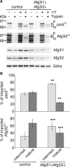
Autocatalytic processing of Afg3l2 in MEFs. (A) Down-regulation of m-AAA protease subunits impairs processing of newly imported paraplegin and Afg3l2. Paraplegin-deficient Spg7−/− MEFs were transfected twice with scrambled siRNA (control) or siRNAs directed against Afg3l1 and Afg3l2. Mitochondria were isolated after ∼2.5 d and 35S-labeled parapleginE575Q (paraEQ) or Afg3l2E574Q (Afg3l2EQ) containing a mutation in the proteolytic center were imported as described in Figure 1B. Import reactions were analyzed by SDS-PAGE and autoradiography. Down-regulation of Afg3l1 and Afg3l2 was monitored by immunoblotting using Afg3l1- and Afg3l2-specific antibodies. Equal loading of the gel was assessed by immunoblotting with antibodies directed against the 70 kDa-subunit of succinate dehydrogenase (Sdha). p, precursor; i, intermediate form; m, mature form; ΔΨ, mitochondrial membrane potential. (B) Quantification of import reactions shown in A. Imported (trypsin-protected) parapleginE575Q and Afg3l2E574Q were quantified by phosphorimaging in control mitochondria and Afg3l1/Afg3l2-depleted mitochondria and are given as percent of the total imported protein. The average of three to four independent experiments (± SD) is shown. ** p < 0.01; *** p < 0.001.

Similar articles

Cited by

References

    1. Arlt H., Tauer R., Feldmann H., Neupert W., Langer T. The YTA10–12-complex, an AAA protease with chaperone-like activity in the inner membrane of mitochondria. Cell. 1996;85:875–885. - PubMed
    1. Cagnoli C., et al. Mutations in AFG3L2 gene (SCA28) in autosomal dominant cerebellar ataxias. Annual Meeting of the American Society of Human Genetics,; Philadelphia, PA. 2008.
    1. Casari G., et al. Spastic paraplegia and OXPHOS impairment caused by mutations in paraplegin, a nuclear-encoded mitochondrial metalloprotease. Cell. 1998;93:973–983. - PubMed
    1. Chan D. C. Mitochondria: dynamic organelles in disease, aging, and development. Cell. 2006;125:1241–1252. - PubMed
    1. Cipolat S., et al. Mitochondrial rhomboid PARL regulates cytochrome c release during apoptosis via OPA1-dependent cristae remodeling. Cell. 2006;126:163–175. - PubMed

Publication types

MeSH terms