Review
. 2009 Aug;10(8):551-64.
doi: 10.1038/nrg2593.

Mechanisms of change in gene copy number

Affiliations
Review

Mechanisms of change in gene copy number

P J Hastings et al. Nat Rev Genet. 2009 Aug.

Abstract

Deletions and duplications of chromosomal segments (copy number variants, CNVs) are a major source of variation between individual humans and are an underlying factor in human evolution and in many diseases, including mental illness, developmental disorders and cancer. CNVs form at a faster rate than other types of mutation, and seem to do so by similar mechanisms in bacteria, yeast and humans. Here we review current models of the mechanisms that cause copy number variation. Non-homologous end-joining mechanisms are well known, but recent models focus on perturbation of DNA replication and replication of non-contiguous DNA segments. For example, cellular stress might induce repair of broken replication forks to switch from high-fidelity homologous recombination to non-homologous repair, thus promoting copy number change.


Figures

Figure 1
Figure 1
An example of a complex genomic rearrangement with microhomology junctions that deleted about 10 kb including exon 4 of the human PMP22 gene. A represents a portion of the normal map of part of PMP22. Blocks of sequence are differentiated by colour. B represents a hypothetical series of 3 template switches that would achieve the rearrangements. The switches could have occurred in the opposite order. Numbers correspond to the junctions detailed below. C shows the rearranged chromosomal region with a deletion of exon 4 joining brown sequence to yellow sequence, followed by an inversion of part of the deleted segment (purple) and a direct duplication of part of the sequence (green). Da shows the nucleotide sequences of the coloured segments in the same colours, where the top line is parental sequence, the second line is the sequence of the rearranged chromosome, and the bottom line is the parental interacting sequence from the other side of the deletion. The junction of brown to yellow sequence shows a 4 bp microhomology (red). b shows the sequences that interacted to make junctions 2 and 3. The second line has the new sequence joining yellow to inverted red sequence (third line) with a 5 bp microhomology, while the fourth line show the sequence that interacted in inverted orientation to make junction 3 with a 3 bp microhomology.
Figure 2
Figure 2
Mechanisms of homologous recombination. A and B Double-strand break repair; C. Double-strand end repair. A. Double Holliday junction recombinational double-strand break repair. At the break (a), 5′ ends are resected (b) to leave 3′ overhanging tails (half arrow heads). These are coated with RecA/Rad51 that catalyzes invasion by one or both ends into homologous sequence forming a D-loop (c). The 3′ end then primes DNA synthesis (dotted lines) that extends it past the position of the original break (d). The second end is incorporated into the D-loop by annealing, and is also extended (e). Following ligation, which forms a double Holliday junction (f), the junctions are resolved by endonuclease (g). The overall effect will be either a non-crossover or a crossover, depending upon whether the two junctions are resolved in the same or different orientations. An alternative resolution pathway is mediated by a helicase and a topoisomerase to converge and undo the double Holliday junction generating only a non-crossover outcome (h). B. Synthesis-dependent strand annealing follows the same pathway as in A through polymerase extension (d). At this point the invading end, together with the newly synthesized DNA is separated from the template by a helicase (e). It now encounters the second end from the double-strand break, and anneals with it by complementary base pairing (f) (dotted arrow). The second end is extended by DNA synthesis (g), and ligated, thus completing repair. C. Break -induced replication occurs at collapsed (broken) replication forks that occur when the replicative helicase at a replication fork encounters a nick in a template strand (solid arrowhead) (a and b). Break induced replication can be understood as a modification of SDSA. As before, a 3′ tail invades a homologue (d), usually the sister from which it broke, and is extended by low processivity polymerization that includes both leading and lagging strands (e). However, the separated extended 3′ end fails to find a complementary second end to which to anneal (f). The end then reinvades (g), and is extended further by a low processivity replication fork. This process of invasion, extension and separation might be repeated several times until a more processive replication fork is formed (h). The fork can now complete replication to the end of the molecule (i). In (g) and (h) we show the Holliday junction following the replication fork, giving conservative segregation of old and new DNA. It is also possible that the Holliday junction is cleaved by an endonuclease, in which case segregation will be semi-conservative. In all parts of the figure, each line shows a single nucleotide chain. Polarity is indicated by half arrows on 3′ ends. New synthesis is shown by dotted lines. The broken DNA molecule is shown in red, a homologue or sister molecule is shown in green.
Figure 3
Figure 3
Change in copy number by homologous recombination. A. Non-allelic homologous recombination (NAHR). (Aa) If a recombination repair event uses a direct repeat (b) as homology, a crossover outcome (shown as an “x”) leads to products that are reciprocally duplicated and deleted for sequence (c) between the repeats. These might segregate from each other at the next cell division, thus changing the copy number in both daughter cells. NAHR can also occur by BIR when the broken molecule uses ectopic homology to restart the replication fork (Ab). BIR will form duplications and deletions in separate events. B. Single-strand annealing. When 5′ end resection on either side of a double-strand break does not lead to invasion of homologous sequence, resection continues. If this resection reveals complementary single-stranded sequence (b) shown by thickened lines, these can anneal. Removal of flaps, gap-filling and ligation complete repair of the double-strand break with deletion of the sequence between the repeats (c) and of one of the repeats. Each line represents a single DNA strand, polarity is indicated by half arrows on 3′ ends, and specific sequences are identified by letters “a” to “d”.
Figure 4
Figure 4
The Breakage-fusion-bridge cycle. (a) An unreplicated chromosome suffers a double-strand break so that it loses a telomere. (b) Upon replication, both sister chromatids lack telomeres. (c) These two ends are proposed to fuse, (d) forming a dicentric chromosome. (e) At anaphase, the two centromeres of the dicentric chromosome are pulled apart, initially forming a bridge between the telophase nuclei. (f) Eventually the bridge is broken in a random position. This inevitably leads to the formation of a large inverted duplication. The chromosome once again has an unprotected end, and upon replication will form two sisters that can fuse to form anew a dicentric chromosome, and so the process is repeated until the end acquires a telomere from another source. Amplification of the large inverted duplication can occur by random breakage in later cycles (not shown). Centromeres are indicated by a blue circle, telomeres by a black block and genomic sequence as red arrows showing orientation. Breakage points are shown as double black lines and fragments that are lost are in grey.
Figure 5
Figure 5
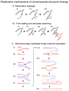
Replicative mechanisms for nonhomologous structural change. A. Replication slippage. (a), during replication, a length of lagging-strand template becomes exposed as a single strand. (b) Whether or not due to secondary structures in the lagging-strand template, the 3′ primer end can move to another sequence showing a short length of homology on the exposed template and (c) continue synthesis after having failed to copy part of the template. As shown, this will produce a deletion. Several variations on this mechanism can also produce a duplication of a length of DNA sequence with or without sister chromatid exchange (reviewed by157). Events occurring by this mechanism are confined to the length of genome to be found in a single replication fork (1 to 2 Kb). B. Fork stalling and template switching (FoSTeS), . Exposed single-stranded lagging strand template (a) might acquire secondary structures (b), which can block the progress of the replication fork. The 3′ primer ends then become free from their templates (c), and might then alight on other exposed single-stranded-template sequence on another replication fork that shares microhomology (d), thus causing duplication, deletion, inversion or translocation depending on the relative position of the other replication fork. Fork stalling can be caused by other situations, such as lesions in the template strand or shortage of deoxynucleotide triphosphates. C. Microhomology-mediated break-induced replication (MMBIR). (a) Replication fork collapse, in which one arm breaks off a replication fork, can occur because the fork encounters a nick on a template strand, or can be caused by endonuclease. (b) the 5′ end of the broken molecule (red) will be recessed from the break, exposing a 3′ tail. When insufficient RecA or Rad51 is present to allow invasion of homologous duplex as shown in Figure 2, the 3′ tail will anneal to any exposed single stranded DNA that shares microhomology. (c) shows the 3′ tail annealing to the lagging-strand template of another replication fork (blue). (d) shows the establishment of a replication fork with both leading and lagging strand synthesis from the microhomology junction. (e), The replication is of low processivity, and the broken end, now extended by a length of a different sequence, shown in blue, is separated from the template and again processed to a 3′ tail, which will then anneal to another single-stranded microhomology sequence. (f), the extended broken end now carrying both the sequence identified in blue, and a length of different sequence identified in green, anneals with single-stranded sequence back onto the red molecule. In this case the single-stranded sequence is shown as a locally melted length of DNA. (g), Another short-processivity fork is established, but this one becomes a fully processive replication fork (h) that can continue to the end of the chromosome or replicon. (i) shows the molecule produced, carrying short sequences from other genomic locations. Whether or not a length of red sequence is duplicated or deleted depends on the position at which synthesis returns to the red chromosome relative to where the initial fork collapse occurred. If the second black sequence is a homologous chromosome instead of the sister chromatid, there will be extensive LOH downstream from the event. Each line represents a single DNA strand, polarity is indicated by half arrows on 3′ ends, and arrowheads show the position of nicks and breaks. Microhomology junctions are indicated by black crosshatching.

Similar articles

Cited by

References

    1. Iafrate AJ, et al. Detection of large-scale variation in the human genome. Nat Genet. 2004;36:949–951. - PubMed
    1. Korbel JO, et al. Paired-end mapping reveals extensive structural variation in the human genome. Science. 2007;318:420–426. - PMC - PubMed
    1. Redon R, et al. Global variation in copy number in the human genome. Nature. 2006;444:444–454. - PMC - PubMed
    2. A survey by SNP array and by aCGH of 270 individuals from the human HapMap samples.

    1. Sebat J, et al. Large-scale copy number polymorphism in the human genome. Science. 2004;305:525–528. - PubMed
    1. Wong KK, et al. A comprehensive analysis of common copy-number variations in the human genome. Am J Hum Genet. 2007;80:91–104. - PMC - PubMed

Publication types

LinkOut - more resources