. 2008 Aug 6;28(32):8063-73.
doi: 10.1523/JNEUROSCI.4487-07.2008.

Targeting of the 5-HT1A serotonin receptor to neuronal dendrites is mediated by Yif1B

Affiliations

Targeting of the 5-HT1A serotonin receptor to neuronal dendrites is mediated by Yif1B

Damien Carrel et al. J Neurosci. .

Abstract

The 5-HT(1A) receptor (5-HT(1A)R) is the most extensively characterized serotonin (5-HT) receptor mainly because of its involvement in the mode of action of antidepressants. The 5-HT(1A)R is confined to the somatodendritic domain of central neurons, where it mediates serotonin-evoked hyperpolarization. Our previous studies underlined the role of the short 5-HT(1A)R C-terminal domain in receptor targeting to dendrites. We used this 17 aa region as bait in a yeast two-hybrid screen, and identified, for the first time, an intracellular protein interacting with the 5-HT(1A)R. This protein is homologous to the yeast Yif1p, previously implicated in vesicular trafficking between the endoplasmic reticulum (ER) and the Golgi apparatus, but not yet characterized in mammals. We confirmed 5-HT(1A)R-Yif1B interaction by glutathione S-transferase pull-down experiments using rat brain extracts and transfected cell lines. Yif1B is highly expressed in the brain, and specifically in raphe 5-HT(1A)R-expressing neurons. Colocalization of Yif1B and 5-HT(1A)R was observed in small vesicles involved in transient intracellular trafficking. Last, inhibition of endogenous expression of Yif1B in primary neuron cultures by small interfering RNA specifically prevented the addressing of 5-HT(1A)R to distal portions of the dendrites, without affecting other receptors, such as sst2A, P2X(2), and 5-HT(3A) receptors. Together, our results provide strong evidence that Yif1B is a member of the ER/Golgi trafficking machinery, which plays a key role in specific targeting of 5-HT(1A)R to the neuronal dendrites. This finding opens up new pathways for the study of 5-HT(1A)R regulation by partner proteins and for the development of novel antidepressant drugs.


Figures
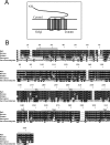
Figure 1.
Figure 1.
Interspecies comparison of Yif1B sequence. A, Schematic predicted topology of the Yif1B protein in Golgi membrane with five transmembrane domains and a large cytoplasmic N terminus. B, The amino acid sequence of Yif1B from rat (Yip1-interacting factor homolog B, Rattus norvegicus, accession numbers: NP_942029 and XP_214879) is aligned with those of homologous proteins in mouse (Yip1-interacting factor homolog B, Mus musculus, accession number: NP_084163.1), human (YIF1B protein, Homo sapiens, accession number: AAH91477.2), Xenopus (LOC443676 protein, Xenopus laevis, accession number: AAH73660.1), and yeast (Yif1p, Saccharomyces cerevisiae, accession number: NP_014136.1). Identical amino acid residues between rat Yif1B and other Yif1Bs or the Yif1p ancestor are in white in black box. Putative transmembrane domains are marked TM1–TM5. The rat sequence (TPRLRKWPSKRRV) represents the peptide used for the production of specific polyclonal anti-Yif1B antibody in rabbit.
Figure 2.
Figure 2.
Yif1B mRNA and protein expression in rat tissues and direct interaction of Yif1B from rat brain homogenates with the 5-HT1AR C-tail. A, Yif1B mRNA expression in rat tissues was analyzed by Northern blot. Each lane represents 3 μg of mRNA from each rat tissue; the human lung is used as a control for the hybridization specificity. The 32P-labeled Yif1B probe consisted of 425 bp in the 5′end of the rat cDNA. B, C, Yif1B protein expression in LLC-PK1 cells untransfected or transfected with Yif1B (B) and in rat tissues (C) was analyzed by Western blot using anti-Yif1B affinity-purified polyclonal antibody (1:1000). Each lane represents 1 μg of total protein, and the same Western blot was also revealed with actin antibody at the bottom. D–G, GST pull-down experiments on Yif1B-transfected LLC-PK1 cells (D) and brain tissue homogenates: cerebellum (E), hippocampus (F), or raphe (G). Extracts were incubated with beads coupled to GST alone or to GST fused with CT1A, I31A, or CT1B. Interaction was analyzed by Western blot with anti-Yif1B affinity-purified polyclonal antibody (1:1000). “Input” represents 2.5 μg of proteins (tissue extract prepared as in C). Results are representative of at least three independent experiments.
Figure 3.
Figure 3.
Yif1B expression in serotoninergic neurons in the dorsal raphe nucleus. A, B, Rat dorsal raphe nucleus sections immunolabeled with anti-TPH antibody (A; red) or with anti-serotonin antibody (B; red) and polyclonal crude anti-Yif1B antiserum (1:5000; in green). Overlay shows the superposition of the two labels. Insets correspond to enlargement of a double-labeled cell. Scale bar, 75 μm.
Figure 4.
Figure 4.
Yif1B subcellular localization. A–C, Transfection of COS-7 cells (A) with Flag-Yif1B or Yif1B and of LLCPK1 cells with Flag Yif1B cDNA (B, C). Yif1B labeling (anti-Yif1B affinity-purified polyclonal antibody; 1:1000) is in red (A1, B1, C1). Flag, calregulin, and CTR433 labeling are shown in green (A2, B2, C2), and colocalized pixels are shown in white on the superposed labeling (A4, B4, C4). Some punctate colocalizations are shown by arrows in insets. Scale bars, 10 μm.
Figure 5.
Figure 5.
Yif1B colocalization with 5-HT1AR. A, Transfection of 5-HT1A-eGFP-stable LLC-PK1 cells with Yif1B. Yif1B immunofluorescence is shown in red (anti-Yif1B affinity-purified polyclonal antibody; 1:1000; A1), and eGFP autofluorescence is shown in green (A2). B, Primary cultures of rat hippocampal neurons (DIV 7) were cotransfected with a plasmid encoding the 5-HT1A-eGFPR and Yif1B. Superposition of labels shown in 1 and 2 is visible in 3. In 4, the colocalized pixels are shown in white. Arrows show Yif1B and 5-HT1AR colocalization in insets. Scale bars, 10 μm.
Figure 6.
Figure 6.
Downregulation of the protein Yif1B by siRNA in primary cultures of rat hippocampal neurons. A1, A2, Yif1B was detected by Western blots (anti-Yif1B affinity-purified polyclonal antibody; 1:1000) of protein extracts, 48 h after transfection of neurons at DIV 7; α-tubulin was detected by Western blots on the same sample (mouse antibody; 1:1000) to normalize the amount of extract. A1, siRNA(Yif1B-1); A2, siRNA(Yif1B-2). All neurons were cotransfected with a control plasmid (encoding eYFP). “Control” corresponds to eYFP alone, “siRNA(Yif1B-1/2)” to eYFP plus siRNA(Yif1B-1/2), and “siRNA(contr-1/2)” to eYFP plus siRNA(contr-1/2). B, Quantification of Yif1B protein expression (normalized with reference to α-tubulin expression) in transfected neurons (n = 5, in 3 independent experiments). Bars represent mean, and error bars represent SEM.
Figure 7.
Figure 7.
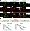
Altered 5-HT1A receptor distribution in the distal part of dendrites after Yif1B RNA interference. A, Primary cultures of rat hippocampal neurons (7 DIV) were transfected with a plasmid encoding the 5-HT1A-eGFPR (Control, left), cotransfected with the 5-HT1A-eGFPR plus the siRNA(Yif1B-1) (middle), or cotransfected with the 5-HT1A-eGFPR plus the siRNA(contr-1) (right). Immunofluorescence was performed with anti-GFP antibody to enhance the GFP signal (green, top) or anti-α-tubulin antibody (red, middle). Bottom, Overlay. Note the drastic reduction of 5-HT1A-eGFPR fluorescence in the distal part of dendrites in siRNA(Yif1B-1)- but not in siRNA(Contr-1)-transfected neurons. B, 5-HT1A-eGFPR (green) and tubulin (red) fluorescence profiles along the longest dendrites (arrows) of the corresponding neurons. C, Cumulated fluorescence profiles for each group (60 neurons analyzed). siRNA(Yif1B-1) reduces 5-HT1A-eGFPR fluorescence in the distal part of dendrites without affecting their average length, as shown by the tubulin fluorescence distribution. Scale bar, 50 μm.
Figure 8.
Figure 8.
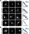
Yif1B RNA interference effect is specific for the 5-HT1A receptor. A, No effect of Yif1B RNA interference on sst2A, P2X2, and 5-HT3A receptor distribution in the dendritic tree. Primary cultures of rat hippocampal neurons (7 DIV) were transfected with plasmids encoding the following receptors: 5-HT1A-eGFP (a), sst2A-eGFP (b), P2X2-eGFP (c), and YFP-5-HT3A (d), and either transfected alone (left), cotransfected with siRNA(Yif1B-1) (middle), or cotransfected with siRNA(Contr-1) (middle right). Immunofluorescence was performed with anti-GFP antibodies to enhance the GFP signal. Cumulated fluorescence profiles along the longest dendrite for each group (15 neurons analyzed) are shown on the right. Note the drastic reduction of fluorescence induced by siRNA(Yif1B-1) in the distal part of dendrites in 5-HT1A-eGFP-transfected neurons but not in sst2A-eGFP-, P2X2-eGFP-, or YFP-5-HT3A-transfected neurons. The higher variability in fluorescence intensities compared with Figure 7 is attributable to the lower number of neurons analyzed. Scale bar, 50 μm. B, Inhibition of 5-HT1A-eGFP receptor distribution in the dendritic tree by another siRNA specific to Yif1B (nucleotides 937–961). Neurons were transfected with a plasmid encoding 5-HT1A-eGFP (left), cotransfected with the siRNA(Yif1B-2) (middle), or cotransfected with the control siRNA, siRNA(Contr-2) (middle right). As above, cumulated fluorescence profiles along the longest dendrite for each group (15 neurons analyzed) are shown on the right. Scale bar, 50 μm.
Figure 9.
Figure 9.
5-HT1A receptor distribution in the dendritic tree is dependent on the C-terminal segment. Primary cultures of rat hippocampal neurons (7 DIV) were transfected with the following plasmids: 5-HT1A-eGFP (A), 5-HT1A-eGFP plus CT1A (B), or 5-HT1AΔ407 (C). Immunofluorescence was performed with anti-GFP antibodies to enhance the GFP signal (A, B) or with anti-5-HT1A antibodies (C). D, Cumulated fluorescence profiles along the longest dendrite for each group (30 neurons analyzed). Note that cotransfection with an excess CT1A reduces the 5-HT1A-eGFP fluorescence in distal dendrites (B), whereas the complete absence of the C-terminal segment virtually abolishes the presence of the mutant receptor in dendrites (C). Scale bar, 50 μm.

Similar articles

Cited by

References

    1. Albert PR, Zhou QY, Van Tol HH, Bunzow JR, Civelli O. Cloning, functional expression, and mRNA tissue distribution of the rat 5-hydroxytryptamine1A receptor gene. J Biol Chem. 1990;265:5825–5832. - PubMed
    1. Altschul SF, Madden TL, Schäffer AA, Zhang J, Zhang Z, Miller W, Lipman DJ. Gapped BLAST and PSI-BLAST: a new generation of protein database search programs. Nucleic Acids Res. 1997;25:3389–3402. - PMC - PubMed
    1. Alvarez VA, Ridenour DA, Sabatini BL. Retraction of synapses and dendritic spines induced by off-target effects of RNA interference. J Neurosci. 2006;26:7820–7825. - PMC - PubMed
    1. Ango F, Robbe D, Tu JC, Xiao B, Worley PF, Pin JP, Bockaert J, Fagni L. Homer-dependent cell surface expression of metabotropic glutamate receptor type 5 in neurons. Mol Cell Neurosci. 2002;20:323–329. - PubMed
    1. Aridor M, Guzik AK, Bielli A, Fish KN. Endoplasmic reticulum export site formation and function in dendrites. J Neurosci. 2004;24:3770–3776. - PMC - PubMed

Publication types

MeSH terms