. 2007 Jun 13;27(24):6581-9.
doi: 10.1523/JNEUROSCI.0338-07.2007.

Leukoencephalopathy upon disruption of the chloride channel ClC-2

Affiliations

Leukoencephalopathy upon disruption of the chloride channel ClC-2

Judith Blanz et al. J Neurosci. .

Abstract

ClC-2 is a broadly expressed plasma membrane chloride channel that is modulated by voltage, cell swelling, and pH. A human mutation leading to a heterozygous loss of ClC-2 has previously been reported to be associated with epilepsy, whereas the disruption of Clcn2 in mice led to testicular and retinal degeneration. We now show that the white matter of the brain and spinal cord of ClC-2 knock-out mice developed widespread vacuolation that progressed with age. Fluid-filled spaces appeared between myelin sheaths of the central but not the peripheral nervous system. Neuronal morphology, in contrast, seemed normal. Except for the previously reported blindness, neurological deficits were mild and included a decreased conduction velocity in neurons of the central auditory pathway. The heterozygous loss of ClC-2 had no detectable functional or morphological consequences. Neither heterozygous nor homozygous ClC-2 knock-out mice had lowered seizure thresholds. Sequencing of a large collection of human DNA and electrophysiological analysis showed that several ClC-2 sequence abnormalities previously found in patients with epilepsy most likely represent innocuous polymorphisms.


Figures

Figure 1.
Figure 1.
Spongiform vacuolation in white matter tracts of ClC-2 KO mice. A, Hematoxylin–eosin-stained brain section of a 22-month-old Clcn2 −/− mouse shows vacuolation in white matter tracts of the cerebellum (a), corpus callosum (b), internal capsule (c), and brainstem (d). B, An age-matched WT brain shows much less vacuolation, which is mostly attributable to capillaries. C–F, Progressive vacuolation in Clcn2 −/− cerebellum from P14 (C) (no vacuolation) to P28 (D), 4 months (E), and 22 months (F). G, Semithin sections of the middle cerebellar peduncle of 2-, 5-, and 14-month-old WT and ClC-2 KO mice used to quantify the area of vacuolation. Abundant vacuoles (asterisks) were visible in cerebellar white matter of 5- and 14-month-old KO but not WT animals, in which capillaries (c) are indicated. H, The white area of randomly taken sections was determined by automated image analysis and shown as the percentage of the total area [5.2 ± 0.6% (KO) vs 1.4 ± 1.3% (WT) at 2 months, 17.8 ± 0.9% (KO) vs 2.1 ± 0.5% (WT) at 5 months, and 21.3 ± 1.2% (KO) vs 3.3 ± 1.2% (WT) at 14 months; 10 sections per mouse, 3 mice each]. Error bars indicate SEM. I, Prominent vacuolation in the white matter of the spinal cord of a 14-month-old KO but not WT mouse. Scale bars: A, B, 1 mm; C–F, 0.25 mm; G, 20 μm; I, 0.5 mm.
Figure 2.
Figure 2.
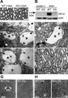
Ultrastructural analysis of vacuoles and myelin. A, Semithin sections of the sciatic nerve show no difference between the genotypes at 14 months of age (14m). B, Western blot analysis detects the ClC-2 protein in brain but not in sciatic nerve (sciatic n.). KO lysates showed the specificity of the ClC-2 antibody. Actin served as a loading control. C–E, Electron micrographs showing typical vacuoles in white matter tracts of ClC-2 KO cerebellum. These vacuoles (asterisks) contained aberrant myelin sheets (arrows) and were surrounded by thin myelin sheaths (arrowheads). a, Axon. D, Myelinated axon surrounded by a vacuole and a normal-appearing oligodendrocyte (n). F, Part of WT white matter cerebellum. G, The structure of myelin sheaths in the area of best compaction revealed no difference in myelin compaction. Distances between MDLs were unchanged in the KO [10.7 ± 0.1 nm (WT) vs 10.8 ± 0.1 nm (KO); n < 90, 3 animals each]. 7w, 7 weeks old. H, Semithin sections of the optic nerve of 7-week-old (7w) WT and Clcn2 −/− mice revealed no vacuolation in the KO (asterisks indicate capillary). Higher magnification is shown in supplemental Fig. S3 (available at www.jneurosci.org as supplemental material). Scale bars: A, 20 μm; C–F, 1 μm; G, 20 nm; H, 10 μm.
Figure 3.
Figure 3.
Inflammation and impaired blood–brain barrier in ClC-2 KO mice and functional tests of motor coordination and central nerve conductance. A, Quantitative PCR (qPCR) analysis of selected transcripts from cerebella of 5-week-old (n = 6, each genotype) and 24-week-old (n = 3, each genotype) animals. Expression levels are given as a percentage of wild type. Genes analyzed by qPCR are as follows: 1, cathepsin-S (accession #NM021281); 2, osteopontin (NM009263); 3, lysozyme (NM013590); 4, F4/80 (NM010130); 5, hexosaminidase (NM010422); 6, leukocyte-derived chemotaxin (NM010701); 7, complement component 1 (NM007572); 8, glutathion peroxidase (NM008161); 9, GFAP (NM010277). Except for GFAP and F4/80, these genes were also found to be upregulated in expression profiling (supplemental Table 1, available at www.jneurosci.org as supplemental material). Rel., Relative. B, C, Red staining by labeled GSA showed microglia activation in 8-month-old Clcn2 −/− (C) but not WT (B) cerebella. D, E, Diaminobenzidine staining (brown) for HRP that was previously injected intravenously into 8-month-old (8m) Clcn2 −/− (E) or WT (D) mice revealed extravasation (arrows), indicative of a disrupted blood–brain barrier only in the KO. Nuclei were counterstained with methylgreen. F, In rotarod tests, ClC-2 KO mice (n = 5) performed as well as WT (n = 4) mice. Motor skills improved with the number of trials in both groups. G, IPIs between waves I and III of auditory brain stem responses were significantly increased in older animals, indicating a slowed nerve conductance in the KO [5 weeks (5w), ***p < 0.1%; 12 weeks (12w), **p < 1%; n = 5–18 mice, same as in supplemental Fig. S7, available at www.jneurosci.org as supplemental material]. 3w, 3 weeks. Error bars indicate SEM. Scale bars: B, C, 0.05 mm; D, E, 0.1 mm.
Figure 4.
Figure 4.
Subcellular localization of ClC-2 in brain. Confocal images of brain sections of 5-week-old WT and ClC-2 KO mice double stained for ClC-2 (green) and marker proteins (red) including GFAP (A–C), NeuN (D), CC1 (E), and Cx47 (F). A, In the hippocampus, ClC-2 colocalized with GFAP in astrocytic endfeet (arrows) surrounding blood vessels (asterisks). B, C, In the cerebellum, ClC-2 was found in GFAP-positive Bergman glia of the molecular layer (B) and in myelinated fiber tracts (C). C, D, In the latter region, ClC-2 did not significantly overlap with either GFAP (C) or NeuN (D). E, Cells with numerous ClC-2 puncta around their cell bodies were identified as oligodendrocytes by staining for CC1. F, Many of these ClC-2-positive puncta also stained for Cx47. Scale bars, 20 μm. Blue indicates TOTO staining of nuclei.
Figure 5.
Figure 5.
Potential role of ClC-2 in epilepsy. A, Two-electrode voltage-clamp analysis of Xenopus oocytes previously injected with 10 ng of WT ClC-2 cRNA (■) or with a construct modeled on the truncating Gins597 mutation (○) from a family with epilepsy (Haug et al., 2003). Injecting half the amount (5 ng) of WT ClC-2 cRNA gave about half the current amplitude (□). Coinjection of 5 ng of WT and 5 ng of Gins597 cRNA (•) yielded similar current amplitudes, indicating that the mutant lacks a dominant-negative effect. Currents were obtained from >15 oocytes from three batches, averaged, and normalized to wild type. B, Two-electrode voltage-clamp analysis of two sequence variants identified in epileptic patients (D'Agostino et al., 2004) in the present screen of patients with leukodystrophy and by Stogmann et al. (2006). Currents from ClC-2_E718D (▾; n = 21 oocytes) and ClC-2_R688Q (○; n = 23) were indistinguishable from WT ClC-2 currents (■; n = 19). Averaged and normalized currents from three batches of oocytes are shown. C, D, Seizure susceptibility of WT, Clcn2 +/− (HET), and Clcn2 −/− (KO) mice as determined by exposure to fluorethyl (applied at 10 ml/min to the chamber; C) or pentylenetetrazol (PTZ; injected at 50 mg/kg body weight; D). C, The apparent slight decrease in threshold in Clcn2 −/− mice (2.34 ± 0.13 min; n = 8 mice) was not statistically different (p = 0.072) from those of WT (2.95 ± 0.25; n = 12) or Clcn2 +/− mice (2.91 ± 0.21 min; n = 3). D, There was no difference between seizure thresholds of WT and KO animals on exposure to PTZ (3.70 ± 1.41 vs 4.04 ± 1.25 min; n = 6 mice each). Error bars indicate SEM.

Similar articles

Cited by

References

    1. Altevogt BM, Paul DL. Four classes of intercellular channels between glial cells in the CNS. J Neurosci. 2004;24:4313–4323. - PMC - PubMed
    1. Bhat RV, Axt KJ, Fosnaugh JS, Smith KJ, Johnson KA, Hill DE, Kinzler KW, Baraban JM. Expression of the APC tumor suppressor protein in oligodendroglia. Glia. 1996;17:169–174. - PubMed
    1. Bösl MR, Stein V, Hübner C, Zdebik AA, Jordt SE, Mukhopadhyay AK, Davidoff MS, Holstein AF, Jentsch TJ. Male germ cells and photoreceptors, both dependent on close cell-cell interactions, degenerate upon ClC-2 Cl− channel disruption. EMBO J. 2001;20:1289–1299. - PMC - PubMed
    1. Butt AM, Kalsi A. Inwardly rectifying potassium channels (Kir) in central nervous system glia: a special role for Kir4.1 in glial functions. J Cell Mol Med. 2006;10:33–44. - PMC - PubMed
    1. Cid LP, Montrose-Rafizadeh C, Smith DI, Guggino WB, Cutting GR. Cloning of a putative human voltage-gated chloride channel (ClC-2) cDNA widely expressed in human tissues. Hum Mol Genet. 1995;4:407–413. - PubMed

Publication types

MeSH terms