. 2002 Oct 1;21(19):5069-78.
doi: 10.1093/emboj/cdf522.

Conversion of PtdIns(4,5)P(2) into PtdIns(5)P by the S.flexneri effector IpgD reorganizes host cell morphology

Affiliations

Conversion of PtdIns(4,5)P(2) into PtdIns(5)P by the S.flexneri effector IpgD reorganizes host cell morphology

Kirsten Niebuhr et al. EMBO J. .

Abstract

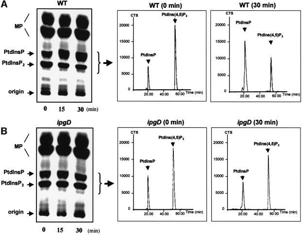
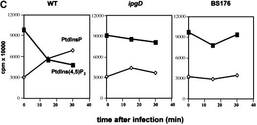
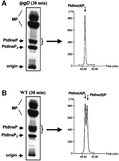
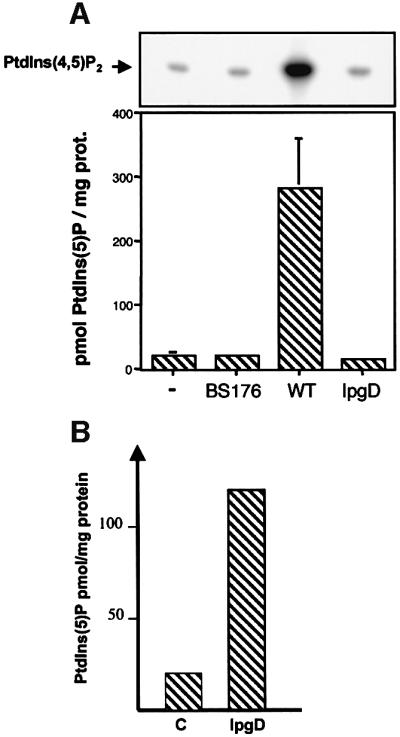
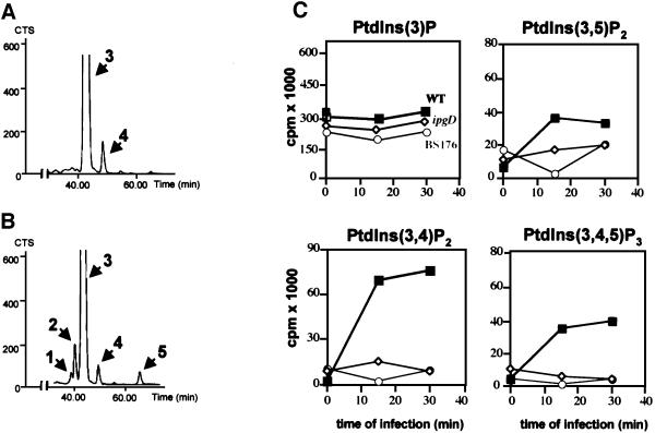
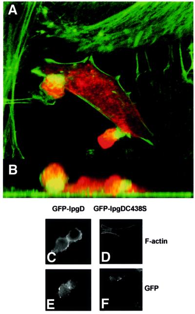
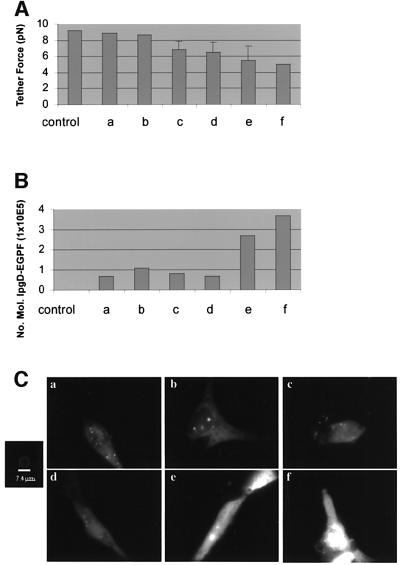
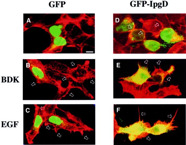
Phosphoinositides play a central role in the control of several cellular events including actin cytoskeleton organization. Here we show that, upon infection of epithelial cells with the Gram-negative pathogen Shigella flexneri, the virulence factor IpgD is translocated directly into eukaryotic cells and acts as a potent inositol 4-phosphatase that specifically dephosphorylates phosphatidylinositol 4,5-bisphosphate [PtdIns(4,5)P(2)] into phosphatidylinositol 5-monophosphate [PtdIns(5)P] that then accumulates. Transfection experiments indicate that the transformation of PtdIns(4,5)P(2) into PtdIns(5)P by IpgD is responsible for dramatic morphological changes of the host cell, leading to a decrease in membrane tether force associated with membrane blebbing and actin filament remodelling. These data provide the molecular basis for a new mechanism employed by a pathogenic bacterium to promote membrane ruffling at the entry site.


Figures

None
Fig. 1. IpgD-dependent PtdIns(4,5)P2 hydrolysis in HeLa cells infected with S.flexneri. HeLa cells were labelled with [32P]orthophosphate and infected with either the wild-type M90T (WT) (A) or the ipgD (ipgD) (B) strains. After infection, cells were washed with PBS and reactions were stopped by adding ice-cold HCl (2.4 M). Cells were recovered by scraping, and lipids were extracted and separated by TLC (left panel). The radiolabelled PtdInsP + PtdInsP2 were recovered by scraping the appropriate bands as indicated, and were deacylated and analysed by HPLC (right panels). MP, major phospholipids; CTS, counts per second. (C) Quantification of [32P]PtdInsP [PtdIns(4)P + PtdIns(5)P which co-elute with the classical HPLC technique] (diamonds) and [32P]PtdIns(4,5)P2 (squares) in HeLa cells infected either with WT, ipgD or the avirulant, non-invasive mutant BS176. Data shown are representative of 4–6 independent experiments with similar results.
None
Fig. 2. Production of PtdIns(5)P without changes in PtdIns(4)P levels during Shigella infection. HeLa cells were labelled with [32P]orthophosphate and infected with either ipgD mutant (ipgD) (A) or wild-type M90T (WT) (B) strains for 30 min. Lipids were then extracted and analysed as described in Figure 1. Radiolabelled PtdInsP was recovered by scraping the corresponding band from the TLC plate as indicated, and was then deacylated and analysed by an appropriate HPLC technique (Tolias et al., 1998) allowing the separation of PtdIns(4)P and PtdIns(5)P (right panel). Peaks corresponding to these phosphoinositides are indicated. Data are from one experiment, representative of three.
None
Fig. 3. Quantification of IpgD-dependent production of PtdIns(5)P by mass assay. (A) The mass level of PtdIns(5)P was measured in non- infected cells (–) or in cells infected with either M90T, ipgD or the avirulant, non-invasive mutant BS176 for 20 min using a specific mass assay as previously described by Morris et al. (2000). Recombinant PIPkinase IIα was used in the presence of [γ-32P]ATP to phosphorylate PtdIns(5)P present in the PtdInsP fraction extracted from cells infected with the different strains. The PtdIns(5)P was transformed specifically to [32P]PtdIns(4,5)P2, which was quantified. A representative TLC illustrating the production of [32P]PtdIns(4,5)P2 from PtdIns(5)P present in the PtdInsP fraction extracted from cells infected with various strains is shown (top panel). Results are also expressed as pmol PtdIns(5)P/mg of HeLa cell proteins and are the mean ± SEM of three independent experiments (lower panel). (B) The mass level of PtdIns(5)P was measured in HeLa cells transfected with GFP (C) or GFP-tagged IpgD (IpgD) after 24 h. Results are representative of three independent experiments.
None
Fig. 4. PI 3-kinase is activated during Shigella infection. HeLa cells were labelled with [32P]orthophosphate and infected with either the ipgD (A) or the wild-type M90T (WT) (B) strains for 30 min. Lipids were then extracted and analysed as described in Figure 1. The radiolabelled PtdInsP2 [PtdIns(3,5)P2, PtdIns(3,4)P2 and PtdIns(4,5)P2] + PtdIns(3,4,5)P3 were recovered by scraping the appropriate bands from the TLC plate, and were deacylated and analysed by HPLC (left panel). Peaks corresponding to PtdIns(3,5)P2 (1), PtdIns(3,4)P2 (2), PtdIns(4,5)P2 (3), ATP (4) and PtdIns(3,4,5)P3 (5) are indicated. (C) Quantification of [32P]D3 phosphoinositides in cells infected with either wild-type M90T (squares), ipgD mutant (diamonds) or the non-invasive mutant BS176 (circles). Data are from one experiment, representative of four.
None
Fig. 5. Expression of IpgD causes formation of membrane blebs. Confocal laser scan analysis of the surface structures elicited on HeLa cells transfected with myc-tagged IpgD after 24 h. For immunofluorescence, transfected cells were visualized using an anti-myc antibody (red), and filamentous actin was stained using FITC-coupled phalloidin. The blebs observed on such cells protruded up to 15 µm above the cells. (A) The sum of all optical sections. (B) The corresponding side view (z-projection). Alternatively, GFP-tagged IpgD (C and E) and GFP-tagged IpgDC438S mutant (D and F) were transfected in NIH-3T3 cells. After 24 h, the filamentous actin was stained using rhodamine-coupled phalloidin (C and D) and the preparations were observed by fluorescence microscopy, using a Zeiss Axioskop microscope equipped with a 63× objective and a Princeton microMAX camera. The data shown are representative of four independent experiments with similar results.
None
Fig. 6. Membrane cytoskeleton adhesion energy is decreased, as seen by tether force in cells expressing IpgD–GFP fusion protein. After calibration of the laser tweezers, the displacement of the bead from the centre of the trap was converted to tether force (i.e. the force needed to hold a tether at a constant length). (A and B) Increasing levels of IpgD–GFP create an inversely proportional decrease in tether force. An expression level of 3.5 × 105 molecules of IpgD decreases the force by half. (C) NIH-3T3 cells transfected with IpgD–GFP show relatively uniform fluorescence with random punctate spots. Hazy fluorescence is due to the high turnover rate of active lamellae. A standardized bead was used to quantitate the number of molecules of IpgD–GFP. Bar = 7.4 µm.
None
Fig. 7. IpgD enhances the effect of Cdc42 and Rac agonists on cytoskeletal remodelling. HEK cells were transfected with GFP (AC) or GFP-tagged IpgD (DF). The duration of transfection was such that cells did not reach the blebbing stage. Cells were then stimulated (B, C, E and F) or not (A and D) with 0.1 µg/ml bradykinin for 20 min (B and E) or 5 nM EGF for 5 min (C and F), and filamentous actin was stained using rhodamine-coupled phalloidin. The preparations were observed by confocal microscopy. Data shown are representative of three independent experiments.

Similar articles

Cited by

References

    1. Adam T., Arpin,M., Prévost,M.-C., Gounon,P. and Sansonetti,P.J. (1995) Cytoskeletal rearrangements and the functional role for T-plastin during entry of Shigella flexneri into HeLa cells. J. Cell Biol., 129, 367–381. - PMC - PubMed
    1. Allaoui A., Ménard,R., Sansonetti,P.J. and Parsot,C. (1993) Characterization of the Shigella flexneri ipgD and ipgF genes, which are located in the proximal part of the mxi locus. Infect. Immun., 61, 1707–1714. - PMC - PubMed
    1. Azuma T., Witke,W., Stossel,T.P., Hartwig J.H. and Kwiatkowski,D.J. (1998) Gelsolin is a downstream effector of rac for fibroblast motility. EMBO J., 17, 1362–1370. - PMC - PubMed
    1. Bligh E.G. and Dyer,W.J. (1959) A rapid method of total lipid extraction and purification. Can. J. Biochem. Physiol., 37, 911–919. - PubMed
    1. Blondeau F., Laporte,J., Bodin,S., Superti-Furga,G., Payrastre,B. and Mandel,J.L. (2000) Myotubularin, a phosphatase deficient in myotubular myopathy, acts on phosphatidylinositol 3-kinase and phosphatidylinositol 3-phosphate pathway. Hum. Mol. Genet., 9, 2223–2229. - PubMed

Publication types

Substances

LinkOut - more resources