Review
. 2016 Apr;22(2 Dementia):510-37.
doi: 10.1212/CON.0000000000000319.

Rapidly Progressive Dementia

Review

Rapidly Progressive Dementia

Michael D Geschwind. Continuum (Minneap Minn). 2016 Apr.

Abstract

Purpose of review: This article presents a practical and informative approach to the evaluation of a patient with a rapidly progressive dementia (RPD).

Recent findings: Prion diseases are the prototypical causes of RPD, but reversible causes of RPD might mimic prion disease and should always be considered in a differential diagnosis. Aside from prion diseases, the most common causes of RPD are atypical presentations of other neurodegenerative disorders, curable disorders including autoimmune encephalopathies, as well as some infections, and neoplasms. Numerous recent case reports suggest dural arterial venous fistulas sometimes cause RPDs.

Summary: RPDs, in which patients typically develop dementia over weeks to months, require an alternative differential than the slowly progressive dementias that occur over a few years. Because of their rapid decline, patients with RPDs necessitate urgent evaluation and often require an extensive workup, typically with multiple tests being sent or performed concurrently. Jakob-Creutzfeldt disease, perhaps the prototypical RPD, is often the first diagnosis many neurologists consider when treating a patient with rapid cognitive decline. Many conditions other than prion disease, however, including numerous reversible or curable conditions, can present as an RPD. This chapter discusses some of the major etiologies for RPDs and offers an algorithm for diagnosis.


Figures

Figure 7-1
Figure 7-1
Major diagnostic categories of patients with rapidly progressive dementia (RPD) referred to, versus evaluated at, the University of California, San Francisco (UCSF) rapidly progressive dementia program over 13 years. A, Diagnostic distribution of patients with RPD referred to UCSF over about a 13-year period, most of whom had extensive medical record review, but only about one-fourth of whom were evaluated in person at UCSF. Almost one-third of cases referred to (as well as evaluated at) UCSF were diagnosed with sporadic Jakob-Creutzfeldt disease. In more than one-fourth of referred cases, although a sporadic Jakob-Creutzfeldt disease diagnosis (potential sporadic Jakob-Creutzfeldt disease) was suspected, not enough information existed to make a probable Jakob-Creutzfeldt disease diagnosis. Acquired Jakob-Creutzfeldt disease includes iatrogenic and infectious forms of prion disease. The genetic prion diseases category included patients who had confirmed mutations (autosomal dominant) in the prion protein gene, PRNP, or were from families with genetic prion disease. Whereas many of the genetic prion diseases presented similarly to sporadic Jakob-Creutzfeldt disease, as an RPD, a significant minority had clinical presentations more similar to other more slowly progressive diseases, such as Alzheimer disease or atypical parkinsonian or ataxic syndromes. One-fourth of cases were diagnosed with a nonprion etiology for their RPD. B, Diagnostic distribution of patients with RPD evaluated in person at UCSF. A larger percentage of nonprion RPDs and genetic prion diseases is evident; the latter is a bias partly because of the UCSF research program in genetic prion diseases and antibody-mediated encephalopathies. JCD = Jakob-Creutzfeldt disease.
Figure 7-2
Figure 7-2
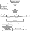
Algorithm for evaluating rapidly progressive dementia. Refer to the text and Table 7-3 for details about the workup for each diagnostic category. CSF = cerebrospinal fluid; IV = intravenous; RPD = rapidly progressive dementia.
Figure 7-3
Figure 7-3
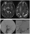
Imaging of a dural arteriovenous fistula (DAVF). A 70-year-old woman developed amnesia, aphasia, incoherent speech, and progressive gait disturbance over 2 weeks, progressing over the next 3 weeks to develop myoclonus with a startle reflex, flaccid tetraparesis with brisk deep tendon reflexes, extensor plantar responses, pronounced primitive reflexes, and akinetic mutism. Initial diagnosis was Jakob-Creutzfeldt disease until brain imaging revealed a DAVF. A, Axial T2-weighted MRI shows multiple dilated vessels in the temporal regions, predominantly in the right temporal region with venous ectasia and hyperintensity of the white matter including the centrum semiovale. B, Digital subtraction angiogram (right common carotid injection [left panel]) and left external carotid injection [right panel]), lateral views, shows multiple DAVFs of the superior sagittal sinus, torcula, and the lateral sinuses.Reprinted with permission from Mendonça N, et al, Neurologist.journals.lww.com/theneurologist/pages/articleviewer.aspx?year=2012&issue=05000&article=00005&type=Abstract.© 2012 Lippincott Williams & Wilkins, Inc.
Figure 7-4
Figure 7-4
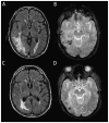
MRI findings of cerebral amyloid angiopathy–related inflammation. An elderly man presented with relatively acute onset left hemiparesis, left homonymous hemianopia, dysarthria, spatial and temporal disorientation, sensory aphasia, and psychomotor slowness. Cerebral amyloid angiopathy–related inflammation was suspected and the finding of APOE genotype ɛ4/ɛ4 supported the diagnosis. Anti–amyloid-β (Aβ) autoantibody concentration in CSF was elevated at 55.9 ng/mL. Physical therapy and corticosteroid therapy with dexamethasone 24 mg/d were started and the patient showed clinical improvement. Initial axial fluid-attenuated inversion recovery (FLAIR) MRI shows bilateral hyperintense lesions (A) and gradient recalled echo (GRE) image shows cortical and subcortical microhemorrhages (B). After 1 month of steroid therapy, FLAIR MRI (C) and GRE (D) sequence show reduction of both cerebral edema and microhemorrhages. Modified with permission from Crosta F, et al, Case Rep Neurol Med.www.hindawi.com/journals/crinm/2015/483020/. © 2015 Francesca Crosta et al.
Figure 7-5
Figure 7-5
Brain MRI of a 68-year-old man with recent hyperintensive encephalopathy leading to subacute diencephalic angioencephalopathy. His first symptoms were hypernasal dysarthria and palatal weakness that resolved over 3 months after treatment of hypertension. Thirteen months after initial onset, he developed headaches, confusion, and speech and language problems due to hyperintensive encephalopathy, which again resolved with treatment. About 15 months after initial onset, he began a downward, but fluctuating course (with treatments), over 8 weeks, of a rapidly progressive neurologic decline leading to his death. Brain autopsy revealed findings consistent with subacute diencephalic angioencephalopathy. Nonenhancing, confluent subcortical axial fluid-attenuated inversion recovery (FLAIR) hyperintensities in the occipital lobes (A) and left temporal lobe (B) at the time of initial onset. Confluent nonenhancing abnormal coronal T2-weighted signal symmetrically in the pons, as well as confluent periventricular FLAIR signal, at 13 months after onset (C). One month later, imaging demonstrated bilaterally symmetric T1 and T2/FLAIR hyperintense abnormal signal in the thalami (D, coronal FLAIR; E, axial FLAIR). Thalamic abnormalities enhanced minimally following gadolinium administration on axial T1-weighted images (F, unenhanced; G, enhanced). Small T1-weighted (H) and T2-weighted hyperintense cortical foci were also seen. Reprinted with permission from Graffeo CS, et al, J Clin Neurosci. www.jocn-journal.com/article/S0967-5868(15)00354-9/abstract. © 2015 Elsevier Ltd.
Figure 7-6
Figure 7-6
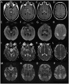
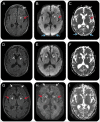
MRI of the patient in Case 7-1. Wernicke encephalopathy (compared to a sporadic Jakob-Creutzfeldt disease case). Fluid-attenuated inversion recovery (FLAIR) (A–D), diffusion-weighted imaging (DWI) (E–H), and apparent diffusion coefficient (ADC) map (I–L) sequences showing FLAIR and DWI hyperintense signal changes involving the periaqueductal gray and midbrain tectum, medial thalami, and perirolandic cortex in the patient with Wernicke encephalopathy. There is relative sparing of the mammillary bodies across all sequences (B,F,J). The ADC sequences (I–L) primarily show subtle hypointensity in the perirolandic cortex (L), corresponding to hyperintensities on FLAIR (D) and DWI (H). This pattern preferentially involving the perirolandic cortex is the opposite of what we typically see in sporadic Jakob-Creutzfeldt disease (M–P; DWI sequences), in which there is generally sparing of the perirolandic region, particularly the primary motor cortex.
Figure 7-7
Figure 7-7
MRI of the patient in Case 7-2. A 66-year-old woman with hypoglycemic encephalopathy. Fluid-attenuated inversion recovery (FLAIR) (A, D, G), diffusion-weighted images (DWI) (B, E, H), and apparent diffusion coefficient (ADC) map (C, F, I) sequences 2 days (A–C), 3 weeks (D–F), and 1 month (G–I) after onset. Initial MRI (A–C) showed left frontal (white arrows), left insular (red arrows), bilateral medial occipital (blue arrows), and left caudate (white arrowhead) FLAIR/DWI hyperintensity with restricted diffusion, which is subtle but definitely appreciable. Repeat MRI about 3 weeks later (D–F) showed possible reduced FLAIR/DWI hyperintensity in the left caudate head and medial occipital regions, and possible increased right caudate FLAIR hyperintensity and restricted diffusion (D–F; white arrowheads). A third MRI 1 week later, 1 month after onset (G–I), revealed more intense FLAIR/DWI insular (G, H; red arrows) and frontal cortical hyperintensities (G, H; white arrows) and possible restricted diffusion and FLAIR hyperintensity still present in the caudate heads (G, H; arrowheads). The resolution of occipital cortical ribboning in such a short time argued against a diagnosis of sporadic Jakob-Creutzfeldt disease. Reprinted with permission from Rosenbloom MH, et al, Neurol Clin Pract. cp.neurology.org/content/5/2/108.full. © 2015 American Academy of Neurology.
Figure 7-8
Figure 7-8
MRI of the patient in Case 7-3. A 50-year-old man with extrapontine myelinolysis. Initial MRI 2 months after onset (A–D) showed symmetric bilateral striatal fluid-attenuated inversion recovery (FLAIR) (A)/diffusion-weighted imaging (DWI) (B) hyperintensities (A, B; white arrows) with corresponding hypointensities on the apparent diffusion coefficient (ADC) map suggesting restricted diffusion (C; black arrows). Bilateral globus pallidus hyperintensities were present on T1-weighted images (D; green arrows). MRI 1 month later, 3 months after onset (E–H), showed resolution of the prior FLAIR (E), DWI (F), and ADC (G) map abnormalities but no change in the globus pallidus T1 hyperintensities (H; green arrows). Reprinted with permission from Rosenbloom MH, et al, Neurol Clin Pract. cp.neurology.org/content/5/2/108.full. © 2015 American Academy of Neurology.
Figure 7-9
Figure 7-9
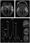
MRI and spectroscopy in a case of mitochondrial myopathy, encephalopathy, lactic acidosis, and strokelike episodes syndrome (MELAS). A 59-year-old woman developed confusion, progressive aphasia, mutism, and fluctuations of alertness over 2 weeks. Diffusion-weighted imaging (DWI) MRI revealed abnormalities overlapping with Jakob-Creutzfeldt disease (A), although the fluid-attenuated inversion recovery (FLAIR) MRI (B) with white and gray matter hyperintensity was not consistent with Jakob-Creutzfeldt disease. CSF showed normal cell counts, negative polymerase chain reaction (PCR) for herpes simplex virus, elevated lactate (4.6 mmol/L), and increased levels of 14-3-3 and tau protein (1300 pg/L), both concerning for Jakob-Creutzfeldt disease. There were no periodic sharp-wave complexes on EEG recordings. Magnetic resonance spectroscopy revealed a lactate signal indicative of mitochondriopathy and genetic analysis confirmed the MELAS A3243G mutation. The DWI (A) displays bitemporal neocortical hyperintense signals. The FLAIR (B) 2 days after the initial MRI scan reveals newly emerging symmetric lesions in the pulvinar thalami. Magnetic resonance spectroscopy (C) displays a strong lactate signal. Cho = choline; Cr = creatine; Cr2 = phosphocreatine; Ins dd1 = myoinositol; LAC = lactate; NAA = N-acetylaspartate; ppm = parts per million.Reprinted with permission from Weiss D, et al, Neurology. www.neurology.org/content/77/9/914.full. © 2011 American Academy of Neurology.

Similar articles

Cited by

References

    1. Grossman M, Irwin DJ. The mental status examination in patients with suspected dementia. Continuum (Minneap Minn) 2016; 22(2 Dementia): 385– 403. - PMC - PubMed
    1. Geschwind MD. Prion diseases. Continuum (Minneap Minn) 2015; 21(6 Neuroinfectious Disease): 1612– 1638. doi:10.1212/CON.0000000000000251. - PMC - PubMed
    1. Gibbs CJ., Jr Spongiform encephalopathies—slow, latent, and temperate virus infections—in retrospect. In: Prusiner SB, Collinge J, Powell J, et al. eds. Prion diseases of humans and animals. London, UK: Ellis Horwood, 1992: 53– 62.
    1. Katscher F. It’s Jakob’s disease, not Creutzfeldt’s. Nature 1998; 393(6680): 11 doi:10.1038/29862. - PubMed
    1. Geschwind MD, Shu H, Haman A, et al. Rapidly progressive dementia. Ann Neurol 2008; 64(1): 97– 108. doi:10.1002/ana.21430. - PMC - PubMed

Publication types