. 1998 Aug 15;12(16):2560-73.
doi: 10.1101/gad.12.16.2560.

Human and mouse homologs of Schizosaccharomyces pombe rad1(+) and Saccharomyces cerevisiae RAD17: linkage to checkpoint control and mammalian meiosis

Affiliations

Human and mouse homologs of Schizosaccharomyces pombe rad1(+) and Saccharomyces cerevisiae RAD17: linkage to checkpoint control and mammalian meiosis

R Freire et al. Genes Dev. .

Abstract

Preventing or delaying progress through the cell cycle in response to DNA damage is crucial for eukaryotic cells to allow the damage to be repaired and not incorporated irrevocably into daughter cells. Several genes involved in this process have been discovered in fission and budding yeast. Here, we report the identification of human and mouse homologs of the Schizosaccharomyces pombe DNA damage checkpoint control gene rad1(+) and its Saccharomyces cerevisiae homolog RAD17. The human gene HRAD1 is located on chromosome 5p13 and is most homologous to S. pombe rad1(+). This gene encodes a 382-amino-acid residue protein that is localized mainly in the nucleus and is expressed at high levels in proliferative tissues. This human gene significantly complements the sensitivity to UV light of a S. pombe strain mutated in rad1(+). Moreover, HRAD1 complements the checkpoint control defect of this strain after UV exposure. In addition to functioning in DNA repair checkpoints, S. cerevisiae RAD17 plays a role during meiosis to prevent progress through prophase I when recombination is interrupted. Consistent with a similar role in mammals, Rad1 protein is abundant in testis, and is associated with both synapsed and unsynapsed chromosomes during meiotic prophase I of spermatogenesis, with a staining pattern distinct from that of the recombination proteins Rad51 and Dmc1. Together, these data imply an important role for hRad1 both in the mitotic DNA damage checkpoint and in meiotic checkpoint mechanisms, and suggest that these events are highly conserved from yeast to humans.


Figures

Figure 1
Figure 1
Sequence comparison of the putative human and mouse Rad1 homologs. Multiple alignment of amino acid sequences of human (hRad1) and mouse Rad1 (mRad1) with Schizosaccharomyces pombe Rad1 (spRad1), Ustilago maydis Rec1 (umRec1), and Saccharomyces cerevisiae Rad17p (scRad17). Amino acid residues, which are identical, are shown by reverse shading and conservative substitutions are indicated by gray shading. Numbers indicate the amino acid position, and residues that align with those of umRec1, which have been proposed to define parts of a nuclease catalytic site (Thelen et al. 1994), are indicated by asterisks. The alignment was obtained with the PILEUP program from the GCG package, and shading was by the BOXSHADE program.
Figure 2
Figure 2
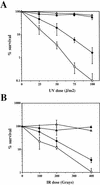
Complementation of radiation sensitivity of the S. pombe rad1 mutant by HRAD1. Viability of wild-type and rad1::ura4+ strains containing pREP3X (WT and rad1, respectively) and the rad1::ura4+ strain containing the pREP3X/sprad1+ plasmid (rad1/sprad1+) or pREP3X/HRAD1 (rad1/HRAD1) in response to different doses of UV light (A) or ionizing radiation (IR) (B) are shown. Each data point is the mean of at least three independent experiments. (▵) Wild type; (▴) rad1/sprad1+; (○) rad1; (•) rad1/HRAD1.
Figure 3
Figure 3
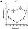
Complementation of the S. pombe rad1 checkpoint defect by HRAD1. Strain designations are as in Fig. 2. Exponentially growing cultures of S. pombe strains [(A) Wild type (WT); (B) rad1; (C) rad1/sprad1+, (D) rad1/HRAD1] were either UV-irradiated with 50 J/m2 (•) or left untreated (○). Samples were taken at the indicated time points and the septation index was measured as described in Materials and Methods. Data shown are the mean ±s.d. of at least three independent experiments, each one conducted in duplicate.
Figure 3
Figure 3
Complementation of the S. pombe rad1 checkpoint defect by HRAD1. Strain designations are as in Fig. 2. Exponentially growing cultures of S. pombe strains [(A) Wild type (WT); (B) rad1; (C) rad1/sprad1+, (D) rad1/HRAD1] were either UV-irradiated with 50 J/m2 (•) or left untreated (○). Samples were taken at the indicated time points and the septation index was measured as described in Materials and Methods. Data shown are the mean ±s.d. of at least three independent experiments, each one conducted in duplicate.
Figure 4
Figure 4
Generation of anti-hRad1 antibodies and their use in immunolocalization studies. (A) Western blot with two different antisera raised against hRad1. Each lane contains 60 μg of HeLa nuclear extract. The two antisera recognize a polypeptide of ∼32 kD. (B) Affinity-purified antibody 1 was used to probe Western blots of the indicated amounts of recombinant tagged bacterially expressed hRad1 (lanes 1–3); 10 μg of extract of the S. pombe rad1 strain expressing hRad1 (lane 4; +) and the same strain with empty vector (lane 5; −); 60 μg of HeLa whole cell extract (lane 6; −) and extract of HeLa cells transfected with Flag-tagged HRAD1 cDNA (lane 7; +). In addition, 60 μg of whole cell extract of HeLa cells transfected with Flag-tagged HRAD1 were also probed with monoclonal anti-Flag antibody (lane 8; +). (C) Immunofluorescence microscopy of HeLa cells with anti-hRad1 affinity-purified antibody using a fluorescein-conjugated secondary antibody (green; left), and staining of nuclear DNA with propidium iodide (red; right). Immunostaining reveals endogenous hRad1 in HeLa cells. (D) Western blot of U2OS cell extracts (50 μg per lane) after UV (50 J/m2) or IR (10 Gy) treatment. Western blots were probed with anti-hRad1, anti-β actin or anti-p53 antibodies, as indicated.
Figure 5
Figure 5
Distribution of Rad1 in different tissues and during spermatocyte development. (A) Western blots of whole cell extracts from various mouse tissues probed with anti-hRad1 affinity-purified antibody. HeLa cell nuclear extract (50 μg) is included on both Western blots. As a control, blots were also probed with anti-β actin antibody. The Rad1 band of ∼32 kD is indicated with an arrow. (B) Northern blot with various human tissues. The blot was probed separately with HRAD1 then with β-actin as a control. The major HRAD1 mRNA species is indicated with an arrow. (C) Western blot of rat testicular cells from 30-day-old rats separated by elutriation. All the lanes contain nuclear extracts normalized with respect to cell number (∼5 × 10 cells per lane). (Fraction I) Small cells, zygotene and early pachytene; (fraction II) cells in leptotene and zygotene some pachytene; (fraction III) mostly full pachytene; (fraction IV) late pachytene, and diplotene; (fraction V) postpachytene.
Figure 5
Figure 5
Distribution of Rad1 in different tissues and during spermatocyte development. (A) Western blots of whole cell extracts from various mouse tissues probed with anti-hRad1 affinity-purified antibody. HeLa cell nuclear extract (50 μg) is included on both Western blots. As a control, blots were also probed with anti-β actin antibody. The Rad1 band of ∼32 kD is indicated with an arrow. (B) Northern blot with various human tissues. The blot was probed separately with HRAD1 then with β-actin as a control. The major HRAD1 mRNA species is indicated with an arrow. (C) Western blot of rat testicular cells from 30-day-old rats separated by elutriation. All the lanes contain nuclear extracts normalized with respect to cell number (∼5 × 10 cells per lane). (Fraction I) Small cells, zygotene and early pachytene; (fraction II) cells in leptotene and zygotene some pachytene; (fraction III) mostly full pachytene; (fraction IV) late pachytene, and diplotene; (fraction V) postpachytene.
Figure 6
Figure 6
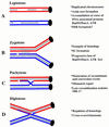
Diagrammatic summary of meiotic prophase stages and events. (A) At leptotene of meiotic prophase, S-phase is completed and chromosomal cores (gray) form in association with the pairs of sister chromatids (red and blue). Rad51/Dmc1 foci appear at the cores at this time. (B) At zygotene, the homologous chromosomes synapse and the cores align in parallel forming the synaptonemal complex (SC; indicated by vertical bars). The gap represents a DNA double-stranded break, which is thought to initiate meiotic recombination. At the onset of zygotene, the number of Rad51/Dmc1 foci reach their maximum and then start to decline. (C) Although the chromosomes are fully synapsed during the pachytene stage, the recombination processes are completed. The chromosomal crossover and heteroduplex DNA are indicted. (D) At the diplotene stage, the homologous chromosomes separate and the points of reciprocal exchanges are visible as chiasmata.
Figure 7
Figure 7
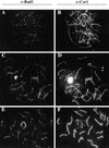
Immunofluorescence microscopy of meiotic chromosomes in different stages of meiosis. Mouse spermatocyte meiotic prophase nuclei were labeled with anti-hRad1 (A,C,E) or anti-Cor1 (B,D,F). (A,B) There are ∼390 hRad1 foci associated with the chromosome cores when synapsis is initiated. (C,D) During synapsis (zygotene), the number of Rad1 foci declines. There are ∼300 foci in this late zygotene nucleus and it is evident that the foci are associated with both unpaired chromosomes and chromosomes that are fully synapsed. (E,F) Extrachromosomal core fragments (f) do not have foci of any kind. There are ∼150 foci at this stage. In contrast, the X–Y chromosome pair remains heavily stained until diplotene.
Figure 8
Figure 8
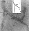
Immunogold localization of Rad1 and Dmc1 at zygotene. Rad1 is labeled with 10-nm, and Dmc1 with 5-nm gold particles. The centromeric ends (15-nm gold grains) are not yet synapsed, and the 10-nm grains on unpaired cores indicate the presence of Rad1 antigen in association with the cores. The SC and the X-chromosome core have both 10- and 5-nm gold clusters, but the two sizes of particle do not colocalize, suggesting distinct functions for Rad1 and Dmc1. A schematic representation of the EM image is shown (top, middle).

Similar articles

Cited by

References

    1. Al-Khodairy F, Carr AM. DNA repair mutants defining G2 checkpoint pathways in Schizosaccharomyces pombe. EMBO J. 1992;11:1343–1350. - PMC - PubMed
    1. Barlow AL, Benson FE, West SC, Hultén MA. Distribution of the Rad51 recombinase in human and mouse spermatocytes. EMBO J. 1997;16:5207–5215. - PMC - PubMed
    1. Barlow, C., M. Liyanage, P.B. Moens, K. Nagahsima, K. Brown, S. Rottinghaus, S.P. Jackson, D. Tagle, T. Ried, and A. Wynshaw-Boris. 1998. ATM deficiency results in severe meiotic disruption as early as leptotenema in prophase I. Development (in press). - PubMed
    1. Baumann P, Benson FE, West SC. Human Rad51 promotes ATP-dependent homologous pairing and strand transfer reactions in vitro. Cell. 1996;87:757–766. - PubMed
    1. Bentley NJ, Holtzman DA, Flaggs G, Keegan KS, DeMaggio A, Ford JC, Hoekstra MF, Carr AM. The Schizosaccharomyces pombe rad3 checkpoint gene. EMBO J. 1996;15:6641–6651. - PMC - PubMed

Publication types

MeSH terms

Associated data