. 2025 Feb 27;17(1):54.
doi: 10.1186/s13195-025-01702-0.

Identification and characterization of variants in PSEN1, PSEN2, and APP genes in Chinese patients with early-onset Alzheimer's disease

Affiliations

Identification and characterization of variants in PSEN1, PSEN2, and APP genes in Chinese patients with early-onset Alzheimer's disease

Haitian Nan et al. Alzheimers Res Ther. .

Abstract

Variants in PSEN1, PSEN2, and APP are major genetic causes of early-onset Alzheimer's disease (EOAD). Our study aimed to identify the genotypic and phenotypic spectrums in a Chinese EOAD cohort and confirm their pathogenicity by functional analysis. This study included 304 unrelated clinically diagnosed EOAD participants of Chinese Han ancestry. Whole-exome sequencing revealed that 26 out of 304 individuals (8.6%) carried rare variants in PSEN1, PSEN2, and APP, including 16 in PSEN1 (5.3%), 6 in PSEN2 (2.0%), and 4 in APP (1.3%). Eight variants were novel, including PSEN1 p.Q56R, PSEN1 p.L174P, PSEN1 p.S289P, PSEN1 p.Y466C, PSEN2 p.R17W, PSEN2 p.F331Y, APP p.D197N, and APP p.D252V. Functional study revealed that the PS1 L174P, S289P, R377M, Y466C, PS2 V214L, and M239T mutants increased Aβ42 levels and Aβ42/Aβ40 ratios. The PS1 L174P, R377M, and Y466C mutants decreased the maturation of presenilin-1. Our findings highlight the prevalence and pathogenic significance of APP /PSENs variants in a Chinese EOAD cohort and expand the phenotypic and genotypic spectrum of EOAD.

Keywords: APP; PSEN1; PSEN2; Alzheimer’s disease; Amyloid β; EOAD.


Conflict of interest statement

Declarations. Ethics approval and consent to participate: The study was approved by the Ethics Committees of the Xuanwu Hospital of Capital Medical University (approval number: 2020026), and it was carried out in compliance with the Declaration of Helsinki’s principles. Written informed consent was obtained from each patient or their guardian. Consent for publication: Written informed consent for publication was obtained from the guardian of each patient. Competing interests: The authors declare no competing interests.

Figures

Fig. 1
Fig. 1
Schematic representation of the frequencies and locations of PSEN1, PSEN2, and APP variants. A Left panel: Pie chart of the percentage of familial and sporadic EOAD patients. Right panel: Schematic diagram of the distribution of APOE allele frequencies in our cohort. B Pie charts representing the percentage of PSEN1, PSEN2, and APP variants represented in our cohort. C This diagram shows the amino acid sequence of PS1 and the distribution of the variants reported in this study. Presenilin 1 contains 467 amino acids with nine potential transmembrane domains. Red circles represent the variants identified in this study. D Distribution of amino acid sequence in presenilin 2. PS2 has a similar structure but contains 448 amino acids. Red circles represent the variants identified in this study. E The structural domain arrangement of the amyloid precursor protein expressed in the APP695 isoform, which contains many functional domains as illustrated. SP: Signal peptide; E1: Ectodomain 1; E2: Ectodomain 2; TM: Transmembrane domain; AcD: acidic domain; JMR: juxtamembrane region. AICD: APP intracellular domain. The APP D197N, p.A235V, D252V, and p.T297M variants were indicated by red arrows
Fig. 2
Fig. 2
Pedigrees of the EOAD families with variants identified in this study
Fig. 3
Fig. 3
A neuroimaging study of EOAD patients with functionally analyzed variants identified in this cohort. The AV45 PET images for Patients 2 and 6 were displayed. The MRI/FDG PET images for the patients with functionally analyzed variants in this study were displayed
Fig. 4
Fig. 4
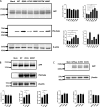
Aβ−40 and Aβ−42 protein expression study. Aβ−40 and Aβ−42 protein expression levels in cell media of each group. WT and indicated mutants were co-expressed with the APP Swedish mutant in HEK239 cells and the conditioned media were harvested 48 h post-transfection for ELISA-determination of Aβ−40 and Aβ−42. Cell lysates were subjected to Western blot for APP, PS1, PS2, and β-actin as an internal standard. A Western blotting of cell lysates and quantification of Aβ42, Aβ40, and ratios of Aβ42 to Aβ40 relative to PS1 WT in conditioned medium of cells expressing PS1 WT and PS1 Q56R, L174P, S289P, R377M, and Y466C mutants. The Aβ levels were normalized to the total protein levels in PSEN1 WT-expressing cells. Quantifications of the full-length PS1 in the corresponding cell lysates relative to PS1 WT were also provided. The PS1 levels were normalized to the β-actin levels. B Western blotting of cell lysates and quantification of Aβ42, Aβ40, and ratios of Aβ42 to Aβ40 relative to PS2 WT in conditioned medium of cells expressing PS2 WT and PS2 V214L, M239T mutants. The Aβ levels were normalized to the total protein levels in PSEN2 WT-expressing cells. C Western blotting of cell lysates and quantification of Aβ42, Aβ40, and ratios of Aβ42 to Aβ40 relative to APPSwe in conditioned medium of cells expressing Mock (empty vector transfected), APPSwe, D197N (APPSwe combined with D197N variant), and D252V (APPSwe combined with D252V variant) mutants. The Aβ levels were normalized to the total protein levels in APPSwe-expressing cells. All Aβ level normalizations were performed relative to the total protein levels, which may reflect the number of cells. This experiment was performed three times with reproducible similar results
Fig. 5
Fig. 5
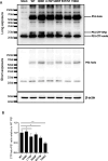
The PS1 maturation analysis. A The PS1 maturation in vitro. Indicated PS1mutants carrying a C-terminal Myc-his tag were expressed in HEK293 cells, and the lysates were blotted for PS1 using a PS1 antibody that recognizes the C terminus of PS1. The endogenous PS1 C-terminal fragment (endo-CTF) and the CTFs derived from the overexpressed PS1 carrying a C-terminally fused Myc-His tag (Myc-CTF) were seen. The Myc-CTFs were detected after relatively long exposure. B The protein bands were quantified using Quantity One (Bio-Rad), and the ratios of CTF/holo-PS1 were plotted. *: p < 0.05, **: p < 0.01

References

    1. McKhann G, Drachman D, Folstein M, Katzman R, Price D, Stadlan EM. Clinical diagnosis of Alzheimer’s disease: report of the NINCDS-ADRDA Work Group under the auspices of Department of Health and Human Services Task Force on Alzheimer’s Disease. Neurology. 1984;34:939–44. - PubMed
    1. Wu L, Rosa-Neto P, Hsiung GY, et al. Early-onset familial Alzheimer’s disease (EOFAD). Can J Neurol Sci. 2012;39:436–45. - PubMed
    1. Alzheimer’s A. 2009 Alzheimer’s disease facts and figures. Alzheimers Dement. 2009;5:234–70. - PubMed
    1. Cacace R, Sleegers K, Van Broeckhoven C. Molecular genetics of early-onset Alzheimer’s disease revisited. Alzheimers Dement. 2016;12:733–48. - PubMed
    1. Shi Z, Wang Y, Liu S, et al. Clinical and neuroimaging characterization of Chinese dementia patients with PSEN1 and PSEN2 mutations. Dement Geriatr Cogn Disord. 2015;39:32–40. - PubMed

LinkOut - more resources