. 2023 May 9;24(10):8475.
doi: 10.3390/ijms24108475.

Alterations in Antibiotic Susceptibility of Staphylococcus aureus and Klebsiella pneumoniae in Dual Species Biofilms

Affiliations

Alterations in Antibiotic Susceptibility of Staphylococcus aureus and Klebsiella pneumoniae in Dual Species Biofilms

Anna V Mironova et al. Int J Mol Sci. .

Abstract

In the last decades, it has been shown that biofilm-associated infections in most cases are caused by rather two or even more pathogens than by single microorganisms. Due to intermicrobial interactions in mixed communities, bacteria change their gene expression profile, in turn leading to alterations in the biofilm structure and properties, as well as susceptibility to antimicrobials. Here, we report the alterations of antimicrobials efficiency in mixed biofilms of Staphylococcus aureus-Klebsiella pneumoniae in comparison with mono-species biofilms of each counterpart and discuss possible mechanisms of these alterations. In cell clumps detached from dual-species biofilms, S. aureus became insensitive to vancomycin, ampicillin, and ceftazidime compared to solely S. aureus cell clumps. In turn, the increased efficiency of amikacin and ciprofloxacin against both bacteria could be observed, compared to mono-species biofilms of each counterpart. Scanning electron microscopy and confocal microscopy indicate the porous structure of the dual-species biofilm, and differential fluorescent staining revealed an increased number of polysaccharides in the matrix, in turn leading to more loose structure and thus apparently providing increased permeability of the dual-species biofilm to antimicrobials. The qRT-PCR showed that ica operon in S. aureus became repressed in mixed communities, and polysaccharides are produced mainly by K. pneumoniae. While the molecular trigger of these changes remains undiscovered, detailed knowledge of the alterations in antibiotic susceptibility to given drugs opens doors for treatment correction options for S. aureus-K. pneumoniae biofilm-associated infections.

Keywords: biochemical composition; dual-species biofilms; extracellular matrix; gene expression; sensitivity to antimicrobials.


Conflict of interest statement

The authors declare no conflict of interest.

Figures
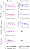
Figure 1
Figure 1
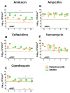
The effect of various antimicrobials on the viability of biofilm-embedded cell S. aureus (A,C,E,G,H) and K. pneumoniae (B,D,F) in mono-(red and blue boxes, respectively), and dual-species (violet boxes) cultures. Biofilms were grown for 48 h under static conditions, washed and antimicrobials were added. After 24 h of incubation, the biofilms were washed, adhered cells were scratched, suspended, and CFUs were counted by plating of 10-fold dilutions. Median values with interquartile ranges from five independent measurements are shown. Dotted lines show the level corresponding to the reduction in the CFUs count by 3 orders of magnitude (death of 99.9% of cells). ND indicated non-determined cases. Asterisks indicate statistically significant in CFUs count in treated mono- and dual-species biofilms according to the non-parametric one-way analysis of variance (Kruskal–Wallis) test, * p < 0.05.
Figure 2
Figure 2
The effect of various antimicrobials on the viability of biofilm-detached cell clumps of S. aureus (A,C,E,G,H), and K. pneumoniae (B,D,F) in mono- (red and blue boxes, respectively), and dual-species (violet boxes) cultures. Biofilms were grown for 48 h under static conditions, washed and antimicrobials were added. After 24 h of incubation, the biofilms were washed, adhered cells were scratched, suspended, and CFUs were counted by plating of 10-fold dilutions. Median values with interquartile ranges from five independent measurements are shown. Dotted lines show the level corresponding to the reduction in the CFUs count by 3 orders of magnitude (death of 99.9% of cells). ND denotes undetermined cases. Asterisks indicate statistically significant in CFUs count in treated mono- and dual-species biofilms according to the non-parametric one-way analysis of variance (Kruskal–Wallis) test, * p < 0.05.
Figure 3
Figure 3
The comparison of susceptibility to various antimicrobials (AE) of S. aureus in biofilm and in detached cell clumps in S. aureus–K. pneumoniae mixed culture. Biofilms were grown 48 h under static conditions, washed and antimicrobials were added. After 24 h of incubation, CFUs were counted in both the biofilm and culture liquid by plating of 10-fold dilutions. Median values with interquartile ranges from five independent measurements are shown. Dotted lines show the level corresponding to the reduction in the CFUs count by 3 orders of magnitude (death of 99.9% of cells). Asterisks indicate statistically significant in CFUs count in treated mono- and dual-species biofilms according to the non-parametric one-way analysis of variance (Kruskal–Wallis) test, * p < 0.05.
Figure 4
Figure 4
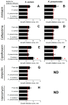
The permeability of S. aureus and K. pneumoniae dual-species biofilm compared to monospecies biofilms of both counterparts. The 48 h old biofilms were established on nitrocellulose discs and then disks were placed on agar containing an antibiotic at a concentration corresponding to 1×MBC in relation to the studied bacteria. The biofilm was covered with another nitrocellulose disc and Whatmann 3M disk was placed to allow diffusing of an antibiotic through biofilm and accumulate in the paper disk. After 24 h, paper disks were transferred to plates with bacterial lawns, and growth inhibition zones of S. aureus (A,C,E,G,H) and K. pneumoniae (B,D,F) were measured after the next 24 h growth. As a control, all manipulations were performed with the disk without an established biofilm. ND denotes undetermined cases. Asterisks indicate statistically significant differences according to the non-parametric one-way analysis of variance (Kruskal–Wallis) test, * p < 0.05.
Figure 5
Figure 5
Comparative analysis of CFUs number (A), total biomass (B), extracellular matrix mass (C), and matrix components (D) in S. aureus and K. pneumoniae mono- and dual-species biofilms. Bacterial biofilms were grown in 24-well adhesive plates for 48 h at 37 °C under static conditions. CFUs were counted by plating of 10-fold dilutions of scratched biofilm (A); the masses of the biofilm (B) and of the extracellular matrix (C) were quantified by crystal violet and Congo red staining, respectively. DNA, proteins, α- and β-polysaccharides of the matrix (D) were differentially stained with SYBR Green, Sypro Orange, ConA-TMR, CFW, respectively. Asterisks indicate statistically significant differences according to non-parametric one-way analysis of variance (Kruskal–Wallis) test, * p < 0.05.
Figure 6
Figure 6
Comparative analysis of the ultrastructure of S. aureus and K. pneumoniae mono- and dual-species biofilms. The 48 h old biofilms were analyzed with scanning electron microscopy (SEM) with magnification of 16,000× (AC) or stained with DAPI and analyzed with confocal laser scanning microscopy (CLSM) on an Olympus IX83 inverted microscope with magnification of 100× (DF) to visualize the biofilm thickness.
Figure 7
Figure 7
Distribution of the main components of mono- (A,B,D,E), and dual-species (C,F) biofilm matrix formed by S. aureus and K. pneumoniae. DNA (green), proteins (orange), α- and β-polysaccharides (pink and white, respectively), of the matrix of 48 h old biofilms were differentially stained with SYBR Green, Sypro Orange (DF), ConA-TMR, CFW (AC), respectively. The microscopy was performed on an Olympus IX83 inverted microscope with magnification of 20×.
Figure 8
Figure 8
Relative expression of icaA (A) and pgaA (B) genes in mono- and dual-species biofilms of S. aureus and K. pneumoniae. Bacterial biofilms were grown at 37 °C on BM medium in polystyrene adhesive dishes (Eppendorf). The 12 h old biofilms were harvested and total RNA was isolated. The qRT-RCR was performed in one-step approach with SYBR Blue with fluorescent detection of the product using FAM filterset. The 16s rRNA gene was used as reference for S. aureus and proC gene for K. pneumoniae. Relative expression of genes in mono-species biofilms was considered as 1×. Asterisks indicate statistically significant differences according to the non-parametric one-way analysis of variance (Kruskal–Wallis) test, * p < 0.05.

Similar articles

Cited by

References

    1. Frieri M., Kumar K., Boutin A. Antibiotic resistance. J. Infect. Public Health. 2017;10:369–378. doi: 10.1016/j.jiph.2016.08.007. - DOI - PubMed
    1. Jiang Y., Geng M., Bai L. Targeting Biofilms Therapy: Current Research Strategies and Development Hurdles. Microorganisms. 2020;8:1222. doi: 10.3390/microorganisms8081222. - DOI - PMC - PubMed
    1. Schulze A., Mitterer F., Pombo J.P., Schild S. Biofilms by bacterial human pathogens: Clinical relevance development, composition and regulation therapeutical strategies. Microb. Cell. 2021;8:28–56. doi: 10.15698/mic2021.02.741. - DOI - PMC - PubMed
    1. Uruen C., Chopo-Escuin G., Tommassen J., Mainar-Jaime R.C., Arenas J. Biofilms as promoters of bacterial antibiotic resistance and tolerance. Antibiotics. 2021;10:3. doi: 10.3390/antibiotics10010003. - DOI - PMC - PubMed
    1. Karygianni L., Ren Z., Koo H., Thurnheer T. Biofilm Matrixome: Extracellular Components in Structured Microbial Communities. Trends Microbiol. 2020;28:668–681. doi: 10.1016/j.tim.2020.03.016. - DOI - PubMed

MeSH terms

Substances