Review
. 2022 Mar 18;13(15):4239-4269.
doi: 10.1039/d2sc01003h. eCollection 2022 Apr 13.

Stimulus-responsive self-assembled prodrugs in cancer therapy

Affiliations
Review

Stimulus-responsive self-assembled prodrugs in cancer therapy

Xiao Dong et al. Chem Sci. .

Abstract

Small-molecule prodrugs have become the main toolbox to improve the unfavorable physicochemical properties of potential therapeutic compounds in contemporary anti-cancer drug development. Many approved small-molecule prodrugs, however, still face key challenges in their pharmacokinetic (PK) and pharmacodynamic (PD) properties, thus severely restricting their further clinical applications. Self-assembled prodrugs thus emerged as they could take advantage of key benefits in both prodrug design and nanomedicine, so as to maximize drug loading, reduce premature leakage, and improve PK/PD parameters and targeting ability. Notably, temporally and spatially controlled release of drugs at cancerous sites could be achieved by encoding various activable linkers that are sensitive to chemical or biological stimuli in the tumor microenvironment (TME). In this review, we have comprehensively summarized the recent progress made in the development of single/multiple-stimulus-responsive self-assembled prodrugs for mono- and combinatorial therapy. A special focus was placed on various prodrug conjugation strategies (polymer-drug conjugates, drug-drug conjugates, etc.) that facilitated the engineering of self-assembled prodrugs, and various linker chemistries that enabled selective controlled release of active drugs at tumor sites. Furthermore, some polymeric nano-prodrugs that entered clinical trials have also been elaborated here. Finally, we have discussed the bottlenecks in the field of prodrug nanoassembly and offered potential solutions to overcome them. We believe that this review will provide a comprehensive reference for the rational design of effective prodrug nanoassemblies that have clinic translation potential.


Conflict of interest statement

The authors declare no competing financial interest.

Figures

Fig. 1
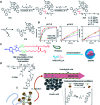
Fig. 1. Schematic illustration of (A) the overall concept of small-molecule prodrug design; (B) physical loading of (pro)-drugs onto a nanoparticle (NP); (C) strategies to improve drug delivery efficiency for tumor (I) monotherapy or (II) combinatorial therapy by using different self-assembled NPs.
Fig. 2
Fig. 2. Schematic illustration of commonly used self-assembly methods for nano-prodrug preparation. (A) Nanoparticle (NP) formation induced by various non-covalent interactions including π–π stacking, hydrogen bonding, van der Waals and Coulomb interactions. (B) Nanoprecipitation-mediated NP formation by kinetic trapping with hydrophobic–hydrophilic interactions.
Fig. 3
Fig. 3. (A) Schematic illustration of the chemical structures of DOM@DOX and its self-assemblies. Reproduced from ref. with permission from Royal Society of Chemistry, Copyright 2020. (B) Illustration of the fabrication of carrier-free, self-assembled prodrug (PEG@D:siRNA) to synergistically induce ICD and reverse tumor immunosuppression. Reproduced from ref. with permission from Elsevier Ltd, Copyright 2020. (C) Synthesis of PEG-Schiff-DOX and the acid-sensitive intracellular drug release. Reproduced from ref. with permission from Frontiers Media SA, Copyright 2021. (D) Schematic illustration of the preparation of DMMA-P-DOX and SA-P-DOX. Reproduced from ref. with permission from Elsevier Ltd, Copyright 2020. (E) Synthesis of zwitterionic BCP-TPZ prodrug conjugates. Reproduced from ref. with permission from American Chemical Society, Copyright 2021. (F) Chemical structure of acid-responsive DOX-ena-PPEG-ena-DOX and its drug activation mechanism. Reproduced from ref. with permission from American Chemical Society, Copyright 2019.
Fig. 4
Fig. 4. (A) Illustration of the design and self-assembly of pH-responsive PEGylated paclitaxel prodrugs (PKPs). (B) PTX release from PKP750, PKP1000 and PKP2000 NPs after 24 h incubation at pH 7.4, 6.8 and 5, respectively. Reproduced from ref. with permission from Elsevier Ltd, Copyright 2020. (C) Molecular structures of pH-responsive gemcitabine (Gem)–polyketal conjugates (PKGems) and nanoparticle formation by nanoprecipitation. Reproduced from ref. with permission from American Chemical Society, Copyright 2020. (D) Chemical design of polymer–GEM conjugates and acid-responsive drug release mechanism under endolysosomal conditions. Reproduced from ref. with permission from Elsevier Ltd, Copyright 2020.
Fig. 5
Fig. 5. (A) Chemical structure of the dopamine-linked amphiphilic PEGylated dendrimer; (B) reaction between dopamine and boric acid on BTZ. Reproduced from ref. with permission from Wiley-VCH, Copyright 2020. (C) Synthesis of the polymeric BTZ prodrug (P1-BTZ) and mechanism of pH-triggered drug release. Reproduced from ref. with permission from Wiley-VCH, Copyright 2020. (D) Illustration of the pH-responsive dynamic conjugation between the mussel-derived cancer-targeting peptide and BTZ. Reproduced from ref. with permission from American Chemical Society, Copyright 2019.
Fig. 6
Fig. 6. (A) Chemical structures of pH-responsive DOX-ADH-DOX-PEG and ADH-(DOX-PEG)2. Reproduced from ref. with permission from WILEY-VCH, Copyright 2019. (B) Synthesis of the homodimeric doxorubicin prodrug (D-DOXcar). Reproduced from ref. with permission from Elsevier Ltd, Copyright 2020. (C) Synthesis of D-DOXMAH and D-DOXMAH-S-PEG. Reproduced from ref. with permission from Elsevier Ltd, Copyright 2020. (D) Chemical structure and the nano-assembly of the proposed ortho-ester-linked indomethacin (IND) dimer. Reproduced from ref. with permission from American Chemical Society, Copyright 2019.
Fig. 7
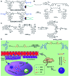
Fig. 7. (A) Chemical design of MPEG-(TK-CPT)-PPa and its self-assembly for ROS-triggered CPT release and tumor therapy under laser irradiation. Reproduced from ref. with permission from WILEY-VCH, Copyright 2020. (B) Schematic illustration of engineered PTX-S-OA/PPa-PEG2k NPs with photodynamic PEG coating for self-enhanced core–shell combination therapy. Reproduced from ref. with permission from Elsevier Ltd, Copyright 2019. (C) Schematic representation of exosome-cloaked sequential-bioactivating paclitaxel prodrug nanoassemblies for cascade-amplified PTX chemotherapy. (D) In vivo fluorescence imaging of DiR NPs and EM@DiR NPs in orthotopic MDA-MB-231 tumor-bearing mice at 2, 8, 12, and 24 h post i.v. injection of NPs. (E) Ex vivo fluorescence imaging of DiR and EM@DiR NPs in the major organs at 24 h after NP injection. Reproduced from ref. with permission from Elsevier Ltd, Copyright 2020.
Fig. 8
Fig. 8. (A) Chemical structure of an RGD-tk-Epo B conjugate and its self-assembly for tumor-targeted drug delivery. (a and b) RGD-mediated tumor targeting and cell internalization of RECNs; (c) ROS-triggered release of Epo B from RECNs upon cleavage of the thioketal linker; (d) Epo B-induced cell apoptosis. Reproduced from ref. with permission from American Chemical Society, Copyright 2020. (B) Schematic illustration of amphiphilic-block-copolymer Lapa@NPs with encapsulation of β-lapachone, which could catalyze the NAD(P)H-dependent increase in intracellular oxidative stress, thus enabling ROS-responsive drug release in tumor cells. Reproduced from ref. with permission from Elsevier Ltd, Copyright 2019. (C) Chemical structure of PPE-TK-DOX and its self-assembly with encapsulation of photosensitizer Ce6 to enable ROS-triggered drug release under laser irradiation. Reproduced from ref. with permission from Elsevier Ltd, Copyright 2019.
Fig. 9
Fig. 9. (A) Chemical structures of the ROS-responsive P3C-Asp prodrug and its self-assembly for TME regulation and enhanced tumor immunotherapy. Reproduced from ref. with permission from Chinese Chemical Society, Copyright 2020. (B) Schematic representation of engineered PTX prodrug-based micelles (PEG-B-PTX) for ROS-triggered drug release in tumor therapy. Reproduced from ref. with permission from Elsevier Ltd, Copyright 2020. (C) Chemical structure of PMPC-b-P[MPA(Cap)-co-TPMA]–PAEMA (PMMTAb-Cap) and its self-assembly for ROS-responsive drug release. Reproduced from ref. with permission from American Chemical Society, Copyright 2019. (D) Chemical structures of PEG–PGA–β-CD, Ada–BODIPY, Ada–PTX and their nanoassemblies at an optimized ratio for combined chemo-dynamic therapy. Reproduced from ref. with permission from Wiley-VCH, Copyright 2019. (E) Chemical design of PEG-PO-PTX and its drug release mechanism in the presence of H2O2. Reproduced from ref. with permission from Elsevier Ltd, Copyright 2019.
Fig. 10
Fig. 10. (A) Schematic illustration of PEGylated PPa-driven nanoassembly of CTX-S-CTX. (B) CTX activation and release mechanism from CTX-S-CTX. Reproduced from ref. with permission from Ivyspring International Publisher, Copyright 2021. (C) Schematic illustration of the construction of PPa-S-PTX-based nanoassemblies for synergistic photo-chemotherapy. Reproduced from ref. with permission from Elsevier Ltd, Copyright 2019. (D) Molecular structures of the ROS-responsive HRC prodrug and the formation of nanoparticles by encapsulating HRC into F127 polymeric micelles for cancer imaging and combinatorial chemo-photodynamic therapy. Reproduced from ref. with permission from American Chemical Society, Copyright 2020.
Fig. 11
Fig. 11. (A) Chemical structure of mitochondrion-targeting prodrug–peptide conjugates. Reproduced from ref. with permission from John Wiley & Sons, Inc., Copyright 2021. (B) Illustration of the engineered redox-sensitive polyHCPT-based NPs and redox-responsive drug release mechanism. Reproduced from ref. with permission from Elsevier Ltd, Copyright 2020. (C) Chemical structure of CPT-S-S-PEG-iRGD. Reproduced from ref. with permission from Elsevier Ltd, Copyright 2020. (D) Schematic illustration of PTX–CIT conjugates linked by different chemical bonds (sulfur/selenium/carbon). Reproduced from ref. with permission from Springer Nature, Copyright 2019. (E) Chemical structure of the polymeric PMPT prodrug and its self-assembly with encapsulation of two AIE photosensitizers (PyTPE and TB) for imaging-guided PDT and drug release. Reproduced from ref. with permission from American Chemical Society, Copyright 2021.
Fig. 12
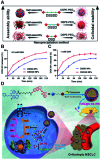
Fig. 12. (A) Nano-prodrug constructed by self-assembly of DSD, DSSD and DSSSD with or without DSPE-PEG2K. In vitro DOX-SH release from DSSD and DSSSD in (B) 0.5 mM or (C) 1 mM GSH. Reproduced from ref. with permission from American Association for the Advancement of Science, Copyright 2020. (D) Chemical structure of a gefitinib linked near-infrared dye and its self-assembly with encapsulation of celastrol for tumor therapy. Reproduced from ref. with permission from Wiley-VCH, Copyright 2019.
Fig. 13
Fig. 13. (A) Chemical design of the amphiphilic imidazoquinoline (IMDQ) prodrug and its self-assembly into vesicular nanostructures. Reproduced from ref. with permission from American Chemical Society, Copyright 2020. (B) Illustration of the F-integrated ASO-b-PEG-b-PCL triblock copolymer and its self-assembly to reverse chemoresistance. Reproduced from ref. with permission from American Chemical Society, Copyright 2021. (C) Preparation of TME-responsive nano-prodrugs by co-self-assembly of a photothermal agent and IDO inhibitor for cancer immunotherapy. Reproduced from ref. with permission from Elsevier Ltd, Copyright 2020. (D) Chemical structures of a cathepsin B-responsive prodrug and its self-assemblies for combinatorial chemo-photodynamic therapy to potentiate the effective checkpoint blockade-based tumor immunotherapy. Reproduced from ref. with permission from American Chemical Society, Copyright 2021.
Fig. 14
Fig. 14. (A) Schematic illustration of a hypoxia-activable semiconducting polymeric nano-prodrug for synergistic chemo-photodynamic therapy. Reproduced from ref. with permission from Wiley-VCH, Copyright 2018. (B) Chemical structures of a hypoxia-responsive nano-prodrug (Ce6/PTX2-Azo) and its self-assembly for synergistic photodynamic-chemotherapy. Reproduced from ref. with permission from Wiley-VCH., Copyright 2020.
Fig. 15
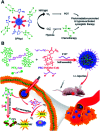
Fig. 15. (A) Chemical design of GGT-activable cationizing PBEAGA-CPT and the non-GGT-activable PEAGA-CPT, and the mechanism for GGT-catalysed γ-glutamylamide hydrolysis. Reproduced from ref. with permission from Springer Nature, Copyright 2019. (B) Molecular structure of the redox/pH dual-responsive CPT-ss-poly(BYP-hyd-DOX-co-EEP). Reproduced from ref. with permission from American Chemical Society, Copyright 2019. (C) Schematic illustration of the PEG-GAx/Pt formation through coordination of the polyphenol and CDDP, and the mechanism for intracellular dual-responsive drug release. Reproduced from ref. with permission from Elsevier Ltd, Copyright 2020. (D) Molecular structures of mPEG-Phe-TK-Phe-hyd-DOX and its self-assemblies for tumor therapy. Reproduced from ref. with permission from Elsevier Ltd, Copyright 2021. (E) Chemical structure of diblock pDHPMA-DOX. Reproduced from ref. with permission from Elsevier Ltd, Copyright 2019.
Fig. 16
Fig. 16. (A) Chemical design and synthetic routes of the PTX-chemogene conjugate and its self-assembly for combination therapy to overcome drug resistance. Reproduced from ref. with permission from Wiley-VCH, Copyright 2020. (B) Schematic representation of sulfur/selenium/tellurium-bridged homodimeric prodrugs and their intracellular ROS/GSH dual-responsive drug release. (C) HPLC analysis of PTX2-S, PTX2-Se and PTX2-Te dimers in the presence of 1 mM H2O2 or 10 mM DTT for 24 h at 37 °C. Reproduced from ref. with permission from Elsevier Ltd, Copyright 2020.
None
Xiao Dong
None
Shao Q. Yao

Similar articles

Cited by

References

    1. Ferlay J. Colombet M. Soerjomataram I. Parkin D. M. Pineros M. Znaor A. Bray F. Int. J. Cancer. 2021;149:778. doi: 10.1002/ijc.33588. - DOI - PubMed
    1. Markham M. J. Wachter K. Agarwal N. Bertagnolli M. M. Chang S. M. Dale W. Diefenbach C. S. M. Rodriguez-Galindo C. George D. J. Gilligan T. D. Harvey R. D. Johnson M. L. Kimple R. J. Knoll M. A. LoConte N. Maki R. G. Meisel J. L. Meyerhardt J. A. Pennell N. A. Rocque G. B. Sabel M. S. Schilsky R. L. Schneider B. J. Tap W. D. Uzzo R. G. Westin S. N. J. Clin. Oncol. 2020;38:1081. doi: 10.1200/JCO.19.03141. - DOI - PubMed
    1. Schirrmacher V. Internet J. Oncol. 2019;54:407–419. - PMC - PubMed
    1. Waks A. G. Winer E. P. JAMA. 2019;321:288–300. doi: 10.1001/jama.2018.19323. - DOI - PubMed
    1. Ko E. C. Raben D. Formenti S. C. Clin. Cancer Res. 2018;24:5792–5806. doi: 10.1158/1078-0432.CCR-17-3620. - DOI - PubMed