. 2022 Apr 12;88(7):e0252921.
doi: 10.1128/aem.02529-21. Epub 2022 Mar 24.

A c-di-GMP Signaling Cascade Controls Motility, Biofilm Formation, and Virulence in Burkholderia thailandensis

Affiliations

A c-di-GMP Signaling Cascade Controls Motility, Biofilm Formation, and Virulence in Burkholderia thailandensis

Zhuo Wang et al. Appl Environ Microbiol. .

Abstract

As a key bacterial second messenger, cyclic di-GMP (c-di-GMP) regulates various physiological processes, such as motility, biofilm formation, and virulence. Cellular c-di-GMP levels are regulated by the opposing activities of diguanylate cyclases (DGCs) and phosphodiesterases (PDEs). Beyond that, the enzymatic activities of c-di-GMP metabolizing proteins are controlled by a variety of extracellular signals and intracellular physiological conditions. Here, we report that pdcA (BTH_II2363), pdcB (BTH_II2364), and pdcC (BTH_II2365) are cotranscribed in the same operon and are involved in a regulatory cascade controlling the cellular level of c-di-GMP in Burkholderia thailandensis. The GGDEF domain-containing protein PdcA was found to be a DGC that modulates biofilm formation, motility, and virulence in B. thailandensis. Moreover, the DGC activity of PdcA was inhibited by phosphorylated PdcC, a single-domain response regulator composed of only the phosphoryl-accepting REC domain. The phosphatase PdcB affects the function of PdcA by dephosphorylating PdcC. The observation that homologous operons of pdcABC are widespread among betaproteobacteria and gammaproteobacteria suggests a general mechanism by which the intracellular concentration of c-di-GMP is modulated to coordinate bacterial behavior and virulence. IMPORTANCE The transition from planktonic cells to biofilm cells is a successful strategy adopted by bacteria to survive in diverse environments, while the second messenger c-di-GMP plays an important role in this process. Cellular c-di-GMP levels are mainly controlled by modulating the activity of c-di-GMP-metabolizing proteins via the sensory domains adjacent to their enzymatic domains. However, in most cases how c-di-GMP-metabolizing enzymes are modulated by their sensory domains remains unclear. Here, we reveal a new c-di-GMP signaling cascade that regulates motility, biofilm formation, and virulence in B. thailandensis. While pdcA, pdcB, and pdcC constitute an operon, the phosphorylated PdcC binds the PAS sensory domain of PdcA to inhibit its DGC activity, with PdcB dephosphorylating PdcC to derepress the activity of PdcA. We also show this c-di-GMP regulatory model is widespread in the phylum Proteobacteria. Our study expands the current knowledge of how bacteria regulate intracellular c-di-GMP levels.

Keywords: Burkholderia thailandensis; biofilm; c-di-GMP; motility; virulence.


Conflict of interest statement

The authors declare no conflict of interest.

Figures
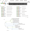
FIG 1
FIG 1
Cotranscription and wide distribution of homologous BTH_II2365BTH_II2363 operon. (A) Gene organization of BTH_II2365BTH_II2363 cluster in B. thailandensis E264. These genes are predicted to encode a single-domain response regulator (BTH_II2365), a phosphatase (BTH_II2364), and a diguanylate cyclase with PAS-GGDEF domain (BTH_II2363). (B) Agarose gel (1%, wt/vol) electrophoresis of the PCR-amplified products. cDNA synthesized from B. thailandensis E264 total RNA was used as a template to determine BTH_II2365BTH_II2363 cotranscription with primers CTF1/CTR1, CTF2/CTR2, and CTF3/CTR3. CTNF and CTNR primers from an unexpressed region were used as a negative control to rule out possible genomic DNA contamination during RNA preparation. (C) Homologous operons of BTH_II2365BTH_II2363 are widespread in betaproteobacteria and gammaproteobacteria. (D) The phylogenetic tree of BTH_II2363 homologs was conducted in MEGA 7 by using the neighbor-joining method (bootstrap, 1,000 replicates). Lines are colored based on the bacterial taxonomy of the corresponding organism. Green, betaproteobacteria; blue, gammaproteobacteria.
FIG 2
FIG 2
BTH_II2363, BTH_II2364, and BTH_II2365 regulate biofilm formation and motility by indirectly modulating intracellular c-di-GMP levels. (A and B) Deletion of BTH_II2363, BTH_II2364, or BTH_II2365 regulates swimming motility (A) and biofilm formation (B) of B. thailandensis. (C and D) Overexpression of BTH_II2363, BTH_II2364, or BTH_II2365 regulates swimming motility (C) and biofilm formation (D) of B. thailandensis. Swimming motility of the indicated strains was tested in the semisolid agar medium (up), and the sizes of swimming zones were measured (down). Biofilm formation of the indicated strains was displayed with crystal violet staining (up) and quantified with optical density measurement (down). Data shown are the averages and standard deviations (SD) from three independent experiments. (E and F) Growth curves of the indicated strains under normal conditions. Growth of the indicated strains in LB (E) or M63 (F) medium was monitored by measuring OD600 at indicated time points at 37°C. (G) The intracellular concentrations of c-di-GMP in WT, Δ2363, Δ2364, Δ2365, and the corresponding complemented strains were detected by LC-MS/MS. Data shown are the averages and SD from six independent experiments. *, P < 0.05; **, P < 0.01; ***, P < 0.001.
FIG 3
FIG 3
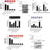
PdcC interacts with PdcA to inhibit c-di-GMP synthesis. (A) Interaction between PdcC and PdcA was assessed using MacConkey plates (up), and the strength of interaction was quantified by measurement of β-galactosidase activity (down). (B) GST pulldown assay confirmed the interaction between PdcC and PdcA. Recombinant protein PdcC-His6 was incubated with GST or GST-PdcA individually, and protein complexes were captured by glutathione beads and detected by Western blotting. (C) PdcC inhibits the DGC activity of PdcA. PdcA was incubated with GTP in the presence or absence of PdcC at 30°C for 0, 30, and 60 min, and the products were analyzed by HPLC (Fig. S2). The levels of synthesized c-di-GMP in the samples were determined from a standard curve established with a serially diluted c-di-GMP solution. To evaluate the effect of the phosphorylation of PdcC on PdcA DGC activity, PdcC was pretreated by 25 mM acetyl phosphate (AcP) for 30 min at 30°C. (D) Swimming motility of the indicated strains was tested in the semisolid agar medium (up), and the diameters of swimming zones were measured (down). (E) Biofilm formation of the indicated strains was displayed with crystal violet staining (up) and quantified with optical density measurement (down). (F) Interactions between PdcC and the PAS domain of PdcA were assessed using MacConkey plates (up), and the strength of interaction was quantified by measurement of β-galactosidase activity (down). (G) GST pulldown assay confirmed the interaction between PdcC and the PAS domain of PdcA. Recombinant protein PdcC-His6 was incubated with GST, GST-PAS, or GST-GGDEF individually, and protein complexes were captured by glutathione beads and detected by Western blotting. Data shown are the averages and SD from three independent experiments. *, P < 0.05; ***, P < 0.001; n.s., not significant.
FIG 4
FIG 4
PdcB dephosphorylates PdcC. (A) Interaction between PdcB and PdcC was assessed using MacConkey plates (up), and the strength of interaction was quantified by measurement of β-galactosidase activity (down). (B) GST pulldown assay confirmed the interaction between PdcB and PdcC. Recombinant protein PdcC-His6 was incubated with GST or GST-PdcB individually, and protein complexes were captured by glutathione beads and detected by Western blotting. (C) PdcB can dephosphorylate PdcC in vitro. The inorganic phosphate released from PdcC was detected by using a phosphate sensor assay kit. The reaction buffer was used as a negative control to exclude the inorganic phosphate in the reaction buffer. Fluorescence signals were measured using a SpectraMax M2 plate reader (Molecular Devices) with excitation/emission wavelengths of 420/450 nm. (D) Swimming motility of the indicated strains was tested in the semisolid agar medium (up) and the diameters of swimming zones were measured (down). (E) Biofilm formation of the indicated strains was displayed with the crystal violet staining (up) and quantified with optical density measurement (down). (F) The swimming motility of WT, mutant, and complemented strains was tested (up) and measured (down). (G) Biofilm formation of the indicated strains was displayed (up) and quantified (down). Data shown are the averages and SD from three independent experiments. **, P < 0.01; ***, P < 0.001; n.s., not significant.
FIG 5
FIG 5
PdcABC signaling cascade contributes to the virulence of B. thailandensis. (A) Virulence survival of relative B. thailandensis strains in G. mellonella larvae. Ordinate represents the mean percent survival rate of G. mellonella infected with different strains after 16 h. (B) Western blot detection of BipD secretion in relative B. thailandensis strains. Proteins in the culture supernatant of relevant B. thailandensis strains were probed for specific anti-BipD rabbit polyclonal antibody. For the pellet fraction, isocitrate dehydrogenase (ICDH) was used as a loading control. (C) The expression of bipD in relative B. thailandensis strains. Data shown are the averages and SD from three independent experiments. *, P < 0.05; **, P < 0.01; ***, P < 0.001.
FIG 6
FIG 6
Proposed model of the PdcABC signaling cascade in B. thailandensis. PdcA, PdcB, and PdcC cooperatively regulate biofilm formation, motility, and virulence in B. thailandensis. Phosphorylated PdcC acts as an inhibitor controlling the DGC activity of PdcA. The levels of phosphorylation of PdcC were affected by the phosphatase PdcB, while the phosphoryl group donors of PdcC remain unknown.

Similar articles

Cited by

References

    1. Hengge R. 2009. Principles of c-di-GMP signalling in bacteria. Nat Rev Microbiol 7:263–273. 10.1038/nrmicro2109. - DOI - PubMed
    1. D'Argenio DA, Miller SI. 2004. Cyclic di-GMP as a bacterial second messenger. Microbiology (Reading) 150:2497–2502. 10.1099/mic.0.27099-0. - DOI - PubMed
    1. Romling U, Galperin MY, Gomelsky M. 2013. Cyclic di-GMP: the first 25 years of a universal bacterial second messenger. Microbiol Mol Biol Rev 77:1–52. 10.1128/MMBR.00043-12. - DOI - PMC - PubMed
    1. Jenal U, Reinders A, Lori C. 2017. Cyclic di-GMP: second messenger extraordinaire. Nat Rev Microbiol 15:271–284. 10.1038/nrmicro.2016.190. - DOI - PubMed
    1. Floyd KA, Lee CK, Xian W, Nametalla M, Valentine A, Crair B, Zhu S, Hughes HQ, Chlebek JL, Wu DC, Hwan Park J, Farhat AM, Lomba CJ, Ellison CK, Brun YV, Campos-Gomez J, Dalia AB, Liu J, Biais N, Wong GCL, Yildiz FH. 2020. c-di-GMP modulates type IV MSHA pilus retraction and surface attachment in Vibrio cholerae. Nat Commun 11:1549. 10.1038/s41467-020-15331-8. - DOI - PMC - PubMed

Publication types

MeSH terms

Supplementary concepts