Ndufs4 knockout mouse models of Leigh syndrome: pathophysiology and intervention
- PMID: 34849584
- PMCID: PMC8967107
- DOI: 10.1093/brain/awab426
Ndufs4 knockout mouse models of Leigh syndrome: pathophysiology and intervention
Abstract
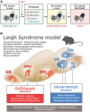
Mitochondria are small cellular constituents that generate cellular energy (ATP) by oxidative phosphorylation (OXPHOS). Dysfunction of these organelles is linked to a heterogeneous group of multisystemic disorders, including diabetes, cancer, ageing-related pathologies and rare mitochondrial diseases. With respect to the latter, mutations in subunit-encoding genes and assembly factors of the first OXPHOS complex (complex I) induce isolated complex I deficiency and Leigh syndrome. This syndrome is an early-onset, often fatal, encephalopathy with a variable clinical presentation and poor prognosis due to the lack of effective intervention strategies. Mutations in the nuclear DNA-encoded NDUFS4 gene, encoding the NADH:ubiquinone oxidoreductase subunit S4 (NDUFS4) of complex I, induce 'mitochondrial complex I deficiency, nuclear type 1' (MC1DN1) and Leigh syndrome in paediatric patients. A variety of (tissue-specific) Ndufs4 knockout mouse models were developed to study the Leigh syndrome pathomechanism and intervention testing. Here, we review and discuss the role of complex I and NDUFS4 mutations in human mitochondrial disease, and review how the analysis of Ndufs4 knockout mouse models has generated new insights into the MC1ND1/Leigh syndrome pathomechanism and its therapeutic targeting.
Keywords: Leigh syndrome; intervention; mouse model; pathomechanism.
© The Author(s) (2021). Published by Oxford University Press on behalf of the Guarantors of Brain.
Figures


Similar articles
-
NDUFS4 deletion triggers loss of NDUFA12 in Ndufs4-/- mice and Leigh syndrome patients: A stabilizing role for NDUFAF2.Biochim Biophys Acta Bioenerg. 2020 Aug 1;1861(8):148213. doi: 10.1016/j.bbabio.2020.148213. Epub 2020 Apr 23. Biochim Biophys Acta Bioenerg. 2020. PMID: 32335026
-
Proteomic and metabolomic analyses of mitochondrial complex I-deficient mouse model generated by spontaneous B2 short interspersed nuclear element (SINE) insertion into NADH dehydrogenase (ubiquinone) Fe-S protein 4 (Ndufs4) gene.J Biol Chem. 2012 Jun 8;287(24):20652-63. doi: 10.1074/jbc.M111.327601. Epub 2012 Apr 25. J Biol Chem. 2012. PMID: 22535952 Free PMC article.
-
The NDUFS4 Knockout Mouse: A Dual Threat Model of Childhood Mitochondrial Disease and Normative Aging.Methods Mol Biol. 2021;2277:143-155. doi: 10.1007/978-1-0716-1270-5_10. Methods Mol Biol. 2021. PMID: 34080150
-
Cellular and animal models for mitochondrial complex I deficiency: a focus on the NDUFS4 subunit.IUBMB Life. 2013 Mar;65(3):202-8. doi: 10.1002/iub.1127. Epub 2013 Feb 3. IUBMB Life. 2013. PMID: 23378164 Review.
-
Mouse models for nuclear DNA-encoded mitochondrial complex I deficiency.J Inherit Metab Dis. 2011 Apr;34(2):293-307. doi: 10.1007/s10545-009-9005-x. Epub 2010 Jan 27. J Inherit Metab Dis. 2011. PMID: 20107904 Review.
Cited by
-
Accelerated differentiation of neo-W nuclear-encoded mitochondrial genes between two climate-associated bird lineages signals potential co-evolution with mitogenomes.Heredity (Edinb). 2024 Nov;133(5):342-354. doi: 10.1038/s41437-024-00718-w. Epub 2024 Aug 22. Heredity (Edinb). 2024. PMID: 39174672 Free PMC article.
-
Reduced expression of mitochondrial complex I subunit Ndufs2 does not impact healthspan in mice.Sci Rep. 2022 Mar 25;12(1):5196. doi: 10.1038/s41598-022-09074-3. Sci Rep. 2022. PMID: 35338200 Free PMC article.
-
An engineered human cardiac tissue model reveals contributions of systemic lupus erythematosus autoantibodies to myocardial injury.bioRxiv [Preprint]. 2024 Mar 12:2024.03.07.583787. doi: 10.1101/2024.03.07.583787. bioRxiv. 2024. Update in: Nat Cardiovasc Res. 2024 Sep;3(9):1123-1139. doi: 10.1038/s44161-024-00525-w. PMID: 38559188 Free PMC article. Updated. Preprint.
-
Interleukin-6-elicited chronic neuroinflammation may decrease survival but is not sufficient to drive disease progression in a mouse model of Leigh syndrome.J Inflamm (Lond). 2024 Jan 11;21(1):1. doi: 10.1186/s12950-023-00369-4. J Inflamm (Lond). 2024. PMID: 38212783 Free PMC article.
-
Animal Models of Mitochondrial Diseases Associated with Nuclear Gene Mutations.Acta Naturae. 2023 Oct-Dec;15(4):4-22. doi: 10.32607/actanaturae.25442. Acta Naturae. 2023. PMID: 38234606 Free PMC article.
References
-
- Mailloux RJ, McBride SL, Harper ME.. Unearthing the secrets of mitochondrial ROS and glutathione in bioenergetics. Trends Biochem Sci. 2013;38(12):592–602. - PubMed
-
- Willems PHGM, Rossignol R, Dieteren CE, Murphy MP, Koopman WJH.. Redox homeostasis and mitochondrial dynamics. Cell Metab. 2015;22(2):207–218. - PubMed
Publication types
MeSH terms
Substances
LinkOut - more resources
Full Text Sources
Medical
Molecular Biology Databases

