

BMP signaling and skeletal development in fibrodysplasia ossificans progressiva (FOP)
- PMID: 34133058
- PMCID: PMC9068236
- DOI: 10.1002/dvdy.387
BMP signaling and skeletal development in fibrodysplasia ossificans progressiva (FOP)
Abstract
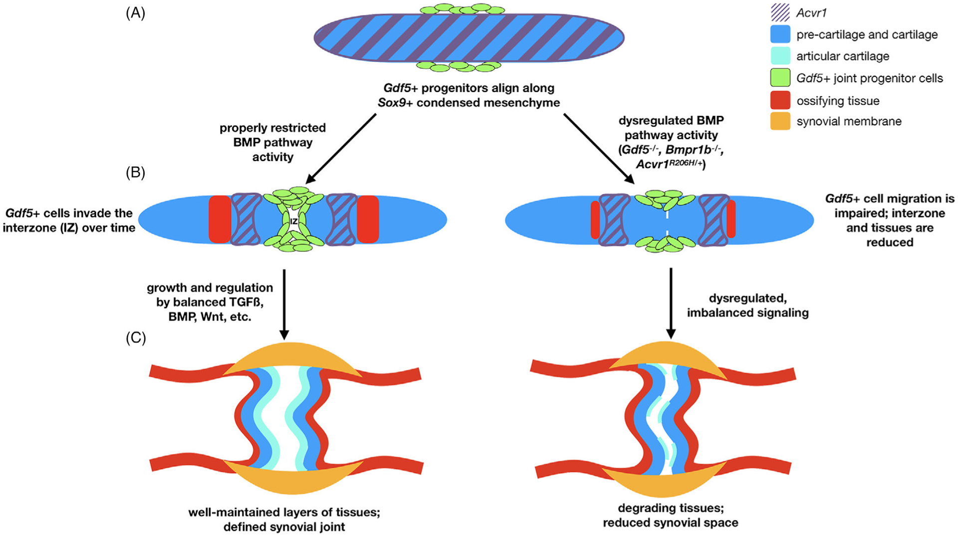
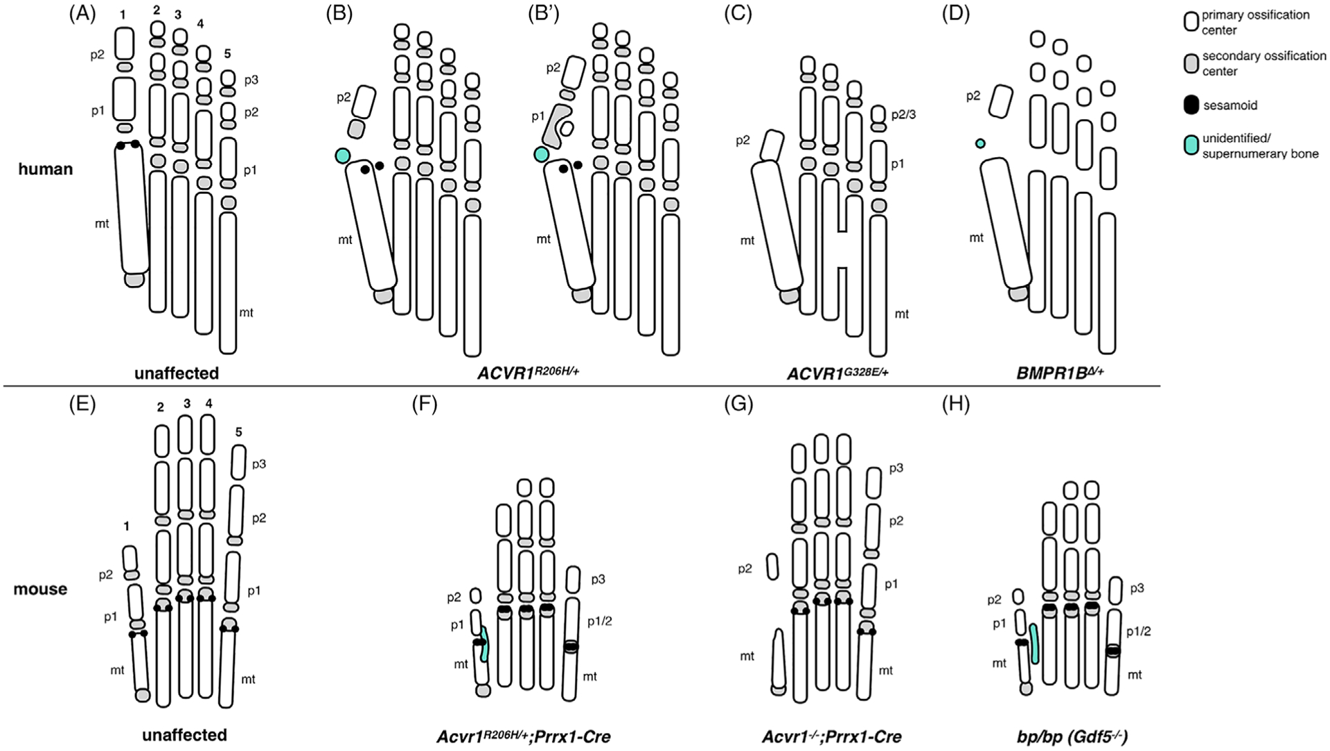
Fibrodysplasia ossificans progressiva (FOP) is an ultra-rare genetic disease caused by increased BMP pathway signaling due to mutation of ACVR1, a bone morphogenetic protein (BMP) type 1 receptor. The primary clinical manifestation of FOP is extra-skeletal bone formation (heterotopic ossification) within soft connective tissues. However, the underlying ACVR1 mutation additionally alters skeletal bone development and nearly all people born with FOP have bilateral malformation of the great toes as well as other skeletal malformations at diverse anatomic sites. The specific mechanisms through which ACVR1 mutations and altered BMP pathway signaling in FOP influence skeletal bone formation during development remain to be elucidated; however, recent investigations are providing a clearer understanding of the molecular and developmental processes associated with ACVR1-regulated skeletal formation.
Keywords: ACVR1; FOP; bone morphogenetic protein; fibrodysplasia ossificans progressiva; heterotopic ossification; joint development; toe/digit malformation.
© 2021 American Association for Anatomy.
Figures
Similar articles
-
Dysregulated BMP signaling through ACVR1 impairs digit joint development in fibrodysplasia ossificans progressiva (FOP).Dev Biol. 2021 Feb;470:136-146. doi: 10.1016/j.ydbio.2020.11.004. Epub 2020 Nov 17. Dev Biol. 2021. PMID: 33217406 Free PMC article.
-
Skeletal malformations and developmental arthropathy in individuals who have fibrodysplasia ossificans progressiva.Bone. 2020 Jan;130:115116. doi: 10.1016/j.bone.2019.115116. Epub 2019 Oct 23. Bone. 2020. PMID: 31655222
-
The ACVR1 R206H mutation found in fibrodysplasia ossificans progressiva increases human induced pluripotent stem cell-derived endothelial cell formation and collagen production through BMP-mediated SMAD1/5/8 signaling.Stem Cell Res Ther. 2016 Aug 17;7(1):115. doi: 10.1186/s13287-016-0372-6. Stem Cell Res Ther. 2016. PMID: 27530160 Free PMC article.
-
Fibrodysplasia ossificans progressiva (FOP): A disorder of osteochondrogenesis.Bone. 2020 Nov;140:115539. doi: 10.1016/j.bone.2020.115539. Epub 2020 Jul 27. Bone. 2020. PMID: 32730934 Free PMC article. Review.
-
Role of altered signal transduction in heterotopic ossification and fibrodysplasia ossificans progressiva.Curr Osteoporos Rep. 2011 Jun;9(2):83-8. doi: 10.1007/s11914-011-0046-3. Curr Osteoporos Rep. 2011. PMID: 21340697 Free PMC article. Review.
Cited by
-
Computational Insights into the Structural and Functional Impacts of nsSNPs of Bone Morphogenetic Proteins.Biomed Res Int. 2022 Jul 4;2022:4013729. doi: 10.1155/2022/4013729. eCollection 2022. Biomed Res Int. 2022. Retraction in: Biomed Res Int. 2024 Jan 9;2024:9891352. doi: 10.1155/2024/9891352. PMID: 35832847 Free PMC article. Retracted.
-
Development of an Animal Model for Traumatic Brain Injury Augmentation of Heterotopic Ossification in Response to Local Injury.Biomedicines. 2023 Mar 18;11(3):943. doi: 10.3390/biomedicines11030943. Biomedicines. 2023. PMID: 36979922 Free PMC article.
-
Rapid Progression of Heterotopic Ossification in Severe Variant of Fibrodysplasia Ossificans Progressiva with p.Arg258Gly in ACVR1: A Case Report and Review of Clinical Phenotypes.Case Rep Genet. 2022 Aug 25;2022:5021758. doi: 10.1155/2022/5021758. eCollection 2022. Case Rep Genet. 2022. PMID: 36060212 Free PMC article.
-
Pay Attention to the Osteochondromas in Fibrodysplasia Ossificans Progressiva.Orthop Surg. 2024 Mar;16(3):781-787. doi: 10.1111/os.13956. Epub 2024 Jan 7. Orthop Surg. 2024. PMID: 38185793 Free PMC article.
-
Lack of evidence that fibrillin1 regulates bone morphogenetic protein 4 activity in kidney or lung.Dev Dyn. 2023 Jun;252(6):761-769. doi: 10.1002/dvdy.578. Epub 2023 Mar 6. Dev Dyn. 2023. PMID: 36825302 Free PMC article.
References
-
- Ashton BA, Allen TD, Howlett CR, Eaglesom CC, Hattori A, Owen M. Formation of bone and cartilage by marrow stromal cells in diffusion chambers in vivo. Clin Orthop Relat Res. 1980;151:294–307. - PubMed
-
- Fell HB. The histogenesis of cartilage and bone in the long bones of the embryonic fowl. J Morphol. 1925;40(3):417–459. 10.1002/jmor.1050400302. - DOI
-
- Thorogood PV, Hinchliffe JR. An analysis of the condensation process during chondrogenesis in the embryonic chick hind limb. J Embryol Exp Morphol. 1975;33(3):581–606. - PubMed
Publication types
MeSH terms
Substances
Grants and funding
LinkOut - more resources
Full Text Sources
Other Literature Sources
