Review
. 2020 Apr 6;48(6):2807-2829.
doi: 10.1093/nar/gkaa112.

Cyclic di-AMP, a second messenger of primary importance: tertiary structures and binding mechanisms

Affiliations
Review

Cyclic di-AMP, a second messenger of primary importance: tertiary structures and binding mechanisms

Jin He et al. Nucleic Acids Res. .

Abstract

Cyclic diadenylate (c-di-AMP) is a widespread second messenger in bacteria and archaea that is involved in the maintenance of osmotic pressure, response to DNA damage, and control of central metabolism, biofilm formation, acid stress resistance, and other functions. The primary importance of c-di AMP stems from its essentiality for many bacteria under standard growth conditions and the ability of several eukaryotic proteins to sense its presence in the cell cytoplasm and trigger an immune response by the host cells. We review here the tertiary structures of the domains that regulate c-di-AMP synthesis and signaling, and the mechanisms of c-di-AMP binding, including the principal conformations of c-di-AMP, observed in various crystal structures. We discuss how these c-di-AMP molecules are bound to the protein and riboswitch receptors and what kinds of interactions account for the specific high-affinity binding of the c-di-AMP ligand. We describe seven kinds of non-covalent-π interactions between c-di-AMP and its receptor proteins, including π-π, C-H-π, cation-π, polar-π, hydrophobic-π, anion-π and the lone pair-π interactions. We also compare the mechanisms of c-di-AMP and c-di-GMP binding by the respective receptors that allow these two cyclic dinucleotides to control very different biological functions.


Figures

Figure 1.
Figure 1.
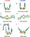
Structures of c-di-GMP and c-di-AMP molecules. (A, B) U-type conformations of c-di-GMP from the PDB entry 2RDE (in complex with the V. cholerae protein PlzD) (A), and c-di-AMP from the PDB entry 4YP1 (in complex with the cation-proton antiporter CpaA from S. aureus) (B); the two bases are oriented in an almost parallel fashion. (C, D) Extended conformations of c-di-GMP from its complex with the EAL domain of Klebsiella pneumoniae photoreceptor BlrP1, PDB: 3GFX (C), and c-di-AMP from its complex with the RECON protein, PDB: 5UXF (D); the two bases are wide apart. (E, F) Widespread conformations of c-di-GMP, a dimer from its complex with the cellulose synthase from Rhodobacter sphaeroides, PDB: 4P02 (E), and c-di-AMP, a V-type monomer from its complex with the YdaO riboswitch, PDB: 4QLN, with two adenine bases pointed in the same direction but inclined by ∼45° from vertical (F). The carbon atoms are in green (in panel E, also in light blue), N atoms are in blue, O in red and P in orange.
Figure 2.
Figure 2.
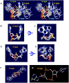
Diversity of c-di-AMP conformations. (A) Stereo view of three main conformations of c-di-AMP: the U-, V- and E-types. (BC) Superposition of U-type c-di-AMP molecules from PDB entries 4QSH1, 4XTT, 4YP1, 5CFN, and 5F29 displayed from two different angles. (D,E) Superposition of V-type c-di-AMP molecules from PDB entries 4RLE, 4RWW, 4WK1, 4D3H and 4S1B displayed in two different angles. (F) E-type c-di-AMP molecules from PDB entries 5XSN, 5UXF and 4QSH2. (G) The O-type c-di-AMP from PDB entry 5KS7.
Figure 3.
Figure 3.
Binding of c-di-AMP to the YdaO riboswitch. (A) Stereo view of two c-di-AMP molecules (shown as spheres) bound to the YdaO riboswitch from B. subtilis (PDB: 4W90), whose sugar-phosphate backbone is shown in cyan (116). C-di-AMP-binding ribose moieties of G27, U62, and C82 are shown in stick representation and indicated by dashed circles. (B–D) Role of the ribose hydroxyl groups of the riboswitch in binding c-di-AMP (carbon atoms in grey). Hoogsteen side hydrogen bonds between N6 and N7 atoms of adenine bases of c-di-AMP and the ribose 2′- and 3′-hydroxyls from riboswitch bases G27 (B), U62 (C), and C82 (D) are coupled with adenine-N1 binding by 2′-hydroxyl groups of G60 (B), G25 (C) and G5(D). The abundance of hydrogen bonds explains the much higher affinity of c-di-AMP binding by the riboswitch than by protein receptors.
Figure 4.
Figure 4.
Binding of c-di-AMP to CpaA. Complex of U-shaped c-di-AMP with two RCK_C domains of S. aureus K+/H+ antiporter CpaA (PDB: 5F29) (99) shows each water molecule (solid blue circles in (A) and magenta spheres in (B)) coordinated by the N7 atom and phosphate oxygen atom of c-di-AMP, as well as the N atom from the side chain of His184. The lone pair electrons of oxygen atom form a water-mediated lone pair–adenine interaction (indicated by a white arrow in B). The N1 and N6 atoms of c-di-AMP adenines bind the backbone nitrogen and carbonyl oxygen atoms of Phe171, respectively, of both RCK_C domains.
Figure 5.
Figure 5.
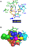
Binding of c-di-AMP to KtrA. (A) Complex of U-shaped c-di-AMP with two RCK_C domains of S. aureus K+ transporter KtrA (PDB: 4XTT) (111) shows an H2O lone pairadenine interaction, in which a water molecule is coordinated by the guanidino nitrogen atom of Arg169 and the phosphate oxygen atom, with its lone pair electrons interacting with the adenine base, as shown in (B). Here, the binding mode is asymmetric and the side-chain guanidino group of Arg169 from the other RCK_C monomer (marked by a blue arrow) is turned sideways to prevent a steric clash with the water oxygen atom.
Figure 6.
Figure 6.
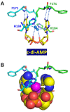
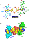
Binding of c-di-AMP to the CBS domains of the carnitine transporter OpuCA. In this structure (PDB: 5KS7), c-di-AMP is in an O-type conformation and both adenine bases are sandwiched between Val280 on one side and Ile355 and Tyr342 on the other. In (A), blue dotted lines indicate the hydrogen bonds between the side-chain hydroxyl group of Thr282 and the N6 and N7 atoms of each adenine base. The bottom part (B) shows this binding site in a space-filling model.
Figure 7.
Figure 7.
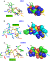
Binding of c-di-AMP to DarA/PstA proteins. Mechanisms of c-di-AMP binding to the DarA/PstA proteins from B. subtilis (PDB: 4RLE, top row, A), S. aureus (PDB: 4D3H, middle row, B), and L. monocytogenes (PDB: 4RWW, bottom row, C) (84–86) are shown in the sticks view on the left and as space-filling models on the right. The c-di-AMP molecules in V-type conformation are shown with carbon atoms in yellow. In (A), the guanidino group of Arg26 stacks with Ade1, while the peptide bond linking it to Val27 stacks with Ade2 (indicated by an orange arrow and a blue arrow, respectively). The dotted blue lines indicate hydrogen bonds between the β-OH group of Thr28 and N3 atom of Ade2 and 2′-OH of its ribose. In (B), the guanidino group and peptide bond of Arg26 also stack with the Ade1 and Ade2 and the β-OH group of Thr28 also forms a hydrogen bond with N3 atom of Ade2. The hydrogen bonds between the N and O atoms of the peptide bond of Gly47 and N1 and N6 atoms of Ade2 are indicated by a red arrow on the left panel. Additional stacking of Ade1 by Phe36 is indicated by a cyan arrow on the right. In (C), the Arg residue of the tripeptide Arg26-Ala/Val27-Thr28 is replaced by Gly, resulting in the loss of cation–π stacking of the guanidino group with Ade1 base. However, this complex retains the polar-π interaction of the peptide bond with Ade2 (indicated by a blue arrow), the hydrogen bonds between backbone atoms of Gly47 and nitrogen atoms of Ade2 (indicated by a red arrow), and the stacking of Ade1 by Phe36 (indicated by a cyan arrow).
Figure 8.
Figure 8.
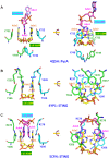
Binding of U-shaped c-di-AMP to the pyruvate carboxylase and STING molecules. (A) In the complex of U-shaped c-di-AMP with pyruvate carboxylase (PycA) dimer from L. monocytogenes (PDB: 4QSH) (69), adenine bases Ade1 and Ade2 form π–π stacking interactions with Tyr722 residues of the two PycA monomers (shown in green and cyan, respectively). This stack also contains an adenine base Ade3 from another c-di-AMP molecule (shown in magenta) that is inserted between Ade1 and Ade2. In addition, oxygen atoms of the central ribose-phosphate ring of c-di-AMP form hydrogen bonds with Tyr749 of both PycA monomers (69). (B) In the mouse STING complex (PDB: 4YP1), a single c-di-AMP molecule (carbon atoms in yellow) is present in the dimeric interface to stabilize the STING dimer formation, with Ade1 stacking with Tyr166 from one subunit (carbon atoms in cyan) and with Arg237 and Tyr239 from another subunit (carbon atoms in green), while Ade2 is stacking with Tyr166 from the second subunit (carbon atoms in green) and Arg237 and Tyr239 from the first subunit (carbon atoms in cyan). The rotated view on the right provides a better view of these four-layer Y166/Ade/R237/Y239 π-π-cation-π stacks. (C) In the STING complex from the sea anemone Nematostella vectensis (PDB: 5CFN), an additional c-di-AMP molecule (carbon atoms in magenta) forms a stack with the first c-di-AMP (carbon atoms in yellow), and, as shown on the right, the four-layer stacks are now formed by R278/Ade4/Ade1/Y206 or R278/Ade3/Ade2/Y206 cation-π-π-π stacking.
Figure 9.
Figure 9.
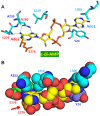
Binding of c-di-AMP to the RECON protein. In its complex with RECON (PDB: 5UXF), c-di-AMP adopts an E-type conformation, with both adenine bases stacked from both sides. Base Ade1 is held by a ππ stacking with Tyr24 from below and a C–H–π stacking with Leu306 from above. Base Ade2 is held by a triple hydrophobic–π interaction with Ala253 and Leu219 from above and an anion–π interaction of the carboxylate of Glu276 from below (indicated by the red arrow in panel B). In (A), dotted blue lines indicate the hydrogen bonds between N and O atoms of the Asn280 side-chain amide group and the N7 and N6 atoms of Ade2, respectively, and between Glu279 and the N6 atom of Ade2.
Figure 10.
Figure 10.
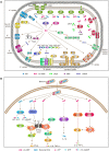
Cyclic di-AMP signaling in bacteria and eukaryotes. (A) In bacteria and archaea, c-di-AMP (magenta double dots) is synthesized by five kinds of diadenylate cyclases (green ovals at the top) and hydrolyzed by three kinds of phosphodiesterases (pink round rectangles on the right). C-di-AMP receptors include, among others, a specific riboswitch that controls expression of a number of K+ uptake proteins, RCK_C domains of K+ transporters KtrA and CpaA, the USP domain of the osmosensitive histidine kinase KdpD that regulates high-affinity K+ uptake system KdpABC, CBS domains of the OpuCA subunit of the carnitine transporter OpuC, K+ transporters KupA and KupB, pyruvate carboxylase PycA, PII-like signal transduction protein DarA (also referred to as PstA), and BusR, transcriptional regulator of osmolyte uptake. (B) In eukaryotic cells, five types of c-di-AMP receptors have been characterized: STING, DDX41, RECON, ERAdP, and NLRP3. Signaling by STING and DDX41 involves protein kinases TBK1 and IKK; TBK1 phosphorylates interferon regulatory factor IRF3 and promotes the production of type I interferons, whereas IKK phosphorylates IκB to release NF-κB, which leads to the production of pro-inflammatory cytokines (interleukin-6, TNF-α and others). RECON and ERAdP modulate the NF-κB to stimulate the production of pro-inflammatory cytokines (interleukin-6, TNF-α and others), while NLRP3 regulates the production of interleukin-1β. The dashed arrows represent steps that remain to be clarified. See the text for references and further details.

Similar articles

Cited by

References

    1. Hsu C.-Y.J., Dennis D., Oger J.. Synthesis and physical characterization of bis 3′→5′ cyclic dinucleotides (NpNp): RNA polymerase inhibitors. Nucleosides Nucleotides. 1985; 4:377–389.
    1. Ross P., Mayer R., Weinhouse H., Amikam D., Huggirat Y., Benziman M., de Vroom E., Fidder A., de Paus P., Sliedregt L.A. et al. .. The cyclic diguanylic acid regulatory system of cellulose synthesis in Acetobacter xylinum. Chemical synthesis and biological activity of cyclic nucleotide dimer, trimer, and phosphothioate derivatives. J. Biol. Chem. 1990; 265:18933–18943. - PubMed
    1. Witte G., Hartung S., Buttner K., Hopfner K.P.. Structural biochemistry of a bacterial checkpoint protein reveals diadenylate cyclase activity regulated by DNA recombination intermediates. Mol. Cell. 2008; 30:167–178. - PubMed
    1. Oppenheimer-Shaanan Y., Wexselblatt E., Katzhendler J., Yavin E., Ben-Yehuda S.. c-di-AMP reports DNA integrity during sporulation in Bacillus subtilis. EMBO Rep. 2011; 12:594–601. - PMC - PubMed
    1. El-Gebali S., Mistry J., Bateman A., Eddy S.R., Luciani A., Potter S.C., Qureshi M., Richardson L.J., Salazar G.A., Smart A. et al. .. The Pfam protein families database in 2019. Nucleic Acids Res. 2019; 47:D427–D432. - PMC - PubMed

Publication types