. 2018 Jul 25;24(42):10646-10652.
doi: 10.1002/chem.201801388. Epub 2018 Jul 4.

Insulin Hexamer-Caged Gadolinium Ion as MRI Contrast-o-phore

Affiliations

Insulin Hexamer-Caged Gadolinium Ion as MRI Contrast-o-phore

Steven K Taylor et al. Chemistry. .

Abstract

High-relaxivity protein-complexes of GdIII are being pursued as MRI contrast agents in hope that they can be used at much lower doses that would minimize toxic-side effects of GdIII release from traditional contrast agents. We construct here a new type of protein-based MRI contrast agent, a proteinaceous cage based on a stable insulin hexamer in which GdIII is captured inside a water filled cavity. The macromolecular structure and the large number of "free" GdIII coordination sites available for water binding lead to exceptionally high relaxivities per one GdIII ion. The GdIII slowly diffuses out of this cage, but this diffusion can be prevented by addition of ligands that bind to the hexamer. The ligands that trigger structural changes in the hexamer, SCN- , Cl- and phenols, modulate relaxivities through an outside-in signaling that is allosterically transduced through the protein cage. Contrast-o-phores based on protein-caged metal ions have potential to become clinical contrast agents with environmentally-sensitive properties.

Keywords: contrast agent; environmentally sensitive; gadolinium; insulin hexamer; proteins.


Figures

Figure 1.
Figure 1.
Assembly of the ‘Ins6Co(III)2Gd’ hexamer. Schematic showing Ins6Co(III)2Gd hexamer self-assembly from six insulin monomers one Gd(III) ion, and two cobalt(II) ions, followed by peroxide oxidation of the cobalt to ‘lock-in’ the structure. Both monomer and hexamer pictures are generated from our X-ray crystallographic structure (cf. Figure 2). Gd(III) ion locations in the structure are shown colored aqua (in monomer GluB13). Each Co(III) ion (pink) is shown bound to HisB10 on one of two ‘tris-α-helix bundles’ (one colored blue and one colored red) which are interdigitated with one another, thus forming a large cavity inbetween.
Figure 2.
Figure 2.
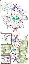
X-ray crystal structure. a) Results showing the amino acids that line the cavity containing the Gd ion that is disordered over six sites: three GdD sites (major) have 66% of the total electron density (at 1/3 of this each); and three GdB sites (minor) equally share the remaining electron density. The Cl ion site is 1/2 occupied, and the Co(III) ion sites are fully occupied. Located water molecules in the central cavity are depicted by red colored spheres. Inset view looks down the C3 axis at the atoms in the cavity. b) Structure depicting only one gadolinium ion-binding site with a Gd ion. The remaining Gd-binding sites are depicted occupied by superimposed waters (transparent red spheres) to depict plausible locations for additional Gd-coordinated water molecules to illustrate the Gd ion is likely to be coordinated to several water molecules within the cavity. Upper inset view looks down the C3 axis at the atoms in the cavity. Lower inset shows an X-ray crystal structure (pdb 1trz) with sodium in analogous positons to the gadolinium. The closest GluB13 residue to the Na(I) ion is orientated in an equatorial positon for binding.
Figure 3.
Figure 3.
1.4T relaxation measurements at 37°C and MRI. a) r1 relaxivites. b) r2 relaxivites. (r1,2 = 1/T1,2 /[hexmer], where 1/T1,2 = 1/T1,2, with contrast – 1/T1,2, without contrast) obtained from slopes of linear fits for 1 (blue), 2 (red), 3 (green), 4 (purple) equivalents of Gd(III) per hexamer. Slopes are virtually identical, showing that the central cavity of the hexamer is capable of binding to no more than one equivalent of Gd(III). Lower insets show slope comparison to the commercial MRI contrast agent Magnevist (orange) r1 (3.2 mM-1 s-1) and r2 (3.7 mM-1s-1) at much higher concentrations. Upper insets show measurements used to calculate a Limit of Detection (LoD) of 500nM (T1) and 200 nM (T2). c) 3T clinical MRI image of phantom samples of Ins6Co(III)2Gd at various concentrations compared to Magnevist (cross-section of samples in plastic tubes surrounded by a waterbath).
Figure 4.
Figure 4.
Stability of gadolinium-hexamers exposed thiocyanate, and to the physiological additives: calcium, phosphate, and ascorbate, over 24 hours in 100mM HEPES at pH 7.4 and 37°C. Y-axis is 1/T2, with contrast and is directly proportional to the gadolinium-hexamer concentration. a) Stability of the hexamer Ins6Co(III)2Gd. b) Stability of hexamer formulations of Ins6Zn(II)2Gd. Arrows indicate the significant increase, then drop, in relaxivity when first thiocyanate (150mM) is added to Ins6Zn(II)2Gd, then resorcinol (100mM), demonstrating that the relaxivity can be modulated allosterically. Addition of thiocyanate and/or resorcinol to Ins6Zn(II)2Gd affords various levels of protection against hexamer degradation by ascorbic acid. Structure of the T6-insulin hexamer Ins6Co(III)2Gd. Entrances to the major tunnels, affording a connection between the Gd(III) ion and the bulk solvent, are indicated by the green arrows. Atoms colored crimson are amino acids 1-9 of the B-chain, and these are extended in T6. (Octahedral Co(III) ion is colored pink). d) R6 insulin hexamer Ins6Zn(II)/resorcinol from X-ray structure (1evr).[42] shows amino acids 1-9 of the B-chain are coiled when the hexamer binds resorcinol, resulting in collapse of the major tunnels. (Cl ion coordinated to tetrahedral Zn(II) ion is colored green). Note: concentrations of additives are higher than their typical physiological levels. 100mM resorcinol; “Ca2+” is 10mM calcium chloride; “PO43−” is 10mM sodium phosphate; 200μM ascorbic acid. Hexamers are at 10μM. Results for the chloride ion are given in the Supporting Information (Figure S7).

Similar articles

References

    1. Gulani V Calamante F Shellock FG, Kanal E, Reeder SB, Lancet Neurol. 2017, 16, 564–570. - PubMed
    1. Pierre VC, Allen MJ, Caravan P, J. Biol. Inorg. Chem 2014, 19, 127–131. - PMC - PubMed
    1. Dumas S, Jacques V, Sun W, Troughton JS, Welch JT, Chasse JM, Schmitt-Willich H, Caravan P, Invest. Radiol 2010, 45(10), 600–612. - PMC - PubMed
    1. Boros E, Gale EM, Caravan P, Dalton Trans, 2015, 44, 4804–4818. - PMC - PubMed
    1. Xue S, Qiao J, Jiang J, Hubbard K, White N, Wei L, Li S, Liu Z, Yang JJ, Med. Res. Rev 2014, 34(5), 1070–1099. - PubMed

LinkOut - more resources