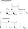
Defining the phenotypic spectrum of SLC6A1 mutations
- PMID: 29315614
- PMCID: PMC5912688
- DOI: 10.1111/epi.13986
Defining the phenotypic spectrum of SLC6A1 mutations
Abstract
Objective: Pathogenic SLC6A1 variants were recently described in patients with myoclonic atonic epilepsy (MAE) and intellectual disability (ID). We set out to define the phenotypic spectrum in a larger cohort of SCL6A1-mutated patients.
Methods: We collected 24 SLC6A1 probands and 6 affected family members. Four previously published cases were included for further electroclinical description. In total, we reviewed the electroclinical data of 34 subjects.
Results: Cognitive development was impaired in 33/34 (97%) subjects; 28/34 had mild to moderate ID, with language impairment being the most common feature. Epilepsy was diagnosed in 31/34 cases with mean onset at 3.7 years. Cognitive assessment before epilepsy onset was available in 24/31 subjects and was normal in 25% (6/24), and consistent with mild ID in 46% (11/24) or moderate ID in 17% (4/24). Two patients had speech delay only, and 1 had severe ID. After epilepsy onset, cognition deteriorated in 46% (11/24) of cases. The most common seizure types were absence, myoclonic, and atonic seizures. Sixteen cases fulfilled the diagnostic criteria for MAE. Seven further patients had different forms of generalized epilepsy and 2 had focal epilepsy. Twenty of 31 patients became seizure-free, with valproic acid being the most effective drug. There was no clear-cut correlation between seizure control and cognitive outcome. Electroencephalography (EEG) findings were available in 27/31 patients showing irregular bursts of diffuse 2.5-3.5 Hz spikes/polyspikes-and-slow waves in 25/31. Two patients developed an EEG pattern resembling electrical status epilepticus during sleep. Ataxia was observed in 7/34 cases. We describe 7 truncating and 18 missense variants, including 4 recurrent variants (Gly232Val, Ala288Val, Val342Met, and Gly362Arg).
Significance: Most patients carrying pathogenic SLC6A1 variants have an MAE phenotype with language delay and mild/moderate ID before epilepsy onset. However, ID alone or associated with focal epilepsy can also be observed.
Keywords: MAE; SLC6A1; epilepsy; epilepsy genetics.
Wiley Periodicals, Inc. © 2018 International League Against Epilepsy.
Conflict of interest statement
ST and KLH are employed by Ambry Genetics. CC was funded by the SNSF Early Postdoc fellowship. YW and IH were funded by DFG We4896/3-1; He5415/6-1. The remaining authors have no conflicts of interest to disclose. added. We confirm that we have read the Journal’s position on issues involved in ethical publication and affirm that this report is consistent with those guidelines.
Figures


Similar articles
-
Phenotypic and genetic spectrum of epilepsy with myoclonic atonic seizures.Epilepsia. 2020 May;61(5):995-1007. doi: 10.1111/epi.16508. Epub 2020 May 29. Epilepsia. 2020. PMID: 32469098
-
Dissecting the phenotypic and genetic spectrum of early childhood-onset generalized epilepsies.Seizure. 2019 Oct;71:222-228. doi: 10.1016/j.seizure.2019.07.024. Epub 2019 Jul 31. Seizure. 2019. PMID: 31401500
-
Concurrent pathogenic variants in SLC6A1/NOTCH1/PRIMPOL genes in a Chinese patient with myoclonic-atonic epilepsy, mild aortic valve stenosis and high myopia.BMC Med Genet. 2020 May 6;21(1):93. doi: 10.1186/s12881-020-01035-9. BMC Med Genet. 2020. PMID: 32375772 Free PMC article.
-
Myoclonic-astatic epilepsy of early childhood--clinical and EEG analysis of myoclonic-astatic seizures, and discussions on the nosology of the syndrome.Brain Dev. 2001 Nov;23(7):757-64. doi: 10.1016/s0387-7604(01)00281-9. Brain Dev. 2001. PMID: 11701290 Review.
-
Epilepsy and electroencephalogram evolution in YWHAG gene mutation: A new phenotype and review of the literature.Am J Med Genet A. 2021 Mar;185(3):901-908. doi: 10.1002/ajmg.a.62026. Epub 2021 Jan 4. Am J Med Genet A. 2021. PMID: 33393734 Review.
Cited by
-
A missense mutation in SLC6A1 associated with Lennox-Gastaut syndrome impairs GABA transporter 1 protein trafficking and function.Exp Neurol. 2019 Oct;320:112973. doi: 10.1016/j.expneurol.2019.112973. Epub 2019 Jun 6. Exp Neurol. 2019. PMID: 31176687 Free PMC article.
-
Insights into cognitive and behavioral comorbidities of SLC6A1-related epilepsy: five new cases and literature review.Front Neurosci. 2023 Aug 28;17:1215684. doi: 10.3389/fnins.2023.1215684. eCollection 2023. Front Neurosci. 2023. PMID: 37700749 Free PMC article.
-
Case report: SLC6A1 mutations presenting with isolated absence seizures: description of 2 novel cases.Front Neurosci. 2023 Jun 29;17:1219244. doi: 10.3389/fnins.2023.1219244. eCollection 2023. Front Neurosci. 2023. PMID: 37457006 Free PMC article.
-
Globus Pallidus Lesion With Iron Deposition and Dopaminergic Denervation in a Patient With a Pathogenic SLC6A1 Variant: A Case Report.Neurol Genet. 2024 Mar 19;10(2):e200136. doi: 10.1212/NXG.0000000000200136. eCollection 2024 Apr. Neurol Genet. 2024. PMID: 38515990 Free PMC article.
-
Common molecular mechanisms of SLC6A1 variant-mediated neurodevelopmental disorders in astrocytes and neurons.Brain. 2021 Sep 4;144(8):2499-2512. doi: 10.1093/brain/awab207. Brain. 2021. PMID: 34028503 Free PMC article.
References
-
- Madsen KK, Hansen GH, Danielsen EM, et al. The subcellular localization of GABA transporters and its implication for seizure management. Neurochem Res. 2015;40:410–9. - PubMed
-
- Dikow N, Maas B, Karch S, et al. 3p25.3 microdeletion of GABA transporters SLC6A1 and SLC6A11 results in intellectual disability, epilepsy and stereotypic behavior. Am J Med Genet A. 2014;164a:3061–8. - PubMed

