TECPR2 Associated Neuroaxonal Dystrophy in Spanish Water Dogs
- PMID: 26555167
- PMCID: PMC4640708
- DOI: 10.1371/journal.pone.0141824
TECPR2 Associated Neuroaxonal Dystrophy in Spanish Water Dogs
Abstract
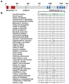
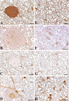
Clinical, pathological and genetic examination revealed an as yet uncharacterized juvenile-onset neuroaxonal dystrophy (NAD) in Spanish water dogs. Affected dogs presented with various neurological deficits including gait abnormalities and behavioral deficits. Histopathology demonstrated spheroid formation accentuated in the grey matter of the cerebral hemispheres, the cerebellum, the brain stem and in the sensory pathways of the spinal cord. Iron accumulation was absent. Ultrastructurally spheroids contained predominantly closely packed vesicles with a double-layered membrane, which were characterized as autophagosomes using immunohistochemistry. The family history of the four affected dogs suggested an autosomal recessive inheritance. SNP genotyping showed a single genomic region of extended homozygosity of 4.5 Mb in the four cases on CFA 8. Linkage analysis revealed a maximal parametric LOD score of 2.5 at this region. By whole genome re-sequencing of one affected dog, a perfectly associated, single, non-synonymous coding variant in the canine tectonin beta-propeller repeat-containing protein 2 (TECPR2) gene affecting a highly conserved region was detected (c.4009C>T or p.R1337W). This canine NAD form displays etiologic parallels to an inherited TECPR2 associated type of human hereditary spastic paraparesis (HSP). In contrast to the canine NAD, the spinal cord lesions in most types of human HSP involve the sensory and the motor pathways. Furthermore, the canine NAD form reveals similarities to cases of human NAD defined by widespread spheroid formation without iron accumulation in the basal ganglia. Thus TECPR2 should also be considered as candidate gene for human NAD. Immunohistochemistry and the ultrastructural findings further support the assumption, that TECPR2 regulates autophagosome accumulation in the autophagic pathways. Consequently, this report provides the first genetic characterization of juvenile canine NAD, describes the histopathological features associated with the TECPR2 mutation and provides evidence to emphasize the association between failure of autophagy and neurodegeneration.
Conflict of interest statement
Figures


Similar articles
-
A tecpr2 knockout mouse exhibits age-dependent neuroaxonal dystrophy associated with autophagosome accumulation.Autophagy. 2021 Oct;17(10):3082-3095. doi: 10.1080/15548627.2020.1852724. Epub 2020 Dec 10. Autophagy. 2021. PMID: 33218264 Free PMC article.
-
A Missense Mutation in the Vacuolar Protein Sorting 11 (VPS11) Gene Is Associated with Neuroaxonal Dystrophy in Rottweiler Dogs.G3 (Bethesda). 2018 Jul 31;8(8):2773-2780. doi: 10.1534/g3.118.200376. G3 (Bethesda). 2018. PMID: 29945969 Free PMC article.
-
TECPR2 Cooperates with LC3C to Regulate COPII-Dependent ER Export.Mol Cell. 2015 Oct 1;60(1):89-104. doi: 10.1016/j.molcel.2015.09.010. Mol Cell. 2015. PMID: 26431026
-
Axonal dystrophies.Handb Clin Neurol. 2013;113:1919-24. doi: 10.1016/B978-0-444-59565-2.00062-9. Handb Clin Neurol. 2013. PMID: 23622415 Review.
-
TECPR2 mutation-associated respiratory dysregulation: more than central apnea.J Clin Sleep Med. 2020 Jun 15;16(6):977-982. doi: 10.5664/jcsm.8434. J Clin Sleep Med. 2020. PMID: 32209221 Free PMC article. Review.
Cited by
-
A tecpr2 knockout mouse exhibits age-dependent neuroaxonal dystrophy associated with autophagosome accumulation.Autophagy. 2021 Oct;17(10):3082-3095. doi: 10.1080/15548627.2020.1852724. Epub 2020 Dec 10. Autophagy. 2021. PMID: 33218264 Free PMC article.
-
An Overview of Canine Inherited Neurological Disorders with Known Causal Variants.Animals (Basel). 2023 Nov 18;13(22):3568. doi: 10.3390/ani13223568. Animals (Basel). 2023. PMID: 38003185 Free PMC article. Review.
-
Lysosomal targeting of autophagosomes by the TECPR domain of TECPR2.Autophagy. 2021 Oct;17(10):3096-3108. doi: 10.1080/15548627.2020.1852727. Epub 2020 Nov 29. Autophagy. 2021. PMID: 33213269 Free PMC article.
-
Developing antisense oligonucleotides for a TECPR2 mutation-induced, ultra-rare neurological disorder using patient-derived cellular models.Mol Ther Nucleic Acids. 2022 Jun 22;29:189-203. doi: 10.1016/j.omtn.2022.06.015. eCollection 2022 Sep 13. Mol Ther Nucleic Acids. 2022. PMID: 35860385 Free PMC article.
-
A Missense Mutation in the Vacuolar Protein Sorting 11 (VPS11) Gene Is Associated with Neuroaxonal Dystrophy in Rottweiler Dogs.G3 (Bethesda). 2018 Jul 31;8(8):2773-2780. doi: 10.1534/g3.118.200376. G3 (Bethesda). 2018. PMID: 29945969 Free PMC article.
References
-
- Sisó S, Hanzlícek D, Fluehmann G, Kathmann I, Tomek A, Papa V, et al. Neurodegenerative diseases in domestic animals: a comparative review. Vet J. 2006;171: 20–38. - PubMed
-
- Summers BA, Cummings JF, de Lahunta A. Degenerative diseases of the central nervous system In: Summers BA, Cummings F, de Lahunta A, editors. Veterinary Neuropathology. New York: Mosby; 1995. pp. 315–317.
-
- Zhou B, Westaway SK, Levinson B, Johnson MA, Gitschier J, Hayflick SJ. A novel pantothenate kinase gene (PANK2) is defective in Hallervorden-Spatz syndrome. Nat Genet. 2001;28: 345–349. - PubMed
Publication types
MeSH terms
Substances
Grants and funding
LinkOut - more resources
Full Text Sources
Other Literature Sources
Miscellaneous

