. 2012 Jul 20;287(30):25060-72.
doi: 10.1074/jbc.M112.387241. Epub 2012 Jun 1.

Intra- and interprotein phosphorylation between two-hybrid histidine kinases controls Myxococcus xanthus developmental progression

Affiliations

Intra- and interprotein phosphorylation between two-hybrid histidine kinases controls Myxococcus xanthus developmental progression

Andreas Schramm et al. J Biol Chem. .

Abstract

Histidine-aspartate phosphorelay signaling systems are used to couple stimuli to cellular responses. A hallmark feature is the highly modular signal transmission modules that can form both simple "two-component" systems and sophisticated multicomponent systems that integrate stimuli over time and space to generate coordinated and fine-tuned responses. The deltaproteobacterium Myxococcus xanthus contains a large repertoire of signaling proteins, many of which regulate its multicellular developmental program. Here, we assign an orphan hybrid histidine protein kinase, EspC, to the Esp signaling system that negatively regulates progression through the M. xanthus developmental program. The Esp signal system consists of the hybrid histidine protein kinase, EspA, two serine/threonine protein kinases, and a putative transport protein. We demonstrate that EspC is an essential component of this system because ΔespA, ΔespC, and ΔespA ΔespC double mutants share an identical developmental phenotype. Neither substitution of the phosphoaccepting histidine residue nor deletion of the entire catalytic ATPase domain in EspC produces an in vivo mutant developmental phenotype. In contrast, substitution of the receiver phosphoaccepting residue yields the null phenotype. Although the EspC histidine kinase can efficiently autophosphorylate in vitro, it does not act as a phosphodonor to its own receiver domain. Our in vitro and in vivo analyses suggest the phosphodonor is instead the EspA histidine kinase. We propose EspA and EspC participate in a novel hybrid histidine protein kinase signaling mechanism involving both inter- and intraprotein phosphotransfer. The output of this signaling system appears to be the combined phosphorylated state of the EspA and EspC receiver modules. This system regulates the proteolytic turnover of MrpC, an important regulator of the developmental program.


Figures

FIGURE 1.
FIGURE 1.
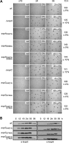
EspA and EspC are components of a single signaling pathway. A, developmental phenotypes of esp null mutants. Wild type (wt; DZ2), ΔespA (DZ4227), ΔespC (PH1044), and ΔespA ΔespC (PH1047) strains were developed under submerged culture in 24-well culture dishes, and pictures were recorded at the indicated hours of development. Heat- and sonication-resistant spores were isolated at the indicated time points and displayed as the percent of wild type spores at 72 h. Spore numbers are the average and associated standard deviation of three biological replicates. Scale bar, 0.5 mm; nd, not determined. B, EspA and EspC developmental protein accumulation patterns. Anti-EspC (left) and anti-EspA (right) immunoblot analysis of cell lysates prepared from the indicated strains developed for the indicated hours under 16 ml of submerged culture format.
FIGURE 2.
FIGURE 2.
Domain organization of the EspA and EspC hybrid histidine kinases. Percent amino acid identity for the indicated domains is represented by shaded areas. The invariant histidine (H) residues in the dimerization and histidine phosphotransfer domains (DHp) and invariant aspartate (D) residues in the receiver (REC) domains are indicated. FHA, forkhead-associated domain; MASE1, predicted integral membrane sensory domain.
FIGURE 3.
FIGURE 3.
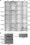
Phosphorylation of EspC receiver domain, but not EspC autophosphorylation, is required for regulation of development. A, developmental phenotypes of espA and espC signal transmission point mutants. Wild type (wt; DZ2), ΔespA (DZ4227), espAH407A (PH1008), espAD696A (PH1009), espAH407A,D696A (PH1029), ΔespC (PH1044), espCH461A (PH1026), espCD749A (PH1027),and espCH461A,D749A (PH1028) strains were developed under submerged culture in 24-well culture dishes, and pictures were recorded at the indicated hours of development. Heat- and sonication-resistant spores were isolated at the indicated time points and displayed as the percent of wild type spores at 72 h. Spore numbers are the average and associated standard deviation of three biological replicates. Scale bar, 0.5 mm; nd, not determined. B, EspC and EspA developmental protein accumulation patterns. Anti-EspC (left panel) and anti-EspA (right panel) immunoblot analyses of cell lysates were prepared from the indicated strains developed for the indicated hours under 16 ml of submerged culture format.
FIGURE 4.
FIGURE 4.
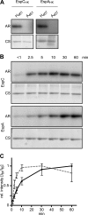
EspC kinase region autophosphorylates in vitro. A, in vitro autophosphorylation of EspAHK-His6, EspCHK-His6, EspAHK H407A-His6, and EspCHK H461A-His6. 10 μm of each recombinant protein was incubated in the presence of [γ-32P]ATP for 60 min, quenched, and resolved by SDS-PAGE, and the radiolabel was detected by exposure to a Storage PhosphoScreen (AR). Total protein was subsequently detected by Coomassie stain (CS). B, relative in vitro autophosphorylation rates of EspC and EspA kinase regions. 10 μm EspAHK-His6 or EspCHK-His6 was incubated in the presence of [γ-32P]ATP; aliquots were removed at the indicated time points and analyzed as above. C, quantification of EspC (dashed line) and EspA (solid line) HK autophosphorylation rates in B. Relative (rel.) signal intensities of the bands from three independent time courses were quantified and normalized to the maximal signal intensity of each protein. The average relative signal intensity and associated standard deviation from each time point was plotted.
FIGURE 5.
FIGURE 5.
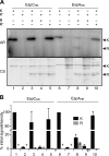
EspAHK, but not EspCHK, efficiently phosphorylates EspCREC. A, in vitro phosphotransfer from autophosphorylated EspC or EspA kinase regions to EspC receiver domain (EspCREC). Lanes 1–5, EspCHK-His6 (K) or EspCHK H461A-His6 (K) was first incubated in the presence of [γ-32P]ATP for 30 min and then incubated with either buffer or equimolar (10 μm) EspCREC-His6 (R) or EspCREC D749A-His6 (R) for 2 min as indicated. +, indicated component present; −, indicated component absent. Lanes 6–10, EspAHK-His6 (K) or EspAHK H407A-His6 (K) analyzed as per lanes 1–5. All samples were quenched and resolved by SDS-PAGE, and radiolabel was detected by exposure to a Storage PhosphoScreen (AR). Total protein was subsequently detected by Coomassie stain (CS). B, quantification of the relative signal intensities of radiolabeled EspC or EspA HK (K, black bars), and EspCREC (R, gray bars). Signal intensities are the average and associated standard deviation of three biological replicates of the reactions represented in A. HK and REC intensity values are reported as a percent of the respective Esp HK autophosphorylation controls (represented by lanes 1 and 6, respectively). *, not determined.
FIGURE 6.
FIGURE 6.
Disruption of a phosphatase motif in EspA but not EspC effects developmental progression. A, developmental phenotypes of espC and espA phosphatase motif mutants. Wild type (wt; DZ2), espCD749A (PH1027), espCN465Y (PH1034), espCN465Y,D749A (PH1035), espCΔCA (PH1032), espCΔCA,D749A (PH1033), and espAN411D (PH1045) strains were developed under submerged culture in 24-well culture dishes, and pictures were recorded at the indicated hours of development. Heat- and sonication-resistant spores were isolated at the indicated time points and displayed as the percent of wild type spores at 72 h. Spore numbers are the average and associated standard deviation of three biological replicates. Scale bar, 0.5 mm; nd, not determined. B, EspC and EspA developmental protein accumulation patterns. Anti-EspC (left panel) and anti-EspA (right panel) immunoblot analyses of cell lysates were prepared from the indicated strains developed for the indicated hours under 16 ml of submerged culture format. *, 5 times longer exposure is required.
FIGURE 7.
FIGURE 7.
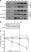
Esp system negatively regulates MrpC protein stability. A, MrpC developmental protein accumulation patterns in the wild type (DZ2, wt), ΔespC (PH1044), ΔespA ΔespC (PH1047), espCH461A (PH1026), espCD749A (PH1027), and espCH461A,D749A (PH1028) strains. 10 μg of total protein lysates developed for the indicated hours in the 16-ml submerged culture format were subject to anti-MrpC immunoblot. B, chloramphenicol chase of MrpC. Wild type (wt) or ΔespA ΔespC cells were developed for 9 h as above and treated with 34 μg ml−1 chloramphenicol for the indicated minutes. Equal proportions of the samples were subject to anti-MrpC immunoblot. C, calculation of the half-life (t½) of MrpC. Triplicate biological replicates of the wild type (black line) and ΔespA ΔespC (gray line) chloramphenicol chase experiments represented in B were performed, and the MrpC band intensity for each time point was normalized to t = 0 of the respective strain and averaged. The natural log of the average intensities was plotted versus min of chloramphenicol treatment and the slope a linear fit of the data were used to calculate the MrpC t½ in wild type cells as described under “Experimental Procedures.” No significant decrease in MrpC signal intensity could be detected in the ΔespA ΔespC strain. Vertical bars, standard deviation of the average from triplicate biological replicates; *, average intensity values are significantly different (p = 0.003).
FIGURE 8.
FIGURE 8.
Model of the Esp signaling system. Two-hybrid histidine kinases EspC (dark gray) and EspA (light gray) regulate the accumulation rate of an important developmental regulator, MrpC, to ensure appropriate and coordinated progression through the M. xanthus developmental program. The combined phosphorylation of EspA and EspC receiver domains (REC) activates an unknown protease or protease targeting factor (×) to stimulate MrpC turnover. EspA histidine kinase region (HK) autophosphorylates and donates a phosphoryl group to both its own and EspC receiver domains. EspA may also act as a phosphatase on EspA and or EspC REC. Autophosphorylation of EspC histidine kinase domain is not required for stimulating MrpC turnover under laboratory developmental conditions. EspA activity is controlled by a signaling module consisting of two serine/threonine kinases, PktA5 (A5) and PktB8 (B8), and a putative transport protein (EspB) predicted to reside in the cytoplasmic membrane (CM) (37, 41). PktA5 and PktB8 are thought to interact with the Forkhead associated (FHA) domain in EspA (41). Two PAS domains in EspA and one in EspC may be involved in sensing internal or membrane-associated redox stimuli (65). EspC is predicted to be anchored in the CM by putative MASE1 sensing domain of unknown function (64). The array of signaling domains may allow cell fate-specific accumulation of MrpC within the developmental program.

Similar articles

Cited by

References

    1. Stock A. M., Robinson V. L., Goudreau P. N. (2000) Two-component signal transduction. Annu. Rev. Biochem. 69, 183–215 - PubMed
    1. Wuichet K., Cantwell B. J., Zhulin I. B. (2010) Evolution and phyletic distribution of two-component signal transduction systems. Curr. Opin. Microbiol. 13, 219–225 - PMC - PubMed
    1. Dutta R., Qin L., Inouye M. (1999) Histidine kinases. Diversity of domain organization. Mol. Microbiol. 34, 633–640 - PubMed
    1. Casino P., Rubio V., Marina A. (2009) Structural insight into partner specificity and phosphoryl transfer in two-component signal transduction. Cell 139, 325–336 - PubMed
    1. Lukat G. S., McCleary W. R., Stock A. M., Stock J. B. (1992) Phosphorylation of bacterial response regulator proteins by low molecular weight phosphodonors. Proc. Natl. Acad. Sci. U.S.A. 89, 718–722 - PMC - PubMed

Publication types

MeSH terms

LinkOut - more resources