. 2009 Apr 28;2(68):ra17.
doi: 10.1126/scisignal.2000118.

Galpha(i1) and Galpha(i3) are required for epidermal growth factor-mediated activation of the Akt-mTORC1 pathway

Affiliations

Galpha(i1) and Galpha(i3) are required for epidermal growth factor-mediated activation of the Akt-mTORC1 pathway

Cong Cao et al. Sci Signal. .

Erratum in

  • Sci Signal. 2010;3(121):er2

Abstract

The precise mechanism whereby epidermal growth factor (EGF) activates the serine-threonine kinase Akt and the mammalian target of rapamycin (mTOR) complex 1 (mTORC1) remains elusive. Here, we report that the alpha subunits of the heterotrimeric guanine nucleotide-binding proteins (G proteins) Galpha(i1) and Galpha(i3) are critical for this activation process. Both Galpha(i1) and Galpha(i3) formed complexes with growth factor receptor binding 2 (Grb2)-associated binding protein 1 (Gab1) and the EGF receptor (EGFR) and were required for the phosphorylation of Gab1 and its subsequent interaction with the p85 subunit of phosphatidylinositol 3-kinase in response to EGF. Loss of Galpha(i1) and Galpha(i3) severely impaired the activation of Akt and of p70 S6 kinase and 4E-BP1, downstream targets of mTORC1, in response to EGF, heparin-binding EGF-like growth factor, and transforming growth factor alpha, but not insulin, insulin-like growth factor, or platelet-derived growth factor. In addition, ablation of Galpha(i1) and Galpha(i3) largely inhibited EGF-induced cell growth, migration, and survival and the accumulation of cyclin D1. Overall, this study suggests that Galpha(i1) and Galpha(i3) lie downstream of EGFR, but upstream of Gab1-mediated activation of Akt and mTORC1, thus revealing a role for Galpha(i) proteins in mediating EGFR signaling.


Figures

Fig. 1
Fig. 1
i1 and Gαi3 are required for the activation of Akt and mTORC1 by EGF. WT and Gαi1, Gαi3 DKO MEFs were treated with different doses of EGF for 15 min (A) or with EGF (100 ng/ml) for the indicated times (B). Gαi1, Gαi3, pEGFR(1173Y), pAkt(473S), pAkt(308T), pGSK-3β(9S), pFoxo3(32T), pp70S6K(389T), pS6 (235/236S), p4E-BP1(65S), Akt1–2, S6, and EGFR were detected by Western blotting analysis. (C) WT, Gαi1-deficient, Gαi3-deficient, or DKO MEFs were treated with EGF (100 ng/ml) for the indicated times. Gαi3, pAkt(473S), pS6(235/236S), p4E-BP1(65S), and Akt1–2 were detected by Western blotting analysis. (D) WT and Gαi2-deficient MEFs were treated with EGF (100 ng/ml) for the indicated times. Gαi2, pAkt(473S), pS6K(389T), p4E-BP1(65S), and AKT1–2 were detected by Western blotting analysis. The experiments were repeated at least three times and similar results were obtained in each case.
Fig. 2
Fig. 2
Loss of Gαi1 and Gαi3 severely impairs activation of Akt and mTORC1 by HB-EGF and TGF-α, but not by insulin, IGF, or PDGF. WT and DKO MEFs were treated with TGF-α (100 ng/ml), HB-EGF (100 ng/ml), PDGF-BB (25 ng/ml) (A), or with insulin (1 μg/ml) or IGF-1 (10 ng/ml) (B) for the indicated times. Gαi3, pAkt(308T), pAkt(473S), pGSK-3β(9/21S), pS6K(389T), pS6(235/236S), p4E-BP1(65S), Akt1–2, and β-actin were detected by Western blotting analysis. (C) EGF-or insulin-induced pSTAT5(694Y) and Akt(473S) in WT and DKO MEFs were detected by Western blotting analysis. (D) EGF-induced phosphorylation of PLC-γ(783Y) and Akt(473S) were determined by Western blotting. The experiments were repeated at least three times and similar results were obtained in each case.
Fig. 3
Fig. 3
i1 and Gαi3 proteins are critical for the phosphorylation of Akt473S and 4E-BP1(65S) in response to EGF. (A) Knockdown of Gαi1 and Gαi3 decreases the activation of Akt by EGF, but not insulin. WT MEFs transfected with siRNA oligos specific for Gαi1 and Gαi3 and DKO MEFs were treated with EGF (100 ng/ml) or insulin (1 μg/ml) for the indicated times. pAkt(308T), pAkt(473S), pGSK-3β(S9), Akt1–2, β-actin, Gαi1, and Gαi3 were detected by Western blotting. (B) Exogenous Gαi1 and Gαi3 rescue the activation of Akt and 4E-BP1 in DKO MEFs in response to EGF. DKO MEFs were transfected with vectors encoding WT Gαi1 or Gαi3 (4 μg). Cells were treated with EGF (100 ng/ml) for the indicated times. pAkt(308T), pAkt(473S), S6K(389T), S6(235/236S), β-actin, Gαi1, and Gαi3 were detected by Western blotting analysis. (C) Cotransfection of DN mutants of Gαi1 and Gαi3 inhibits the activation of Akt by EGF. WT MEFs were cotransfected with DN, EE-tagged Gαi1 and Gαi3 expression vectors (2 μg each) or were untransfected as controls followed by treatment with EGF (100 ng/ml) for the indicated times. pAkt(473S), pGSK-3β(9S), pS6K(389T), Akt1–2, EE tag, and Gαi3 were detected by Western blotting. (D) Constitutively active (CA) Gαi3(Q204L) induces the phosphorylation of Akt(473S) and S6(235/236S). DKO cells were transfected with a vector expressing Gαi3(Q204L). pAkt(473S), pS6(235/236S), Gαi3, and β-actin were determined by Western blotting analysis. (E) WT MEFs were pretreated overnight with pertussis toxin (PTX, 100 ng/ml) followed by treatment with EGF. pAkt(473S), pGSK-3β(9S), p4E-BP1(65S), pS6K(389T), and β-actin were detected by Western blotting. All transfection experiments were repeated at least twice and similar results were obtained in each case.
Fig. 4
Fig. 4
EGF-induced phosphorylation of Gab1 depends on Gαi1 and Gαi3. (A) Gab1 is required for the activation of Akt by EGF and TGF-α, but not by insulin, IGF, and PDGF. pAkt(308T) and pAkt(473S) were detected in WT and Gab1-deficient MEFs treated with EGF (100 ng/ml), TGF-α (100 ng/ml), PDGF-BB (25 ng/ml), insulin (1 μg/ml), or IGF-1 (10 ng/ml). (B) Gαi1 and Gαi3 are required for the phosphorylation of Gab1 in response to EGF. pGab1(307Y) and pGab1(627Y) in WT and DKO MEFs treated with EGF (100 ng/ml) were detected by Western blotting analysis. (C) Loss of Gαi1 and Gαi3 largely impairs phosphorylation of Akt and Gab1 in response to EGF and TGF-α, but not insulin. WT and DKO MEFs were treated with EGF (100 ng/ml), insulin (1 μg/ml), or TGF-α (100 ng/ml) for 5 min. pAkt(308T), pAkt(473S), pGab1(627Y), Gab1, PI3Kp85, and ERK1–2 were detected by Western blotting analysis.
Fig. 5
Fig. 5
EGF induces the association of Gab1 with Gαi1 and Gαi3, and the EGF-induced interaction of Gab1 with PI3K p85 depends on Gαi1 and Gαi3. (A) EGF induces the association of Gαi3 with Gab1. WT and DKO MEFs were treated with EGF (100 ng/ml) for the indicated times and were then lysed. The precleared cell lysates (0.6 mg) were incubated with anti-EGFR or anti-Gαi3 antibodies overnight at 4°C, followed by incubation with protein A/G beads for another 2 hours at 4°C. Beads were washed, boiled, resolved by 10% SDS-PAGE, and proteins were transferred onto a PVDF membrane followed by Western blotting analysis with anti-EGFR, anti-Gαi3, and anti-Gab1, respectively. (B and C) EGF induces the association of Gab1 with Gαi1 and Gαi3. WT and DKO MEFs were treated with EGF (100 ng/ml) for the indicated times. The precleared 600-μg aliquots of cell lysates were incubated with anti-Gab1 (B), or with anti-Gab1 and anti-Gαi1 (C), respectively, followed by Western blotting analysis with anti-Gab1, anti-pGab1(627Y), anti-Gαi1, and anti-Gαi3, respectively. (D) HEK 293 cells were treated with EGF for 5 min and then lysed. The precleared, 700-μg cell lysates were incubated with anti-EGFR, anti-Gab1, or anti-Gαi3 and 20 μl of protein A/G beads at 4°C overnight. Beads were washed four times with lysis buffer, boiled, resolved by SDS-PAGE, and transferred onto a PVDF membrane, followed by Western blotting analysis to detect EGFR, Gαi3, and Gab1. (E) The same cell lysates used for Fig. 4C were precleared and incubated overnight with anti-p85 and 20 μl of protein A/G beads at 4°C. Beads were washed four times with lysis buffer, boiled, loaded onto an SDS-PAGE gel and transferred onto a PVDF membrane followed by Western blotting analysis to detect Gab1, pGab1(627Y), and PI3K p85. All experiments were repeated at least two or three times and similar results were obtained in each case.
Fig. 6
Fig. 6
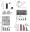
Roles for Gαi1 and Gαi3 proteins in EGF-mediated cell proliferation, growth, migration, survival, and the production of cyclin D1. (A) Gαi1 and Gαi3 are important for EGF-induced cell proliferation. WT and DKO MEFs were treated with EGF (100 ng/ml) for 24 hours. Cell proliferation was determined by the MTT assay. (B) Gαi1 and Gαi3 are involved in FBS-induced cell growth. FBS-induced cell growth in both WT and DKO cells was determined by counting cell numbers under a microscope. (C) WT and DKO MEFs were treated with EGF (100 ng/ml) for 4, 8, or 24 hours. The abundance of cyclin D1 was determined by Western blotting analysis with an antibody to cyclin D1. (D and E) Gαi1 and Gαi3 are required for EGF-induced cell migration. WT and DKO MEFs were incubated with serum-free medium in the presence or absence of EGF (100 ng/ml) or 10% FBS for 36 hours. In vitro cell migration was determined by the phagokinetic track motility assay (D) or the scratch assay (E). The image represents a minimum of 10 random fields for each group. Magnification: 200× for phagokinetic track motility assay and 100× for scratch assay. (F) Gαi proteins are required for EGF-induced cell survival. WT and DKO MEFs were treated with different doses of H2O2 in the presence or absence of EGF (100 ng/ml). After 24 hours, cell viability was measured by the MTT assay. Mitomycin C (10 μg/ml) was always present in the media to prevent cell proliferation from occurring. The data represent the mean ± SE of at least triplicate experiments. *P < 0.05 versus WT groups.
Fig. 7
Fig. 7
Model of Gαi-mediated activation of the PI3K-Akt-mTORC1 pathway in response to EGF. EGF induces formation of a complex between EGFR, Gαi, and Gab1. Recruitment of Gαi to the receptor may occur directly or through an as yet unidentified protein. Subsequently, Gαi interacts with Gab1, which promotes its phosphorylation by the EGFR or a non-RTK. Activated Gab1 interacts with the regulatory subunit of PI3K (p85), which leads to the activation of the catalytic subunits of PI3K (p110). Active PI3K phosphorylates PIP2 (phosphatidylinositol 4,5-bisphosphate) to generate PIP3 (phosphatidylinositol 3,4,5-trisphosphate), which in turn interacts with the pleckstrin homology domain of Akt and PDK1, and PDK1 phosphorylates Akt on Thr308. Activated Gab1 uses an unidentified mechanism to trigger the activation of mTORC2, which leads to the phosphorylation of Akt on Ser473. When Akt is activated, it phosphorylates FOXO, GSK-3α, and GSK-3β and triggers the activation of mTORC1, which results in phosphorylation of 4E-BP1 and S6K, which in turn phosphorylates S6.

Similar articles

Cited by

References

    1. Rozengurt E. Mitogenic signaling pathways induced by G protein-coupled receptors. J Cell Physiol. 2007;213:589–602. - PubMed
    1. Steelman LS, Abrams SL, Whelan J, Bertrand FE, Ludwig DE, Bäsecke J, Libra M, Stivala F, Milella M, Tafuri A, Lunghi P, Bonati A, Martelli AM, McCubrey JA. Contributions of the Raf/MEK/ERK, PI3K/PTEN/Akt/mTOR and Jak/STAT pathways to leukemia. Leukemia. 2008;22:686–707. - PubMed
    1. Sabatini DM. mTOR and cancer: Insights into a complex relationship. Nat Rev Cancer. 2006;6:729–734. - PubMed
    1. Greer EL, Brunet A. FOXO transcription factors in ageing and cancer. Acta Physiol (Oxf) 2008;192:19–28. - PubMed
    1. Lam EW, Francis RE, Petkovic M. FOXO transcription factors: Key regulators of cell fate. Biochem Soc Trans. 2006;34:722–726. - PubMed

MeSH terms

LinkOut - more resources