. 2009 May 15;284(20):13696-13704.
doi: 10.1074/jbc.M807366200. Epub 2009 Mar 23.

Coexpression and heteromerization of two neuronal K-Cl cotransporter isoforms in neonatal brain

Affiliations

Coexpression and heteromerization of two neuronal K-Cl cotransporter isoforms in neonatal brain

Pavel Uvarov et al. J Biol Chem. .

Abstract

The neuron-specific K-Cl cotransporter KCC2 maintains the low intracellular chloride concentration required for the fast hyperpolarizing actions of inhibitory neurotransmitters. The KCC2 gene codes for two isoforms, KCC2a and KCC2b, which differ in their N termini. The relative expression and cellular distribution of the two KCC2 protein isoforms are unknown. Here, we characterize an antibody against the KCC2a isoform and show that a previously described antibody against KCC2 is specific for the KCC2b isoform (Hubner, C. A., Stein, V., Hermans-Borgmeyer, I., Meyer, T., Ballanyi, K., and Jentsch, T. J. (2001) Neuron 30, 515-524). Immunostaining of dissociated hippocampal cultures confirms that both KCC2 isoforms are neuron-specific. Immunoblot analysis indicates that KCC2b is the major KCC2 isoform in the adult brain, whereas in the neonatal mouse central nervous system, half of total KCC2 protein is KCC2a. At this stage, the two KCC2 isoforms are largely colocalized and show similar patterns of distribution in the brain. When coexpressed in HEK293 cells, KCC2a and KCC2b proteins form heteromeric complexes. Moreover, the two isoforms can be coimmunoprecipitated from the neonatal brain, suggesting the presence of endogenous KCC2a-KCC2b heteromers. Consistent with this, native gel analysis shows that a substantial part of endogenous KCC2 isoforms in the neonatal brain constitute dimers.


Figures

FIGURE 1.
FIGURE 1.
Specific antibodies against the KCC2a and KCC2b isoforms. A, whole-brain lysates obtained from P2 mice were analyzed by standard SDS-PAGE and immunoblotting (IB) with affinity-purified KCC2a antiserum (see “Experimental Procedures”). The KCC2a antiserum (diluted 1: 2000) detects a major product of ∼140 kDa (arrowhead, corresponding to KCC2a monomer), and occasionally (cf. B), a minor band of ∼200 kDa. A similar, higher molecular mass band observed with other KCC2 antibodies was suggested to represent the interaction of KCC2 with other proteins (28). These bands are not observed when the antiserum is preadsorbed with 1 μg/ml immunizing peptide. As a control, the membrane was reblotted with an antibody against GAPDH. B, whole-brain lysates of embryonic day 17.5 (E17) wild-type (WT) mice and their KCC2 null mutant (KO) littermates were analyzed as above. The KCC2a antiserum detected the expected product of ∼140 kDa from the WT but not from the KO lysate. C, lysates of HEK293 cells, transiently transfected with KCC2a or KCC2b expression plasmids, were analyzed as above with KCC2a, KCC2b, or KCC2pan antibodies (see “Experimental Procedures”). The KCC2a antiserum detected a product of ∼140 kDa from lysates of HEK293 cells transfected with the KCC2a plasmid but not with the KCC2b expression vector. Similarly, the KCC2b antiserum stained a product of ∼140 kDa from lysates of KCC2b-expressing but not KCC2a-expressing cells. The KCC2b antibody was raised against a 15-amino-acid peptide corresponding to the N terminus of the KCC2b isoform (11). Because the last five amino acids in the peptide are common to both KCC2 isoforms, the specificity of the antibody for the KCC2b isoform is notable.
FIGURE 2.
FIGURE 2.
CNS-specific expression of KCC2a protein in adult mouse tissues. Equal amounts (20 μg) of total protein lysates obtained from different CNS regions, as well as from several non-neural tissues of adult (48-week-old) mice, were subjected to standard immunoblotting (IB) procedure with the KCC2a antibody. A product of ∼140 kDa was detected in all CNS but not in non-neural tissues. Similar results were obtained in two independent experiments.
FIGURE 3.
FIGURE 3.
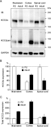
Although total KCC2 expression is increased in the cortex during postnatal development, KCC2a levels remain constant. A, equal amounts (20 μg) of total protein lysates obtained from brainstems, cortices, and spinal cords of P2 or adult (48-week-old) mice were analyzed by standard SDS-PAGE and detected sequentially with the KCC2a (upper panel) and KCC2pan (middle panel) antibodies. A protein of 140 kDa, close to the predicted molecular mass for KCC2a, was observed in all samples analyzed. IB, immunoblot. B, shown is the result of quantification of four independent experiments, one of which is presented in A. Expression of KCC2a in the mouse brainstem and spinal cord was about 2-fold higher in P2 than in adult mice. By contrast, KCC2a expression in the cortex was about 2-fold lower in P2 than in adult mice. Values are mean ± S.E. (error bars) of four independent experiments. C, total KCC2 expression (detected with KCC2pan antibody) was dramatically up-regulated in the cortex between P2 and adulthood but increased only moderately in the brainstem. In the spinal cord, total KCC2 expression did not change significantly between the P2 and adult time points. Values are mean ± S.E. (error bars) of four independent experiments.
FIGURE 4.
FIGURE 4.
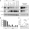
KCC2a contributes at least half of the total KCC2 protein in the neonatal mouse, whereas KCC2b is the predominant isoform in the adult brain. A, equal amounts (20 μg) of total protein lysates obtained from spinal cords and different brain regions of P2 or adult (48-week-old) mice were analyzed in parallel with KCC2a protein standards by immunoblotting. Membrane was first immunoblotted (IB) with the KCC2a antibody (upper panel), stripped, and then processed with KCC2pan (middle panel) antibody. GAPDH expression in the samples is shown as a loading control (lower panel). B, relative (percentage) contribution of KCC2a to total KCC2 protein expression in different CNS regions of neonatal (P2) and adult mice. Shown is the result of quantification of five independent experiments, one of which is presented in A. Band intensities of the ∼140-kDa protein product, corresponding to the predicted molecular mass for KCC2a and KCC2b, were measured, and the ratio of KCC2a to total KCC2 was calculated by using standard curves (panel C, see “Experimental Procedures”). KCC2a contributes about 50–65% of the total KCC2 expression in the neonatal mouse brainstem (Bs), cortex (Cx), and spinal cord (Sp). By contrast, KCC2a is a minor KCC2 isoform in the adult CNS; the percentage of KCC2a of the total KCC2 ranges from 4% in the cerebellum (Cb) to 17–18% in the brainstem and spinal cord. Intermediate values are seen in the cortex (8%), hippocampus (Hc, 10%), and olfactory bulb (Ob, 11%). Values are mean ± S.E. (error bars) of five independent experiments. C, standard curves for the KCC2a and KCC2pan antibodies were made by plotting the concentration of the KCC2a standard (= different dilutions, from undiluted up to 1/20, of a KCC2a-expressing HEK293 cell lysate) on the x axis and the measured band intensities on the y axis and then fitting a line to the data.
FIGURE 5.
FIGURE 5.
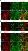
Localization of the KCC2a isoform in cultured neurons. Shown are maximum intensity projections of confocal optical images. A, hippocampal neurons, derived from E17 WT or KCC2 null mutant (KO) mice and cultured for 11 days in vitro (DIV11), were double-immunostained (see “Experimental Procedures”) with antibodies against KCC2a (red) and neuron-specific β-III tubulin (TUJ-1) (green). WT and KO cultures were treated in parallel, and the same settings were applied during the imaging procedures, thus allowing a direct comparison of the KCC2a staining in WT and KO neurons. KCC2a antibody revealed a strong KCC2a protein expression in WT neurons, whereas only residual background staining was found in KO neurons. B, DIV11 hippocampal cultures from WT mice were double-stained with antibodies against KCC2a (red) and GFAP (green). Non-overlapping patterns revealed by these antibodies confirmed an exclusively neuron-specific expression of the KCC2a protein. C, mouse hippocampal neuronal cultures, prepared similar to A and cultured for 5 or 19 days, were double-immunostained with KCC2a (red) and TUJ-1 (green) antibodies. KCC2a expression was exclusively neuronal at all time points analyzed. KCC2a immunostaining was present in cell bodies and dendritic shafts already at DIV5 and remained similar in DIV19 neurons. Scale bar is 10 μmin A and B and 20 μmin C.
FIGURE 6.
FIGURE 6.
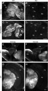
KCC2a and KCC2b protein isoforms have a similar pattern of expression in E18 mouse brain. Sagittal (A–D) or coronal (E–H) cryosections of E18 WT (A, C, and E–H) or KCC2 null mutant (KO, B and D) mice were analyzed by immunohistochemistry (see “Experimental Procedures”) with KCC2a (A, B, E, and G), KCC2pan (C and D), and KCC2b (F and H) antibodies. Immunoreactivity for KCC2a was widely distributed in non-cortical brain areas (A) of wild-type mice and was absent in the KCC2 null mutant littermates (B). The KCC2pan antibody produced a pattern similar to KCC2a antibody in wild-type mice (C) and no staining in sections from KCC2 null mutant embryos (D). Immunostaining of adjacent sections with the KCC2a and KCC2b antibodies revealed a similar distribution of the two KCC2 isoforms in the olfactory bulb (compare E and F) and thalamus (compare G and H). All three KCC2 antibodies detected only a weak signal in the cerebral cortex and hippocampus. The signal in the skin (asterisk in A and G) was unspecific. Cb, cerebellum; Cx, neocortex; Hc, hippocampus; Hy, hypothalamus; Me, medulla; Ob, olfactory bulb; Th, thalamus. Scale bar is 1 m min A–D; 0.5 mm in E and F, and 0.2 mm in G and H.
FIGURE 7.
FIGURE 7.
KCC2a and KCC2b are colocalized in E18 mouse brain neurons. Shown are confocal optical section images of a coronal cryosection through E18 mouse midbrain double-stained with the KCC2a (green) and KCC2b (red) antibodies (see “Experimental Procedures”). C is the merged image of A and B. Most neurons in this area coexpress KCC2a and KCC2b, although the distribution is not identical. 4V, fourth ventricle. Scale bar is 50 μm.
FIGURE 8.
FIGURE 8.
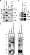
KCC2a can interact with KCC2b in vitro. HEK293 cells were transfected with the indicated plasmids and lysed 48 h later. A and B, the lysates were immunoprecipitated (IP) with the indicated antibodies and analyzed for interacting partners using standard SDS-PAGE and immunoblotting (IB). A, after precipitation with the KCC2a antibody, immunoblotting with the KCC2b antibody detected a band of ∼140 kDa (corresponding to KCC2b) from cells coexpressing KCC2a and KCC2b but not from the combined lysate of cells expressing KCC2a and KCC2b separately (upper panel of IP:KCC2a part). Immunoblotting with the SPAK antibody confirmed that KCC2a is able to interact with SPAK (lower panel of IP: KCC2a part). Analysis of the lysates (Input) ensured that the proteins were successfully expressed. B, after precipitation with an anti-Myc antibody, immunoblotting with the KCC2a antibody detected a band corresponding to KCC2a from cells coexpressing KCC2a and Myc-KCC2b fusion construct but not from the combined lysate of cells expressing KCC2a and Myc-KCC2b separately (upper panel of IP:Myc part). Expression of KCC2a in the cell lysates is also shown (Input). For simplicity, only the monomer bands of KCC2 are shown in A and B. C, the lysates were separated in native PFO gel and analyzed by immunoblotting sequentially with KCC2pan (left panel) and KCC2a (right panel) antibodies. Bands corresponding to the expected size of monomeric (band 1) and homo-oligomeric (band 2–4) forms of KCC2a are numbered. The corresponding bands for the GFP-KCC2b fusion protein are larger due to the additional GFP moiety (∼30 kDa). Bands corresponding to the expected size of hetero-oligomers between KCC2a and GFP-KCC2b (marked by arrows) are seen between the KCC2a and GFP-KCC2b homo-oligomeric bands, and they appear more prominent in the left than in the right panel because the KCC2pan antibody can bind both KCC2a and GFP-KCC2b in the hetero-oligomers.
FIGURE 9.
FIGURE 9.
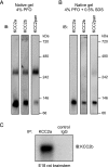
The two KCC2 isoforms form dimers and interact with each other in vivo. A, native PFO-PAGE analysis of protein complexes present in neonatal (P2) mouse brain lysates (see “Experimental Procedures”). Immunoblotting (IB) with antibodies to KCC2a and KCC2b (directed against their unique N termini) indicates that a substantial part of both KCC2 isoforms exists as dimers in the neonatal brain. Note that the relative amount of KCC2 dimers versus monomers appears to be higher when detected with the KCC2pan antibody (directed against the common C terminus). B, the same sample as in A, but 0.5% SDS was included in the sample 20 min before loading to PFO-PAGE. C, a lysate of E18 rat brainstem was subjected to coIP with the KCC2a or a control antibody (normal rabbit IgG), and the precipitate was analyzed by immunoblotting with the KCC2b antibody. A band of 140 kDa, corresponding to the KCC2b, was detected in the sample after coIP with KCC2a but not with the control antibody.

Similar articles

Cited by

References

    1. Blaesse, P., Airaksinen, M. S., Rivera, C., and Kaila, K. (2009) Neuron 61 820-838 - PubMed
    1. Payne, J. A., Stevenson, T. J., and Donaldson, L. F. (1996) J. Biol. Chem. 271 16245-16252 - PubMed
    1. Gamba, G. (2005) Physiol. Rev. 85 423-493 - PubMed
    1. Blaesse, P., Guillemin, I., Schindler, J., Schweizer, M., Delpire, E., Khiroug, L., Friauf, E., and Nothwang, H. G. (2006) J. Neurosci. 26 10407-10419 - PMC - PubMed
    1. Casula, S., Shmukler, B. E., Wilhelm, S., Stuart-Tilley, A. K., Su, W., Chernova, M. N., Brugnara, C., and Alper, S. L. (2001) J. Biol. Chem. 276 41870-41878 - PubMed

Publication types