Insights into the dynamics of hereditary diffuse leukoencephalopathy with axonal spheroids
- PMID: 18794495
- PMCID: PMC2843529
- DOI: 10.1212/01.wnl.0000325916.30701.21
Insights into the dynamics of hereditary diffuse leukoencephalopathy with axonal spheroids
Abstract
Objective: To report a new American family with hereditary diffuse leukoencephalopathy with spheroids (HDLS), including serial, presymptomatic and symptomatic, cranial MRIs from the proband.
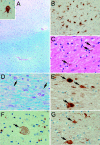
Methods: We report clinical and genealogic investigations of an HDLS family, sequential brain MRIs of the proband, and autopsy slides of brain tissue from the proband's father.
Results: We identified seven affected family members (five deceased). The mean age at symptomatic disease onset was 35 years (range: 20-57), and the mean disease duration was 16 years (range: 3-46). Five affected individuals initially manifested memory disturbance and behavioral changes, whereas two experienced a mood disorder as their presenting symptom. Our proband's father had been diagnosed clinically with vascular dementia, but his brain autopsy was consistent with HDLS. The proband had a cranial MRI prior to symptom onset, with two subsequent MRIs performed during follow-up. These serial images reveal a progressive, confluent, frontal-predominant leukoencephalopathy with symmetric cortical atrophy.
Conclusions: The proband of our newly identified hereditary diffuse leukoencephalopathy with spheroids (HDLS) kindred had subtle evidence of an incipient leukoencephalopathy on a presymptomatic cranial MRI. Conceivably, MRI may facilitate identifying affected presymptomatic individuals within known HDLS kindreds, increasing the likelihood of isolating the causative genes.
Figures


Similar articles
-
Autosomal dominant diffuse leukoencephalopathy with neuroaxonal spheroids.Neurology. 2000 Jan 25;54(2):463-8. doi: 10.1212/wnl.54.2.463. Neurology. 2000. PMID: 10668715
-
Hereditary diffuse leukoencephalopathy with axonal spheroids (HDLS): a misdiagnosed disease entity.J Neurol Sci. 2012 Mar 15;314(1-2):130-7. doi: 10.1016/j.jns.2011.10.006. Epub 2011 Nov 1. J Neurol Sci. 2012. PMID: 22050953 Free PMC article.
-
Discriminative clinical and neuroimaging features of motor-predominant hereditary diffuse leukoencephalopathy with axonal spheroids and primary progressive multiple sclerosis: A preliminary cross-sectional study.Mult Scler Relat Disord. 2019 Jun;31:22-31. doi: 10.1016/j.msard.2019.03.008. Epub 2019 Mar 12. Mult Scler Relat Disord. 2019. PMID: 30901701
-
Adult-onset leukoencephalopathy with axonal spheroids and pigmented glia (ALSP): Integrating the literature on hereditary diffuse leukoencephalopathy with spheroids (HDLS) and pigmentary orthochromatic leukodystrophy (POLD).J Clin Neurosci. 2018 Feb;48:42-49. doi: 10.1016/j.jocn.2017.10.060. Epub 2017 Nov 6. J Clin Neurosci. 2018. PMID: 29122458 Review.
-
Leukoencephalopathy with spheroids (HDLS) and pigmentary leukodystrophy (POLD): a single entity?Neurology. 2009 Jun 2;72(22):1953-9. doi: 10.1212/WNL.0b013e3181a826c0. Neurology. 2009. PMID: 19487654 Free PMC article. Review.
Cited by
-
A clinicopathological and genetic study of sporadic diffuse leukoencephalopathy with spheroids: a report of two cases.Neuropathol Appl Neurobiol. 2013 Dec;39(7):837-43. doi: 10.1111/nan.12046. Neuropathol Appl Neurobiol. 2013. PMID: 23521113 Free PMC article. Review. No abstract available.
-
Imaging features in conventional MRI, spectroscopy and diffusion weighted images of hereditary diffuse leukoencephalopathy with axonal spheroids (HDLS).J Neurol. 2014 Dec;261(12):2351-9. doi: 10.1007/s00415-014-7509-2. Epub 2014 Sep 20. J Neurol. 2014. PMID: 25239393
-
Adult-onset leukoencephalopathy with neuroaxonal spheroids and pigmented glia: report of five cases and a new mutation.J Neurol. 2013 Feb;260(2):558-71. doi: 10.1007/s00415-012-6680-6. Epub 2012 Sep 30. J Neurol. 2013. PMID: 23052599 Review.
-
Asymmetric focal cortical atrophy in CSF1R-related leukoencephalopathy; case report.Acta Neurol Belg. 2023 Oct;123(5):2001-2003. doi: 10.1007/s13760-022-02065-1. Epub 2022 Aug 18. Acta Neurol Belg. 2023. PMID: 35980505 No abstract available.
-
Sporadic diffuse leucoencephalopathy with axonal spheroids: report of a profuse and rapid cortical-spinal degeneration.Neurol Sci. 2012 Aug;33(4):905-9. doi: 10.1007/s10072-011-0817-8. Epub 2011 Oct 18. Neurol Sci. 2012. PMID: 22005946
References
-
- Baba Y, Ghetti B, Baker MC, et al. Hereditary diffuse leukoencephalopathy with spheroids: clinical, pathologic and genetic studies of a new kindred. Acta Neuropathol (Berl) 2006;111:300–311. - PubMed
-
- Axelsson R, Roytta M, Sourander P, Akesson HO, Andersen O. Hereditary diffuse leucoencephalopathy with spheroids. Acta Psychiatr Scand Suppl 1984;314:1–65. - PubMed
-
- Marotti JD, Tobias S, Fratkin JD, Powers JM, Rhodes CH. Adult onset leukodystrophy with neuroaxonal spheroids and pigmented glia: report of a family, historical perspective, and review of the literature. Acta Neuropathol (Berl) 2004;107:481–488. - PubMed
-
- Terada S, Ishizu H, Yokota O, et al. An autopsy case of hereditary diffuse leukoencephalopathy with spheroids, clinically suspected of Alzheimer’s disease. Acta Neuropathol (Berl) 2004;108:538–545. - PubMed
Publication types
MeSH terms
Grants and funding
LinkOut - more resources
Full Text Sources
