Hypomyelination and congenital cataract: neuroimaging features of a novel inherited white matter disorder
- PMID: 17974614
- PMCID: PMC8118974
- DOI: 10.3174/ajnr.A0792
Hypomyelination and congenital cataract: neuroimaging features of a novel inherited white matter disorder
Abstract
Background and purpose: Hypomyelination and congenital cataract (HCC) is an autosomal recessive white matter disease caused by deficiency of hyccin, a membrane protein implicated in both central and peripheral myelination. We aimed to describe the neuroimaging features of this novel entity.
Materials and methods: A systematic analysis of patients with unclassified leukoencephalopathies admitted to our institutions revealed 10 children with congenital cataract, slowly progressive neurologic impairment, and diffuse white matter abnormalities on neuroimaging. Psychomotor developmental delay was evident after the first year of life. Peripheral neuropathy was demonstrated by neurophysiologic studies in 9 children. The available neuroimaging studies were retrospectively reviewed.
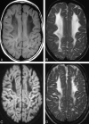
Results: In all patients, neuroimaging revealed diffuse involvement of the supratentorial white matter associated with preservation of both cortical and deep gray matter structures. Supratentorial white matter hypomyelination was detected in all patients; 7 patients also had evidence of variably extensive areas of increased white matter water content. Deep cerebellar white matter hypomyelination was found in 6 patients. Older patients had evidence of white matter bulk loss and gliosis. Proton MR spectroscopy showed variable findings, depending on the stage of the disease. Sural nerve biopsy revealed hypomyelinated nerve fibers. Mutations in the DRCTNNB1A gene on chromosome 7p15.3, causing complete or severe deficiency of hyccin, were demonstrated in all patients.
Conclusions: HCC is characterized by a combined pattern of primary myelin deficiency and secondary neurodegenerative changes. In the proper clinical setting, recognition of suggestive neuroimaging findings should prompt appropriate genetic investigations.
Figures


Similar articles
-
Phenotypic characterization of hypomyelination and congenital cataract.Ann Neurol. 2007 Aug;62(2):121-7. doi: 10.1002/ana.21175. Ann Neurol. 2007. PMID: 17683097
-
Hypomyelination and congenital cataract: identification of novel mutations in two unrelated families.Eur J Paediatr Neurol. 2013 Jan;17(1):108-11. doi: 10.1016/j.ejpn.2012.06.004. Epub 2012 Jun 30. Eur J Paediatr Neurol. 2013. PMID: 22749724
-
Hypomyelination and congenital cataract: broadening the clinical phenotype.Arch Neurol. 2011 Sep;68(9):1191-4. doi: 10.1001/archneurol.2011.201. Arch Neurol. 2011. PMID: 21911699 Review.
-
Hyccin, the molecule mutated in the leukodystrophy hypomyelination and congenital cataract (HCC), is a neuronal protein.PLoS One. 2012;7(3):e32180. doi: 10.1371/journal.pone.0032180. Epub 2012 Mar 26. PLoS One. 2012. PMID: 22461884 Free PMC article.
-
Imaging manifestations of the leukodystrophies, inherited disorders of white matter.Radiol Clin North Am. 2014 Mar;52(2):279-319. doi: 10.1016/j.rcl.2013.11.008. Radiol Clin North Am. 2014. PMID: 24582341 Review.
Cited by
-
The chemistry and biology of phosphatidylinositol 4-phosphate at the plasma membrane.Bioorg Med Chem. 2021 Jun 15;40:116190. doi: 10.1016/j.bmc.2021.116190. Epub 2021 May 1. Bioorg Med Chem. 2021. PMID: 33965837 Free PMC article. Review.
-
Mitochondrial hsp60 chaperonopathy causes an autosomal-recessive neurodegenerative disorder linked to brain hypomyelination and leukodystrophy.Am J Hum Genet. 2008 Jul;83(1):30-42. doi: 10.1016/j.ajhg.2008.05.016. Epub 2008 Jun 19. Am J Hum Genet. 2008. PMID: 18571143 Free PMC article.
-
Cockayne syndrome: a diffusion tensor imaging and volumetric study.Br J Radiol. 2016 Nov;89(1067):20151033. doi: 10.1259/bjr.20151033. Epub 2016 Sep 19. Br J Radiol. 2016. PMID: 27643390 Free PMC article.
-
Magnetic resonance imaging pattern recognition in hypomyelinating disorders.Brain. 2010 Oct;133(10):2971-82. doi: 10.1093/brain/awq257. Brain. 2010. PMID: 20881161 Free PMC article.
-
Late-onset spastic ataxia phenotype in a patient with a homozygous DDHD2 mutation.Sci Rep. 2014 Nov 24;4:7132. doi: 10.1038/srep07132. Sci Rep. 2014. PMID: 25417924 Free PMC article.
References
-
- van der Knaap MS, Breiter SN, Naidu S, et al. Defining and categorizing leukoencephalopathies of unknown origin: MR imaging approach. Radiology 1999;213:121–33 - PubMed
-
- Schiffmann R, van der Knaap MS. The latest on leukodystrophies. Curr Opin Neurol 2004;17:187–92 - PubMed
-
- Zara F, Biancheri R, Bruno C, et al. Deficiency of hyccin, a newly identified membrane protein, causes hypomyelination and congenital cataract. Nat Genet 2006;38:1111–13. Epub 2006 Sep 3 - PubMed
-
- Provencher SW. Estimation of metabolite concentrations from localized in vivo proton MR spectra. Magn Reson Med 1993;30:672–79 - PubMed
MeSH terms
LinkOut - more resources
Full Text Sources
Medical
