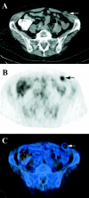
Positron emission tomography/computed tomography influences on the management of resectable pancreatic cancer and its cost-effectiveness
- PMID: 16041214
- PMCID: PMC1357729
- DOI: 10.1097/01.sla.0000172095.97787.84
Positron emission tomography/computed tomography influences on the management of resectable pancreatic cancer and its cost-effectiveness
Abstract
Objective: We sought to determine the impact of positron emission tomography/computed tomography (PET/CT) on the management of presumed resectable pancreatic cancer and to assess the cost of this new staging procedure.
Summary background data: PET using 18F-fluorodeoxyglucose (FDG) is increasingly used for the staging of pancreatic cancer, but anatomic information is limited. Integrated PET/CT enables optimal anatomic delineation of PET findings and identification of FDG-negative lesions on computed tomography (CT) images and might improve preoperative staging.
Material and methods: Patients with suspected pancreatic cancer who had a PET/CT between June 2001 to April 2004 were entered into a prospective database. Routine staging included abdominal CT, chest x-ray, and CA 19-9 measurement. FDG-PET/CT was conducted according to a standardized protocol, and findings were confirmed by histology. Cost benefit analysis was performed based on charged cost of PET/CT and pancreatic resection and included the time frame of staging and surgery.
Results: Fifty-nine patients with a median age of 61 years (range, 40-80 years) were included in this analysis. Fifty-one patients had lesions in the head and 8 in the tail of the pancreas. The positive and negative predictive values for pancreatic cancer were 91% and 64%, respectively. PET/CT detected additional distant metastases in 5 and synchronous rectal cancer in 2 patients. PET/CT findings changed the management in 16% of patients with pancreatic cancer deemed resectable after routine staging (P = 0.031) and was cost saving.
Conclusions: PET/CT represents an important staging procedure prior to pancreatic resection for cancer, since it significantly improves patient selection and is cost-effective.
Figures


Comment in
-
Positron emission tomography/computed tomography influences on the management of resectable pancreatic cancer and its cost-effectiveness.Ann Surg. 2006 May;243(5):709-10; author reply 710. doi: 10.1097/01.sla.0000216766.93589.34. Ann Surg. 2006. PMID: 16633011 Free PMC article. No abstract available.
-
Canadian Association of General Surgeons and American College of Surgeons evidence based reviews in surgery. 22. The use of PET/CT scanning on the management of resectable pancreatic cancer.Can J Surg. 2007 Oct;50(5):400-2. Can J Surg. 2007. PMID: 18031643 Free PMC article. No abstract available.
Similar articles
-
A prospective diagnostic accuracy study of 18F-fluorodeoxyglucose positron emission tomography/computed tomography, multidetector row computed tomography, and magnetic resonance imaging in primary diagnosis and staging of pancreatic cancer.Ann Surg. 2009 Dec;250(6):957-63. doi: 10.1097/SLA.0b013e3181b2fafa. Ann Surg. 2009. PMID: 19687736
-
[Benefit of PET/CT in the preoperative staging in pancreatic carcinomas].Rozhl Chir. 2010 Aug;89(7):433-40. Rozhl Chir. 2010. PMID: 20925260 Czech.
-
How useful is integrated PET and CT for the management of pancreatic cancer?Nat Clin Pract Gastroenterol Hepatol. 2006 Feb;3(2):74-5. doi: 10.1038/ncpgasthep0407. Nat Clin Pract Gastroenterol Hepatol. 2006. PMID: 16456571 No abstract available.
-
18Fluorodeoxyglucose-positron emission tomography in the management of patients with suspected pancreatic cancer.Ann Surg. 1999 May;229(5):729-37; discussion 737-8. doi: 10.1097/00000658-199905000-00016. Ann Surg. 1999. PMID: 10235532 Free PMC article. Review.
-
Preoperative intrathoracic lymph node staging in patients with non-small-cell lung cancer: accuracy of integrated positron emission tomography and computed tomography.Eur J Cardiothorac Surg. 2009 Sep;36(3):440-5. doi: 10.1016/j.ejcts.2009.04.003. Epub 2009 May 22. Eur J Cardiothorac Surg. 2009. PMID: 19464906 Review.
Cited by
-
Impact of 18-fluorodeoxyglucose positron emission tomography on the management of pancreatic cancer.J Gastrointest Surg. 2010 Jul;14(7):1151-8. doi: 10.1007/s11605-010-1207-x. Epub 2010 May 5. J Gastrointest Surg. 2010. PMID: 20443074
-
Diagnostic impact of 18F-FDG PET-CT evaluating solid pancreatic lesions versus endosonography, endoscopic retrograde cholangio-pancreatography with intraductal ultrasonography and abdominal ultrasound.Eur J Nucl Med Mol Imaging. 2008 Oct;35(10):1775-85. doi: 10.1007/s00259-008-0818-x. Epub 2008 May 15. Eur J Nucl Med Mol Imaging. 2008. PMID: 18481063 Clinical Trial.
-
A prospective study of the impact of fluorodeoxyglucose positron emission tomography with concurrent non-contrast CT scanning on the management of operable pancreatic and peri-ampullary cancers.HPB (Oxford). 2015 Jul;17(7):624-31. doi: 10.1111/hpb.12418. Epub 2015 Apr 30. HPB (Oxford). 2015. PMID: 25929273 Free PMC article.
-
Ductal adenocarcinoma of the pancreatic head: a focus on current diagnostic and surgical concepts.World J Gastroenterol. 2012 Jun 28;18(24):3058-69. doi: 10.3748/wjg.v18.i24.3058. World J Gastroenterol. 2012. PMID: 22791941 Free PMC article. Review.
-
Accuracy of various criteria for lymph node staging in ductal adenocarcinoma of the pancreatic head by computed tomography and magnetic resonance imaging.World J Surg Oncol. 2020 Aug 18;18(1):213. doi: 10.1186/s12957-020-01951-3. World J Surg Oncol. 2020. PMID: 32811523 Free PMC article.
References
-
- Ferlay J, Bray F, Pisani P GLOBOCAN 2000: cancer incidence, mortality and prevalence worldwide. In: IARC, ed. Cancer Base No. 5. Lyon: IARC; 2001.
-
- Li D, Xie K, Wolff R, et al. Pancreatic cancer. Lancet. 2004;363:1049–1057. - PubMed
-
- Raut C, Grau A, Staerkel G, et al. Diagnostic accuracy of endoscopic ultrasound-guided fine-needle aspiration in patients with presumed pancreatic cancer. J Gastrointest Surg. 2003;7:118–128. - PubMed
MeSH terms
Substances
LinkOut - more resources
Full Text Sources
Other Literature Sources
Medical
Miscellaneous

