. 2002 Mar 15;21(6):1427-36.
doi: 10.1093/emboj/21.6.1427.

Ccr4p is the catalytic subunit of a Ccr4p/Pop2p/Notp mRNA deadenylase complex in Saccharomyces cerevisiae

Affiliations

Ccr4p is the catalytic subunit of a Ccr4p/Pop2p/Notp mRNA deadenylase complex in Saccharomyces cerevisiae

Morgan Tucker et al. EMBO J. .

Abstract

The major pathways of mRNA turnover in eukaryotic cells are initiated by shortening of the poly(A) tail. Recent work has identified Ccr4p and Pop2p as components of the major cytoplasmic deadenylase in yeast. We now demonstrate that CCR4 encodes the catalytic subunit of the deadenylase and that Pop2p is dispensable for catalysis. In addition, we demonstrate that at least some of the Ccr4p/Pop2p-associated Not proteins are cytoplasmic, and lesions in some of the NOT genes can lead to defects in mRNA deadenylation rates. The Ccr4p deadenylase is inhibited in vitro by addition of the poly(A) binding protein (Pab1p), suggesting that dissociation of Pab1p from the poly(A) tail may be rate limiting for deadenylation in vivo. In addition, the rapid deadenylation of the COX17 mRNA, which is controlled by a member of the Pumilio family of deadenylation activators Puf3p, requires an active Ccr4p/Pop2p/Not deadenylase. These results define the Ccr4p/Pop2p/Not complex as the cytoplasmic deadenylase in yeast and identify positive and negative regulators of this enzyme complex.


Figures
None
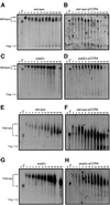
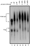
Fig. 1. Comparison of deadenylation end-points. Shown are poly acrylamide northern gels examining the deadenylation end-points for the PGK1pG transcript in wild-type, ccr4Δ and pop2Δ strains containing either a Flag-Ccr4p over-expression plasmid (pRP1045) or vector control as indicated. Here, and in other figures, the full-length (FL) mRNA and decay fragment (Frag.) are indicated on the left. Steady-state mRNA samples were resolved on 6% polyacrylamide/8 M urea northern gels, with or without removal of poly(A) tails with RNase H and oligo(dT) (lane 1). Here, and in other figures, to allow for size resolution of the poly(A) tail the 3′ 319 nucleotides of the 1.4 kb PGK1pG mRNA were cleaved by hybridizing to oRP70 followed by cleavage with RNase H prior to loading on the gel.
None
Fig. 2. Transcriptional pulse–chase analysis of the MFA2pG and PGK1pG transcripts. Shown are polyacrylamide northern gels examining the decay of MFA2pG (AD) and PGK1pG (EH) in wild-type, ccr4Δ and pop2Δ strains containing either a Flag-Ccr4p over-expression plasmid (pRP1045) or vector control as indicated. Numbers above the lanes are minutes after transcriptional repression by the addition of glucose following an 8 min induction of transcription (Decker and Parker, 1993). The 0 min time point was treated with RNase H and oligo(dT) to indicate the position of the deadenylated mRNA. Here, and in all subsequent figures, poly(A) tail lengths were determined by comparison of bands to size standards and the poly(A) mRNA species generated by cleavage of RNase H and oligo(dT) (data not shown).
None
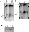
Fig. 3. Flag-Ccr4p co-purifies with deadenylase activity. Analysis of deadenylation activity in Flag-Ccr4p purified fractions from ccr4Δ and ccr4Δ/pop2Δ strains on a capped, polyadenylated substrate. Numbers above the lanes indicate time points taken after addition of substrate to the reaction. The arrow represents the fully deadenylated form of the substrate based on migration of substrate after cleavage with RNase H and oligo(dT) (data not show). The asterisk indicates the position of the radiolabeled phosphates.
None
Fig. 4. Comparison of deadenylation end-points in ccr4 point mutants. Shown are polyacrylamide northern gels for the MFA2pG transcript in (Accr4Δ strains or (Bccr4Δ/pan2Δ strains, containing either a vector control, Flag-Ccr4p wild-type plasmid (pRP1045) or Flag-Ccr4p plasmids containing specific point mutants (pRP1046–1049) as indicated (see Materials and methods). Steady-state mRNA samples were resolved on 6% polyacrylamide/8 M urea northern gels, with or without removal of poly(A) tails with RNase H and oligo(dT) (lane 1). (C) Western analysis of extracts from ccr4Δ strains harboring both wild-type and mutant forms of the CCR4 plasmid. Probing with anti-Flag antibodies indicates that each mutant produces a full-length Ccr4p.
None
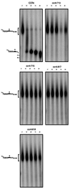
Fig. 5. The Ccr4p nuclease motif is required for deadenylase activity. Analysis of deadenylation activity in Flag-Ccr4p purified fractions on a capped, polyadenylated substrate from a ccr4Δ strain containing either a Flag-Ccr4p wild-type plasmid (pRP1045) or Flag-Ccr4p plasmids containing specific point mutants (pRP1046–1049) as indicated (see Materials and methods). Numbers above the lanes indicate time points taken after addition of substrate to the reaction. The asterisk indicates the position of the radiolabeled phosphates.
None
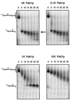
Fig. 6. Pab1p inhibits Ccr4p deadenylase activity in vitro. Analysis of deadenylation activity in Flag-Ccr4p purified fractions with addition of increasing amounts of purified Pab1p. Purified Pab1p was added to each time course in molar amounts relative to Flag-Ccr4p as indicated. Numbers above the lanes indicate time points taken after addition of substrate to the reaction. The asterisk indicates the position of the radiolabeled phosphate.
None
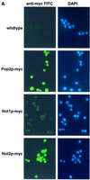
Fig. 7. The Not protein complex functions in deadenylation. (A) Localization of Not1p and Not2p by indirect immunofluorescence. Strains expressing myc-tagged Not1p (yRP1668), myc-tagged Not2p (yRP1669), myc-tagged Pop2p (yRP1622), or wild-type control (yRP841), were stained with anti-myc antibodies and anti-mouse (FITC) secondary antibody and DAPI as indicated. It is critical to note that all photographs were taken under identical conditions. The localization of myc-tagged Not1p was consistently cytoplasmic; however, the signal often appeared weaker. This observation is presumably a result of the Not1p myc-epitope being less accessible to the anti-myc antibodies. (B) Transcriptional pulse–chase analysis of the MFA2pG in not2Δ, not3Δ, not4Δ and not5Δ strains. Shown are polyacrylamide northern gels examining the decay of MFA2pG in the indicated strains. Numbers above the lanes are minutes after transcriptional repression by the addition of glucose following an 8 min induction of transcription (Decker and Parker, 1993). The 0 min time points were treated with RNase H and oligo(dT) to indicate the position of the deadenylated mRNA.
None
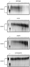
Fig. 8. Transcriptional pulse–chase analysis of the COX17 transcript. Shown are polyacrylamide northern gels examining the decay of COX17 in wild-type, ccr4Δ and ccr4Δ/pan2Δ strains. Numbers above the lanes are minutes after transcriptional repression by the addition of glucose following an 8 min induction of transcription (Decker and Parker, 1993). The 0 min time point was treated with RNase H and oligo(dT) to indicate the position of the deadenylated mRNA. To allow for size resolution of the poly(A) tail the 3′ 160 nucleotides of the 390 nucleotide COX17 mRNA were cleaved by hybridizing to oRP808 followed by treatment with RNase H prior to loading on the gel.

Similar articles

Cited by

References

    1. Amrani N., Minet,M., Le Gouar,M., Lacroute,F. and Wyers,F. (1997) Yeast Pab1 interacts with Rna15 and participates in the control of the poly(A) tail length in vitro. Mol. Cell. Biol., 17, 3694–3701. - PMC - PubMed
    1. Anderson J.S.J. and Parker,R.P. (1998) The 3′ to 5′ degradation of yeast mRNAs is a general mechanism for mRNA turnover that requires the SKI2 DEVH box protein and 3′ to 5′ exonucleases of the exosome complex. EMBO J., 17, 1497–1506. - PMC - PubMed
    1. Badarinarayana V., Chiang,Y.C. and Denis,C.L. (2000) Functional interaction of CCR4–NOT proteins with TATAA-binding protein (TBP) and its associated factors in yeast. Genetics, 155, 1045–1054. - PMC - PubMed
    1. Bai Y., Salvadore,C., Chiang,Y.C., Collart,M.A., Liu,H.Y. and Denis,C.L. (1999) The CCR4 and CAF1 proteins of the CCR4–NOT complex are physically and functionally separated from NOT2, NOT4 and NOT5. Mol. Cell. Biol., 19, 6642–6651. - PMC - PubMed
    1. Beelman C.A. and Parker,R. (1995) Degradation of mRNA in eukaryotes. Cell, 81, 179–183. - PubMed

Publication types

MeSH terms