Inherited susceptibility to uterine leiomyomas and renal cell cancer
- PMID: 11248088
- PMCID: PMC30663
- DOI: 10.1073/pnas.051633798
Inherited susceptibility to uterine leiomyomas and renal cell cancer
Abstract
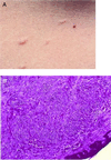
Herein we report the clinical, histopathological, and molecular features of a cancer syndrome with predisposition to uterine leiomyomas and papillary renal cell carcinoma. The studied kindred included 11 family members with uterine leiomyomas and two with uterine leiomyosarcoma. Seven individuals had a history of cutaneous nodules, two of which were confirmed to be cutaneous leiomyomatosis. The four kidney cancer cases occurred in young (33- to 48-year-old) females and displayed a unique natural history. All these kidney cancers displayed a distinct papillary histology and presented as unilateral solitary lesions that had metastasized at the time of diagnosis. Genetic-marker analysis mapped the predisposition gene to chromosome 1q. Losses of the normal chromosome 1q were observed in tumors that had occurred in the kindred, including a uterine leiomyoma. Moreover, the observed histological features were used as a tool to diagnose a second kindred displaying the phenotype. We have shown that predisposition to uterine leiomyomas and papillary renal cell cancer can be inherited dominantly through the hereditary leiomyomatosis and renal cell cancer (HLRCC) gene. The HLRCC gene maps to chromosome 1q and is likely to be a tumor suppressor. Clinical, histopathological, and molecular tools are now available for accurate detection and diagnosis of this cancer syndrome.
Figures


Similar articles
-
Familial multiple cutaneous and uterine leiomyomas associated with papillary renal cell cancer.Clin Exp Dermatol. 2005 Jan;30(1):75-8. doi: 10.1111/j.1365-2230.2004.01675.x. Clin Exp Dermatol. 2005. PMID: 15663510
-
Conventional renal cancer in a patient with fumarate hydratase mutation.Hum Pathol. 2007 May;38(5):793-6. doi: 10.1016/j.humpath.2006.10.011. Epub 2007 Jan 31. Hum Pathol. 2007. PMID: 17270241
-
Few FH mutations in sporadic counterparts of tumor types observed in hereditary leiomyomatosis and renal cell cancer families.Cancer Res. 2002 Aug 15;62(16):4554-7. Cancer Res. 2002. PMID: 12183404
-
[From gene to disease; cutaneous leiomyomatosis].Ned Tijdschr Geneeskd. 2007 Feb 3;151(5):300-4. Ned Tijdschr Geneeskd. 2007. PMID: 17326474 Review. Dutch.
-
Clinical and molecular genetic aspects of hereditary multiple cutaneous leiomyomatosis.Eur J Dermatol. 2009 Nov-Dec;19(6):545-51. doi: 10.1684/ejd.2009.0749. Epub 2009 Jul 10. Eur J Dermatol. 2009. PMID: 19939761 Review.
Cited by
-
Mitochondrial dysfunction and risk of cancer.Br J Cancer. 2015 Mar 17;112(6):1134-40. doi: 10.1038/bjc.2015.66. Br J Cancer. 2015. PMID: 25742477 Free PMC article.
-
A novel fumarate hydratase-deficient HLRCC kidney cancer cell line, UOK268: a model of the Warburg effect in cancer.Cancer Genet. 2012 Jul-Aug;205(7-8):377-90. doi: 10.1016/j.cancergen.2012.05.001. Cancer Genet. 2012. PMID: 22867999 Free PMC article.
-
Reed's Syndrome.Indian J Dermatol. 2012 Mar;57(2):156-7. doi: 10.4103/0019-5154.94296. Indian J Dermatol. 2012. PMID: 22615521 Free PMC article. No abstract available.
-
TCA Cycle Defects and Cancer: When Metabolism Tunes Redox State.Int J Cell Biol. 2012;2012:161837. doi: 10.1155/2012/161837. Epub 2012 Jul 19. Int J Cell Biol. 2012. PMID: 22888353 Free PMC article.
-
Glutamine reliance in cell metabolism.Exp Mol Med. 2020 Sep;52(9):1496-1516. doi: 10.1038/s12276-020-00504-8. Epub 2020 Sep 17. Exp Mol Med. 2020. PMID: 32943735 Free PMC article. Review.
References
-
- Latif F, Tory K, Gnarra J, Yao M, Duh F M, Orcutt M L, Stackhouse T, Kuzmin I, Modi W, Geil L, et al. Science. 1993;260:1317–1320. - PubMed
-
- Schmidt L, Duh F M, Chen F, Kishida T, Glenn G, Choyke P, Scherer S W, Zhuang Z, Lubensky I, Dean M, et al. Nat Genet. 1997;16:68–73. - PubMed
-
- Cohen A J, Li F P, Berg S, Marchetto D J, Tsai S, Jacobs C S, Brown R S. N Engl J Med. 1979;301:592–595. - PubMed
-
- Zbar B, Lerman M. Adv Cancer Res. 1998;75:163–201. - PubMed
-
- Iliopoulos O, Eng C. Semin Oncol. 2000;27:138–149. - PubMed
Publication types
MeSH terms
Substances
LinkOut - more resources
Full Text Sources
Other Literature Sources
Medical
Molecular Biology Databases

