October 15, 2019

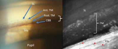
The left image shows the trabecular meshwork as it can be seen in a doctor's office. The right image shows a portion of the same part of the eye in much greater detail using methods developed at IU. Images courtesy the Indiana University School of Optometry
Using methods originally developed by astronomers to view stars more clearly through Earth's atmosphere, optometry researchers at Indiana University have taken the first undistorted microscopic images of a part of the eye involved in glaucoma.