. 2024 Nov 7;15(1):9653.
doi: 10.1038/s41467-024-53942-7.

Structures of the multi-domain oxygen sensor DosP: remote control of a c-di-GMP phosphodiesterase by a regulatory PAS domain

Affiliations

Structures of the multi-domain oxygen sensor DosP: remote control of a c-di-GMP phosphodiesterase by a regulatory PAS domain

Wenbi Wu et al. Nat Commun. .

Abstract

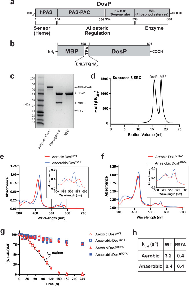
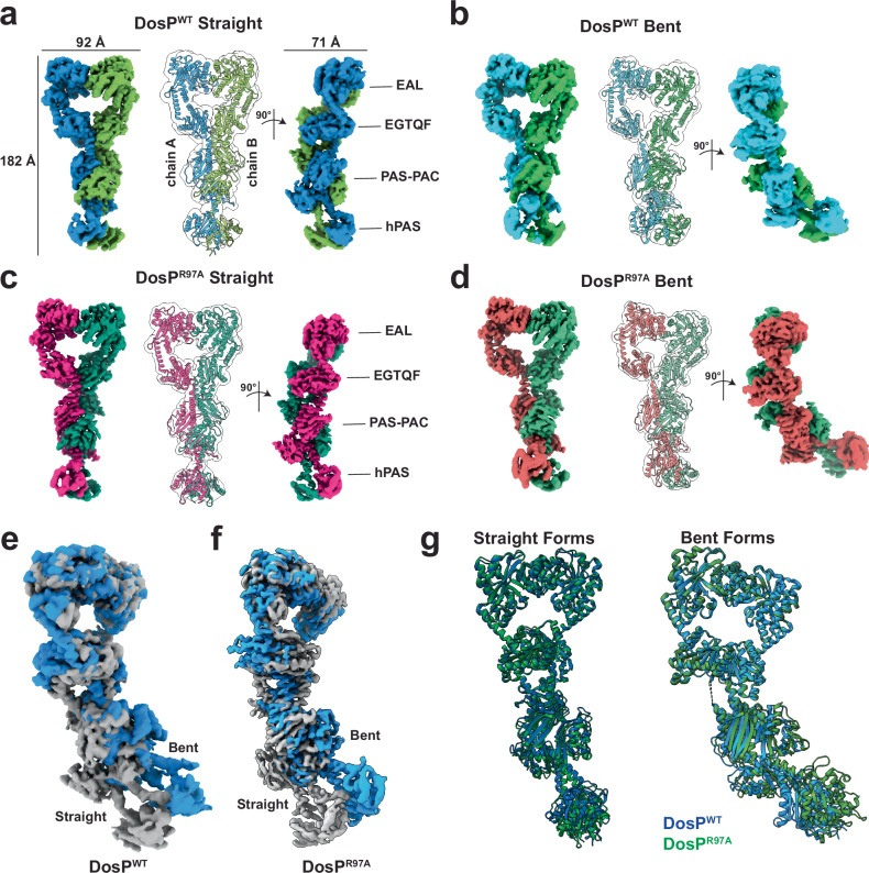
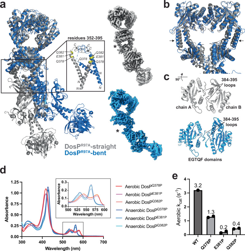
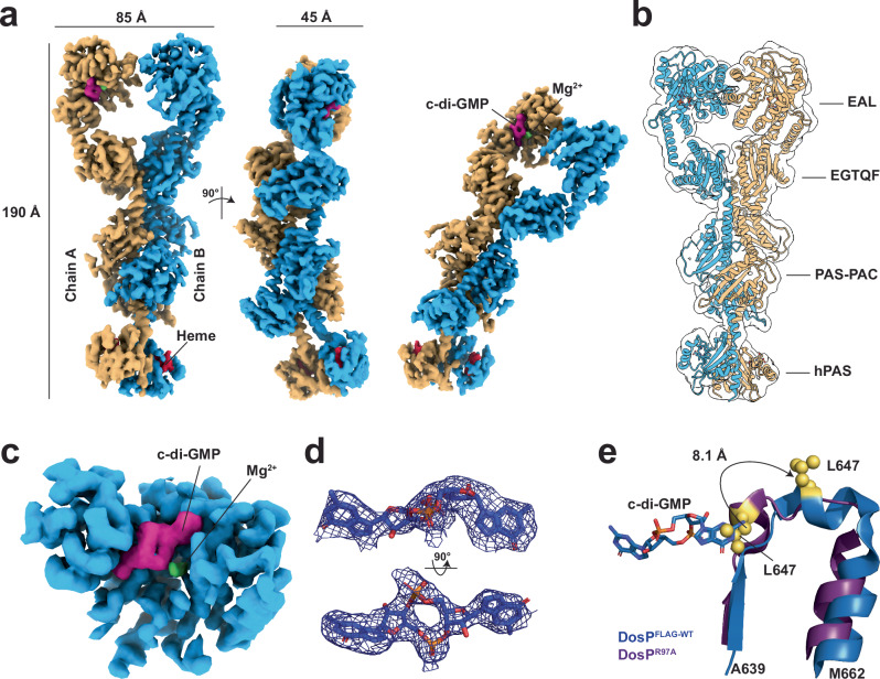
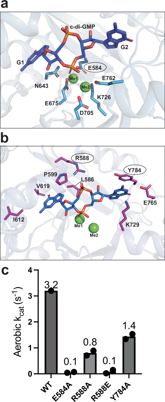
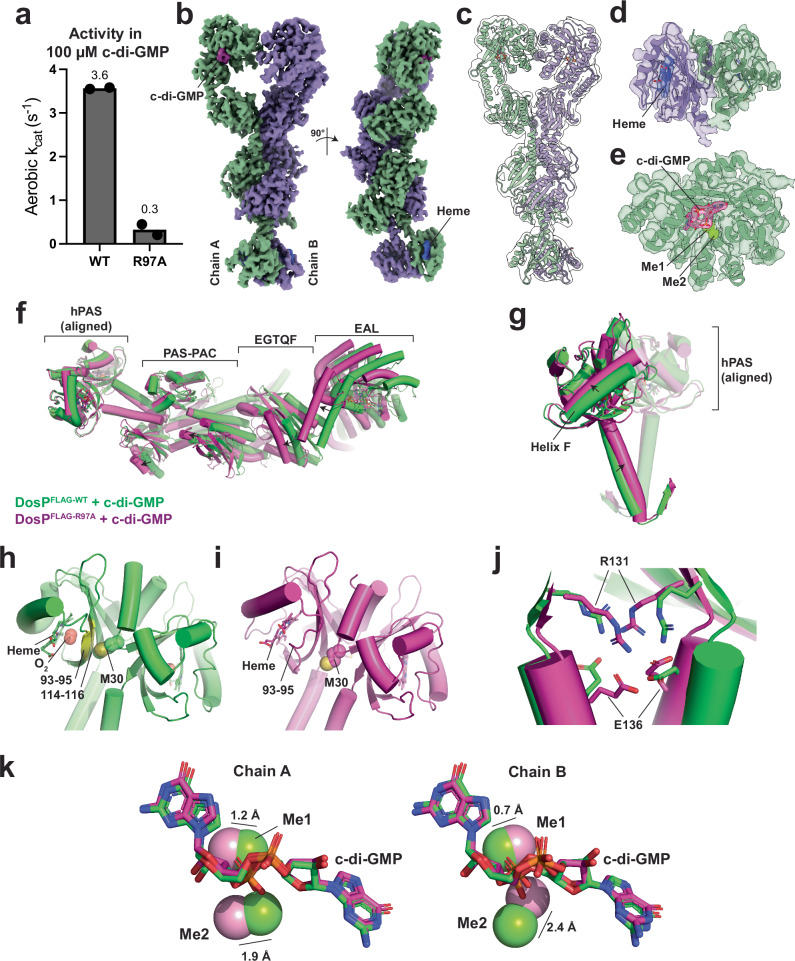
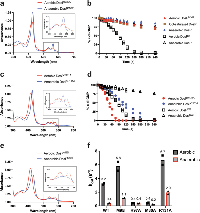
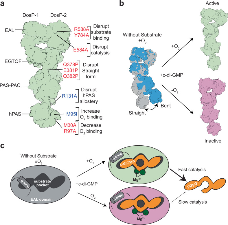
The heme-based direct oxygen sensor DosP degrades c-di-GMP, a second messenger nearly unique to bacteria. In stationary phase Escherichia coli, DosP is the most abundant c-di-GMP phosphodiesterase. Ligation of O2 to a heme-binding PAS domain (hPAS) of the protein enhances the phosphodiesterase through an allosteric mechanism that has remained elusive. We determine six structures of full-length DosP in its aerobic or anaerobic conformations, with or without c-di-GMP. DosP is an elongated dimer with the regulatory heme containing domain and phosphodiesterase separated by nearly 180 Å. In the absence of substrate, regardless of the heme status, DosP presents an equilibrium of two distinct conformations. Binding of substrate induces DosP to adopt a single, ON-state or OFF-state conformation depending on its heme status. Structural and biochemical studies of this multi-domain sensor and its mutants provide insights into signal regulation of second-messenger levels.


Conflict of interest statement

The authors declare no competing interests.

Figures

Fig. 1
Fig. 1. Purification and biochemical characterization of DosP.
a Domain organization of DosP over its linear amino acid sequence. b Schematic for the MBP-DosP expression construct. TEV protease recognition sequence is shown under brackets. Cleavage site marked by arrow and resulting N-terminal methionine corresponds to DosP M1. c Coomassie-stained 4-15% SDS-PAGE analysis of purified DosP from the indicated steps. Migration positions of the proteins are indicated with arrows. Molecular weight standards are labeled. d SEC purification of free DosP, MBP, and TEV protease following proteolysis of MBP-DosP using a 10/300 Superose 6 gel filtration column as described in Methods. e Absorption spectra of DosPWT under aerobic conditions (red), compared to anaerobic conditions (blue). f Absorption spectra of DosPR97A under aerobic conditions (red), compared to anaerobic DosPR97A (blue). g Turnover of 2 µM c-di-GMP at 25 °C by 10 nM protein: aerobic DosPWT (open red triangles), anaerobic DosPWT (open blue squares), aerobic DosPR97A (filled red triangles), and anaerobic DosPR97A (filled blue squares). Black line indicates time regime used to determine kcat. Each data point represents a measurement from an independent experiment (n = 2). The assay buffer was 20 mM Tris-HCl, 2.5 mM MgCl2, 2.0 mM DTT, pH 8.0. h kcat values determined from g. Values are the averages of Kcat measurements from two independent experiments. Source data are provided as a Source Data file.
Fig. 2
Fig. 2. Structures of DosPWT and DosPR97A.
a Cryo-EM map of the DosPWT straight conformer. Left: map is shown at a contour of 0.0335 and colored by chain. Lines and labels indicate axial dimension. Middle: Atomic model depicted as cartoon and overlaid with a map that was gaussian filtered to level 1.5. Right: Map colored as in left panel and rotated 90°. Domains of DosP are labeled. b Cryo-EM map of the DosPWT bent conformer. Left: map is shown at a contour of 0.026 and colored by chain. Middle: Atomic model depicted as cartoon and overlaid with a map that was gaussian filtered to level 1.5. Right: Map colored as in left panel and rotated 90°. c Cryo-EM map of the DosPR97A straight conformer. Left: map is shown at a contour of 0.0142 and colored by chain. Middle: Atomic model depicted as cartoon and overlaid with a map that was gaussian filtered to level 1.5. Right: Map colored as in left panel and rotated 90°. Domains are labeled. d Cryo-EM map of the DosPR97A bent conformer. Left: map is shown at a contour of 0.075 and colored by chain. Middle: Atomic model depicted as cartoon and overlaid with a map that was gaussian filtered to level 1.5. Right: Map colored as in left panel and rotated 90°. e DosPWT bent form and straight form maps superposed through their EAL domains. The straight map is colored grey, and the bent form map is colored blue. f DosPR97A bent form and straight form maps superposed through their EAL domains. The straight map is colored grey, and the bent form map is colored blue. g Atomic models of DosPWT and DosPR97A are superimposed. DosPWT is depicted as blue cartoon and DosPR97A is depicted as green cartoon. Bent and Straight forms are labeled. All panels are generated using ChimeraX. hPAS, heme-binding Per-Arnt-Sim; EAL, c-di-GMP phosphodiesterase domain; EGTQF- diguanylate cyclase domain.
Fig. 3
Fig. 3. Investigation of the PAS-PAC/EGTQF hinge of DosP.
a Atomic models of the straight and bent forms of DosPR97A depicted as grey cartoon or blue cartoon, respectively. Box highlights the PAS-PAC/EGTQF hinge helix (residues 352-382). Middle inset: Atomic models of residues 352-395 of the bent and straight forms of DosPR97A. NH2 and COOH termini are labeled with N and C, respectively. Positions of residues interrogated by mutagenesis are labeled yellow on the model of DosPR97A straight. Top right inset: Cryo-EM map of DosPR97A straight form. Asterisk denotes the well-resolved hinge helix. Bottom right inset: Cryo-EM map of DosPR97A bent form. Asterisk denotes the unresolved hinge helix. b Atomic models of the EAL and EGTQF domains of the straight and bent forms of DosPR97A depicted as grey cartoon or blue cartoon, respectively. Arrows indicate changes of the helix between the EAL and EGTQF domains. c The EGTQF dimer interface, viewed as if the position in b was rotated 90° upwards towards the reader. Top: the straight form of DosPR97A shown as grey cartoon. The two chains and dimer interface loops are labeled. Bottom: the bent form of DosPR97A shown as blue cartoon. d Absorption spectra of DosPQ378P, DosPE381P, and DosPQ382P under aerobic conditions (red, magenta, and orange, respectively), compared to anaerobic conditions (dark blue, blue, and cyan, respectively). e Bar graph showing the kcat values determined from the turnover of 2 µM c-di-GMP at 25°C by 10 nM aerobic: DosPWT, DosPQ378P, DosPE381P, and DosPQ382P. Value for DosPWT is taken from Fig. 1g, values for mutants are the average of two independent experiments. In d and e the assay buffer was 20 mM Tris-HCl, 2.5 mM MgCl2, 2.0 mM DTT, pH 8.0. Source data are provided as a Source Data file. Cartoons depicted in a and b generated using PyMOL. Inset maps in a and cartoons in c generated using ChimeraX. EAL, c-di-GMP phosphodiesterase domain; EGTQF- diguanylate cyclase domain.
Fig. 4
Fig. 4. Structural details of DosPWT with bound c-di-GMP.
a Composite map of DosPFLAG-WT with the bound c-di-GMP from three orientations. Density for the two chains are shown as cyan and yellow density maps at a contour level of 0.064. The density for c-di-GMP and heme is shown in magenta and red, respectively. The density of magnesium is shown in green. Axial dimensions are labeled. b Atomic model depicted as cartoon and overlaid with a map that was gaussian filtered to level 1.5. Domains of DosP are labeled. c close view of a single EAL domain with bound c-di-GMP. Density for the EAL domain is colored cyan, c-di-GMP density is colored magenta, and magnesium density is colored green. d Density for c-di-GMP shown in two orientations. The c-di-GMP is represented as sticks and the density map displayed as a blue mesh carved at 2.0 Å and sigma=10. e DosPFLAG-WT and DosPR97A residues 639-662 superposed and displayed with blue and purple cartoon, respectively. L647 is depicted with yellow spheres and c-di-GMP is depicted in sticks. Arrow indicates the change in position of L647 between the two forms of DosP. Panels a-c generated using ChimeraX. Panels d-e generated using PyMOL. hPAS, heme-binding Per-Arnt-Sim; EAL, c-di-GMP phosphodiesterase domain; EGTQF- diguanylate cyclase domain.
Fig. 5
Fig. 5. The DosP catalytic pocket.
a Bound c-di-GMP is shown as blue sticks. The guanine bases are labeled G1 and G2. The two magnesium ions are shown as green spheres and labeled Me1 and Me2. Metal-coordinating residues are depicted as cyan sticks. The rest of the EAL domain is depicted as a transparent cartoon. b Residues interacting with c-di-GMP are shown as magenta sticks. The rest of the EAL domain is depicted as transparent cartoon. c Bar graph showing the kcat values determined from the turnover of 2 µM c-di-GMP at 25 °C by 10 nM aerobic DosPWT, DosPE584A, DosPR588A, DosPR588E, and DosPY784A. Value for DosPWT was taken from Fig. 1g, values for mutants are the mean of two independent experiments. The buffer for the assays was 20 mM Tris-HCl, 2.5 mM MgCl2, 2.0 mM DTT, pH 8.0. Residues whose substitutions were tested in c are highlighted with circles in a and b. Panels a and b generated with PyMOL. Source data are provided as a Source Data file.
Fig. 6
Fig. 6. Structural details of DosPR97A with bound c-di-GMP.
a Turnover of 100 µM c-di-GMP at 25 °C by 10 nM aerobic DosPWT or DosPR97A. The buffer for the assays was 20 mM Tris-HCl, 2.5 mM MgCl2, 2.0 mM DTT, pH 8.0. Values are the mean of two independent experiments. b Cryo-EM map of substrate-bound DosPFLAG-R97A in complex with c-di-GMP (colored magenta) with the two protein chains colored green and lavender. c Atomic model depicted as cartoon and overlaid with a map that was gaussian filtered to level 1.5. d Density and atomic model for the hPAS domains with their bound heme. e Density and atomic model for one of the EAL domains with its bound c-di-GMP and magnesium ions. f Atomic models of substrate-bound DosPFLAG-WT and DosPFLAG-R97A aligned by their hPAS domains and colored green and magenta, respectively. Domains are labeled with brackets. Arrows indicate differences in conformation. g The hPAS domains of substrate-bound DosPFLAG-WT and DosPFLAG-R97A aligned and colored as in f. One monomer is set partially transparent. Arrows label differences in conformation. h The hPAS dimer of DosPFLAG-WT depicted as green cartoon. Heme depicted as sticks. Residues 93-95 and 114−114 of one monomer are highlighted as yellow cartoon and the side chain atoms of M30 from the other monomer are depicted with spheres. i The hPAS dimer of DosPFLAG-R97A depicted as magenta cartoon. Heme depicted as sticks. Residues 93-95 of one monomer are highlighted as yellow cartoon and the side chain atoms of M30 from the other monomer are depicted with spheres. j A hydrogen bond between R131 and E136 links the hPAS domain to the helix leading to the PAS-PAC domain. DosPFLAG-WT and DosPFLAG-R97A are colored green and magenta, respectively. k Changes in the position of Me1 and Me2 between DosPFLAG-WT and DosPFLAG-R97A. The c-di-GMP and DosP are depicted as in f. The magnesium ions shown as green or pink spheres correspond to DosPFLAG-WT and DosPFLAG-R97A, respectively. Source data are provided as a Source Data file. Panels b-k Generated using ChimeraX and PyMOL. hPAS, heme-binding Per-Arnt-Sim; EAL, c-di-GMP phosphodiesterase domain; EGTQF- diguanylate cyclase domain.
Fig. 7
Fig. 7. Effect of allosteric hPAS domain mutations.
a Absorption spectra of DosPM30A under aerobic conditions (red), compared to anaerobic conditions (blue). b Turnover of 2 µM c-di-GMP at 25 °C by 10 nM aerobic DosPM30A (red triangles), compared to anaerobic DosPM30A (blue triangles) and CO-saturated DosPM30A (orange squares). Each data point represents a measurement from an independent experiment (DosPM30A aerobic and anaerobic n = 2. DosPM30A with CO n = 1). c Absorption spectra of DosPR131A under aerobic conditions (red), compared to anaerobic conditions (blue). d Turnover of 2 µM c-di-GMP at 25 °C by 10 nM aerobic DosPR131A (red circles), compared to anaerobic DosPR131A (blue circles). Each data point represents a measurement from an independent experiment (n = 2). For comparison, the enzymatic activities of aerobic DosPWT (open squares, n = 2) and anaerobic DosPWT (open triangles, n = 2) are re-plotted from Fig. 1g in b and d. e absorption spectra of DosPM95I under aerobic conditions (red), compared to anaerobic conditions (blue). f Bar graph showing the kcat values determined from the turnover of 2 µM c-di-GMP at 25 °C by 10 nM aerobic DosPWT, DosPM95I, DosPR97A, DosPM30A, and DosPR131A. Values for DosPWT and DosPR97A are taken from Fig. 1. Other mutant values are the mean of two independent experiments with each individual experiment represented as a dot. The assay buffer was 20 mM Tris-HCl, 2.5 mM MgCl2, 2.0 mM DTT, pH 8.0. Source data are provided as a Source Data file.
Fig. 8
Fig. 8. Model for DosP allostery.
a Schematic of DosP derived from the atomic model for DosPFLAG-WT bound to c-di-GMP. The two DosP monomers and domains are labeled. Sites interrogated through mutagenesis are highlighted with brackets. Mutations colored red reduce the catalytic activity of DosP, and mutations colored blue increase the catalytic activity of DosP. b Model for DosP allostery. In the absence of substrate, DosP exists in two conformations. Substrate binding stabilizes an oxy-state ON or deoxy-state OFF form. c Schematic for the state of the EAL domain in the oxy and deoxy-states without and with substrate.

Update of

Similar articles

References

    1. Tuckerman, J. R. et al. An Oxygen-Sensing Diguanylate Cyclase and Phosphodiesterase Couple for c-di-GMP Control. Biochemistry48, 9764–9774 (2009). - PubMed
    1. Schmidt Andrew, J., Ryjenkov Dmitri, A. & Gomelsky, M. The Ubiquitous Protein Domain EAL Is a Cyclic Diguanylate-Specific Phosphodiesterase: Enzymatically Active and Inactive EAL Domains. J. Bacteriol.187, 4774–4781 (2005). - PMC - PubMed
    1. Tanaka, A., Takahashi, H. & Shimizu, T. Critical Role of the Heme Axial Ligand, Met95, in Locking Catalysis of the Phosphodiesterase from Escherichia coli (Ec DOS) toward Cyclic diGMP*. J. Biol. Chem.282, 21301–21307 (2007). - PubMed
    1. Shimizu, T. The Heme-Based Oxygen-Sensor Phosphodiesterase Ec DOS (DosP): Structure-Function Relationships. Biosensors3, 211–237 (2013). - PMC - PubMed
    1. Delgado-Nixon, V. M., Gonzalez, G. & Gilles-Gonzalez, M.-A. Dos, a Heme-Binding PAS Protein from Escherichia coli, Is a Direct Oxygen Sensor. Biochemistry39, 2685–2691 (2000). - PubMed

MeSH terms

LinkOut - more resources