

Potential Medicinal Efficacy of Alkaline Extract of Pine Seed Shell: Anti-UVC Activity and Macrophage Activation
- PMID: 39477410
- PMCID: PMC11535912
- DOI: 10.21873/invivo.13739
Potential Medicinal Efficacy of Alkaline Extract of Pine Seed Shell: Anti-UVC Activity and Macrophage Activation
Abstract
Background/aim: Alkaline extracts of several plants which contain lignin degradation products have several unique biological activities. In order to search for new biological activities of alkaline extracts of pine seed shell (APs), their anti-ultraviolet C (UVC) and macrophage stimulation activity were investigated.
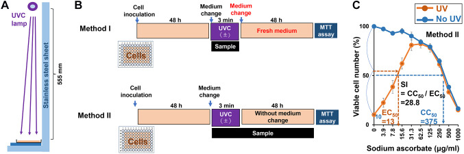
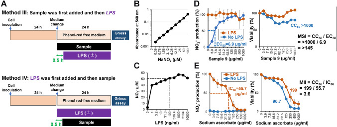
Materials and methods: Anti-UVC activity was determined by the ratio of the 50% cytotoxic concentration against human melanoma cell line COLO679 to the 50% UVC-protective concentration. Extracellularly secreted nitrite (NO2 -) by unstimulated and lipopolysaccharide (LPS)-stimulated mouse macrophage-like cells RAW264.7 was determined by Griess method.
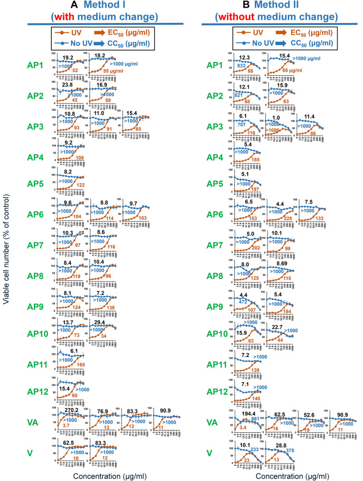
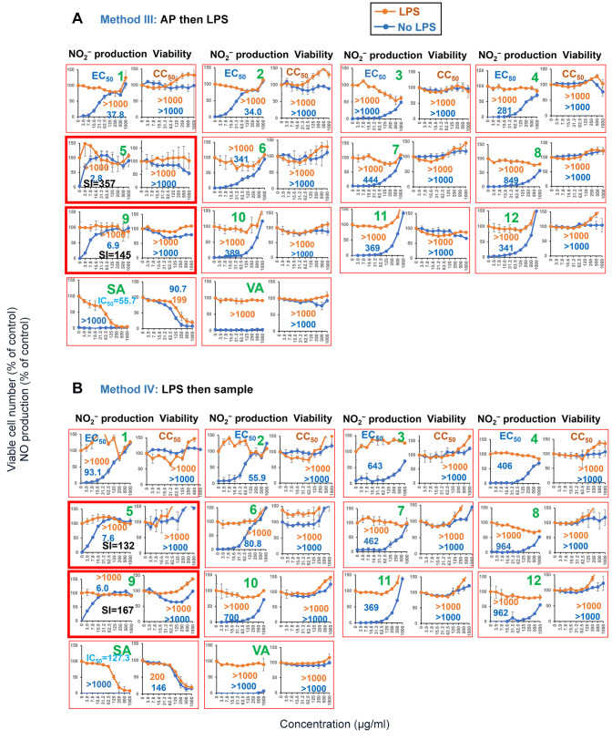
Results: APs showed significantly higher anti-UVC activity than previously reported hot-water extracts of medical herbs. Anti-UVC activity of AP and vanillic acid was maintained for much longer than that of sodium ascorbate and vanillin. APs enhanced the production of NO2 - to the level induced by LPS. Simultaneous addition of AP and LPS did not further increase NO2 - production, suggesting their mechanisms of action overlap.
Conclusion: The present study suggests the possible application of APs as UVC protectors and immunopotentiators via macrophage activation.
Keywords: UVC protection; alkaline extract; macrophage activation; pine seed shell.
Copyright © 2024, International Institute of Anticancer Research (Dr. George J. Delinasios), All rights reserved.
Conflict of interest statement
MY is a representative director of Nippon Sunshow Medicine Manufacture Co. Ltd. and provided all pine seed shells used in this study. SU is a visiting researcher from Nippon Sunshow Medicine Manufacture Co. Ltd. AMA is a visiting researcher from the Autonomous University of the State of Mexico (UAEMex), Toluca, Mexico. However, it was confirmed that such support did not influence the outcome of the experimental study. The Authors wish to confirm that there are no known conflicts of interest associated with this publication and there has been no significant financial support for this work that could have influenced its outcome.
Figures
Similar articles
-
Anti-UV activity of lignin-carbohydrate complex and related compounds.In Vivo. 2013 Jan-Feb;27(1):133-9. In Vivo. 2013. PMID: 23239862
-
Comprehensive Study of Anti-UVC Activity and Cytotoxicity of Hot-water Soluble Herb Extracts.In Vivo. 2023 Jul-Aug;37(4):1540-1551. doi: 10.21873/invivo.13239. In Vivo. 2023. PMID: 37369486 Free PMC article.
-
Cerbera manghas methanol extract exerts anti-inflammatory activity by targeting c-Jun N-terminal kinase in the AP-1 pathway.J Ethnopharmacol. 2016 Dec 4;193:387-396. doi: 10.1016/j.jep.2016.08.033. Epub 2016 Aug 22. J Ethnopharmacol. 2016. PMID: 27562319
-
Anti-inflammatory properties of fermented pine (Pinus morrisonicola Hay.) needle on lipopolysaccharide-induced inflammation in RAW 264.7 macrophage cells.J Food Biochem. 2019 Nov;43(11):e12994. doi: 10.1111/jfbc.12994. Epub 2019 Jul 28. J Food Biochem. 2019. PMID: 31659812
-
Prominent Anti-UVC Activity of Lignin Degradation Products.In Vivo. 2022 Nov-Dec;36(6):2689-2699. doi: 10.21873/invivo.13004. In Vivo. 2022. PMID: 36309360 Free PMC article.
References
-
- Ohtake N, Nakai Y, Yamamoto M, Sakakibara I, Takeda S, Amagaya S, Aburada M. Separation and isolation methods for analysis of the active principles of Sho-saiko-to (SST) oriental medicine. J Chromatogr B Analyt Technol Biomed Life Sci. 2004;812(1-2):135–148. doi: 10.1016/j.jchromb.2004.06.051. - DOI - PMC - PubMed
MeSH terms
Substances
LinkOut - more resources
Full Text Sources
Research Materials
Miscellaneous