. 2024 Sep 19;187(19):5238-5252.e20.
doi: 10.1016/j.cell.2024.07.050. Epub 2024 Aug 28.

Structural insights into the diversity and DNA cleavage mechanism of Fanzor

Affiliations

Structural insights into the diversity and DNA cleavage mechanism of Fanzor

Peiyu Xu et al. Cell. .

Abstract

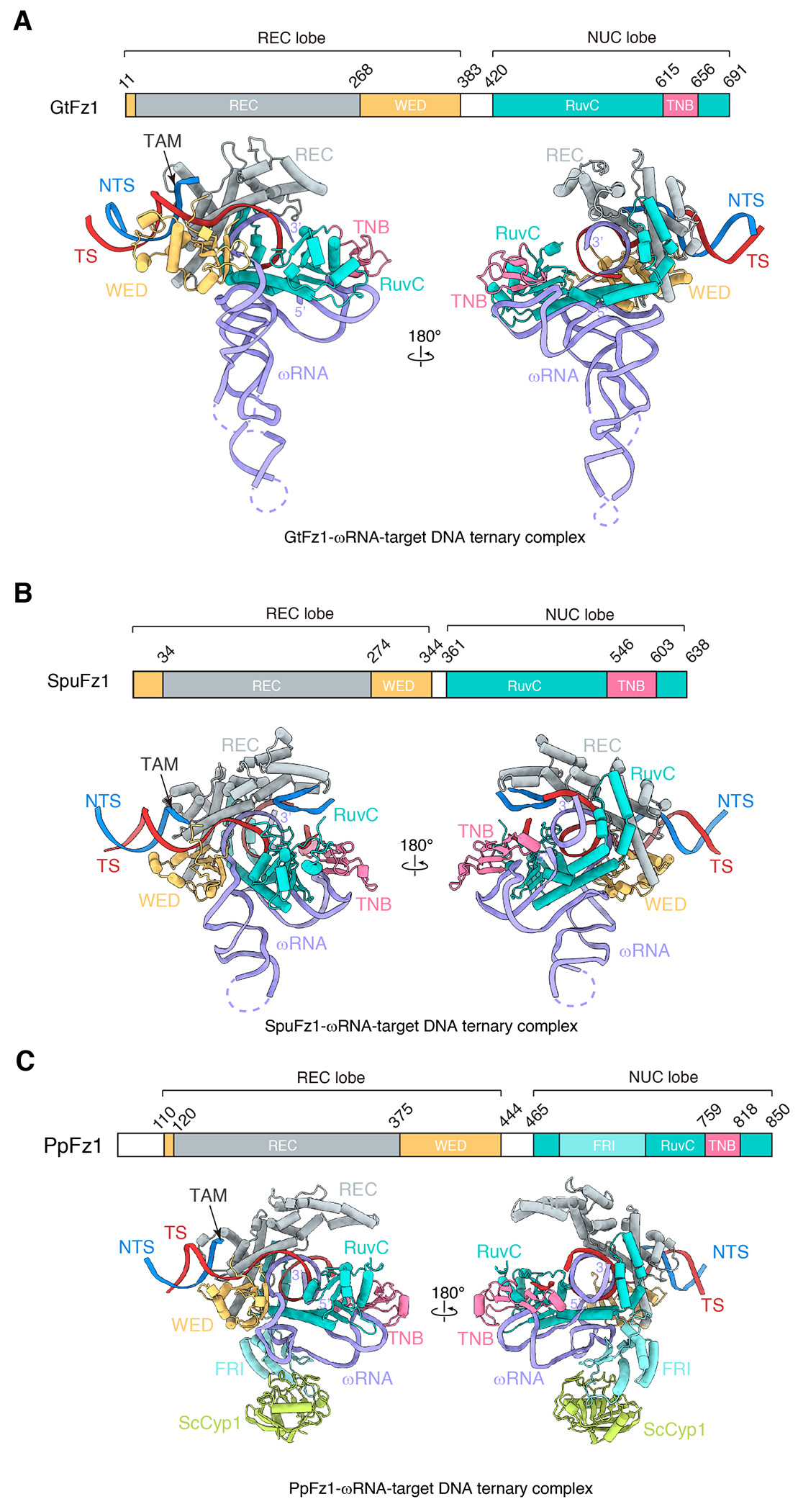
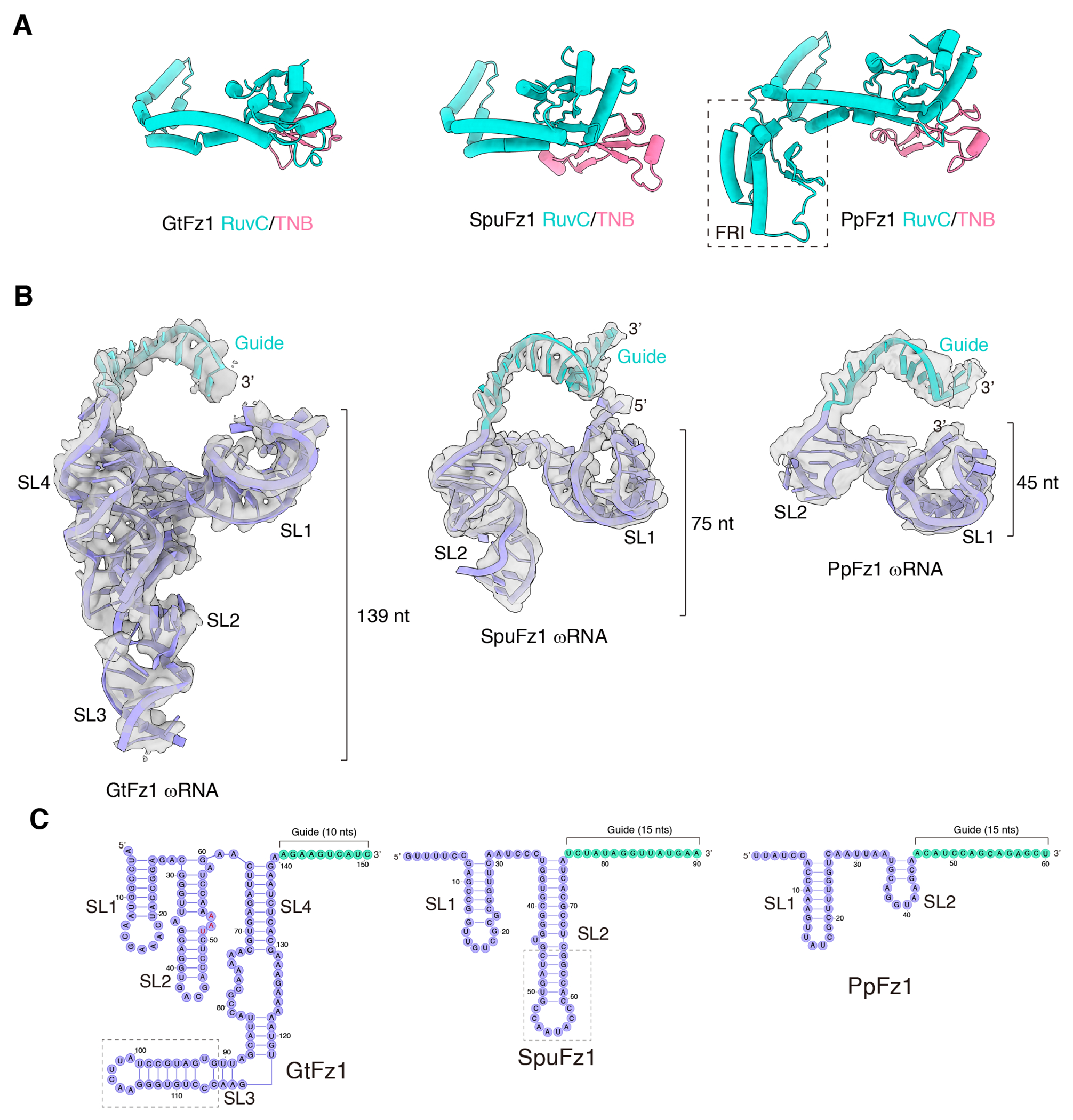
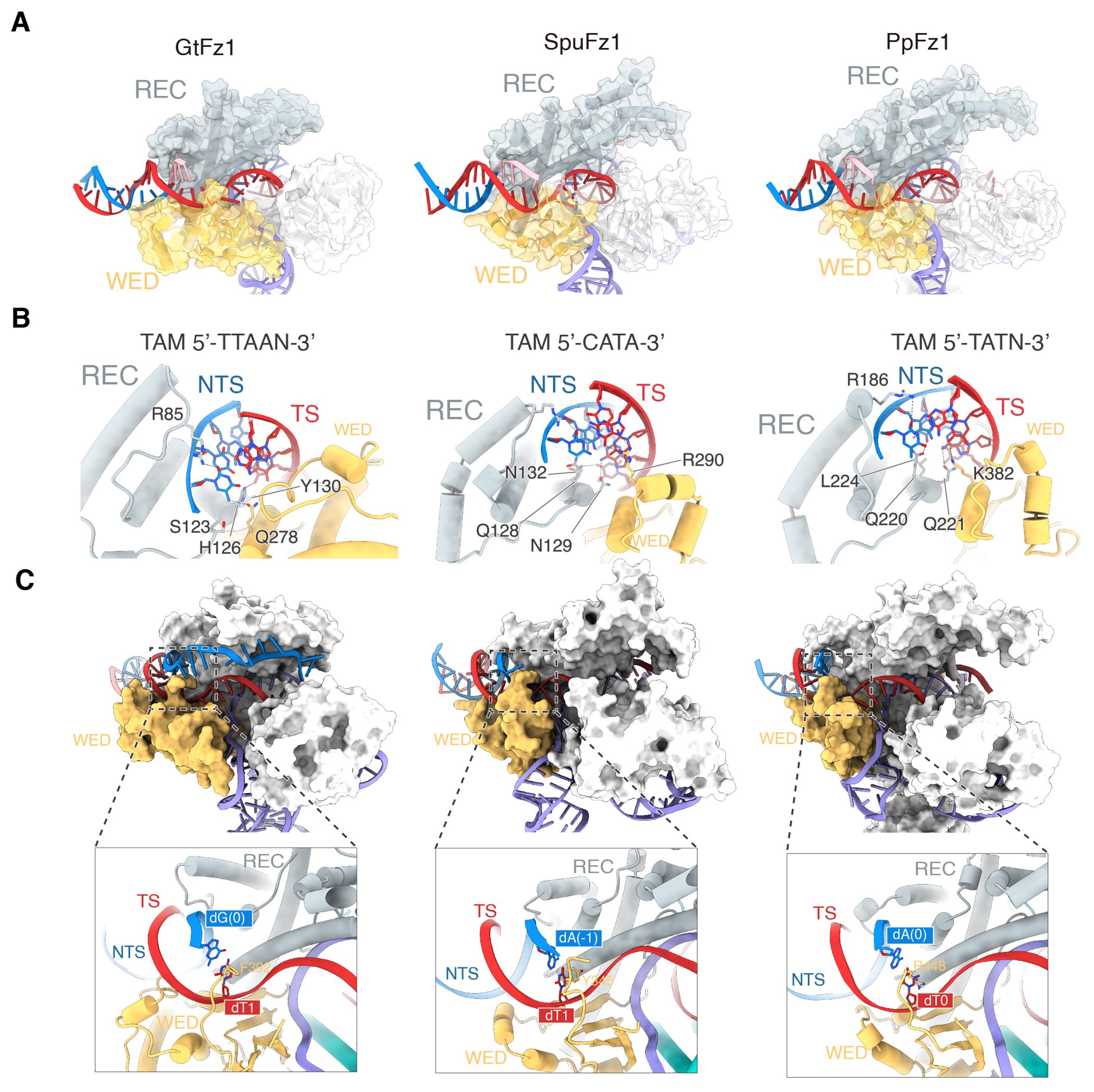
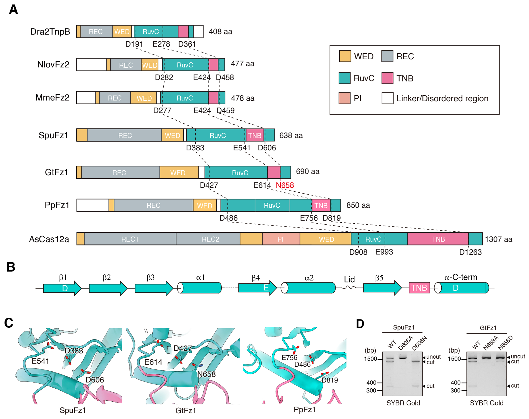
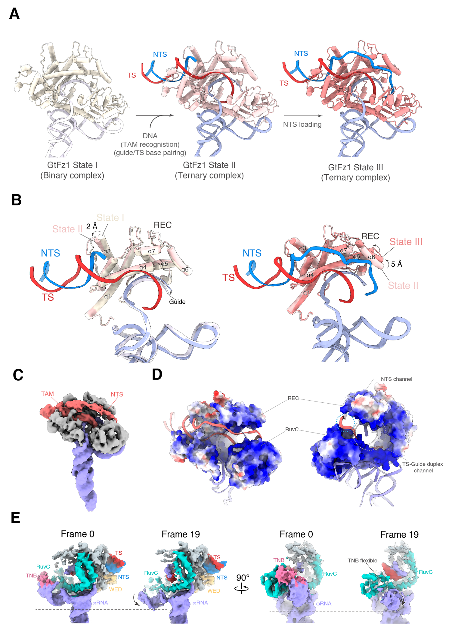
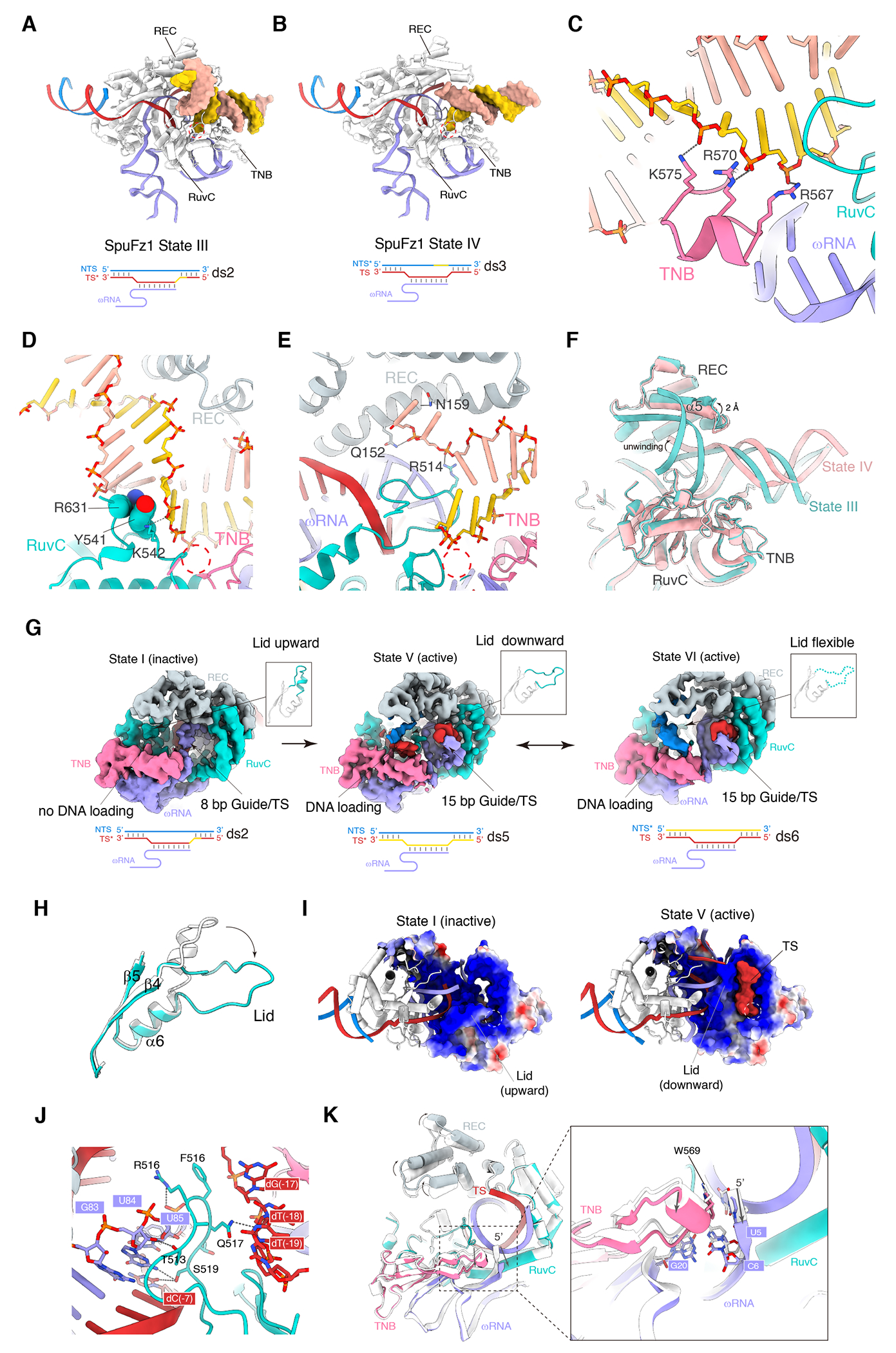
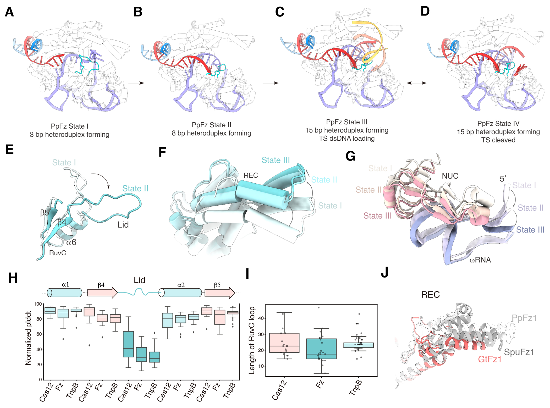
Fanzor (Fz) is an ωRNA-guided endonuclease extensively found throughout the eukaryotic domain with unique gene editing potential. Here, we describe the structures of Fzs from three different organisms. We find that Fzs share a common ωRNA interaction interface, regardless of the length of the ωRNA, which varies considerably across species. The analysis also reveals Fz's mode of DNA recognition and unwinding capabilities as well as the presence of a non-canonical catalytic site. The structures demonstrate how protein conformations of Fz shift to allow the binding of double-stranded DNA to the active site within the R-loop. Mechanistically, examination of structures in different states shows that the conformation of the lid loop on the RuvC domain is controlled by the formation of the guide/DNA heteroduplex, regulating the activation of nuclease and DNA double-stranded displacement at the single cleavage site. Our findings clarify the mechanism of Fz, establishing a foundation for engineering efforts.

Keywords: DNA cleavage mechanisms; Fanzor; R-loop structure; activation mechanisms; catalytic site; eukaryotic RNA-guided DNA endonuclease; gene editing; structural diversity.


Conflict of interest statement

Declaration of interests P.X., M.S., G.F., and F.Z. are coinventors on a patent application (PCT/US2022/081593) related to this work filed by the Broad Institute and MIT. F.Z. is a scientific advisor and cofounder of Editas Medicine, Beam Therapeutics, Pairwise Plants, Arbor Biotechnologies, Aera Therapeutics, and Moonwalk Biosciences. F.Z. is a scientific advisor for Octant.

Figures

Figure 1.
Figure 1.. Structural overview of Fanzor1 complexes
(A-C) Schematic locus and cryo-EM structure of the Fz1-ωRNA-target DNA complex from Spizellomyces punctatus (SpuFz1) (A), Guillardia theta (GtFz1) (B), and Parasitella parasitica (PpFz1) (C). REC domain is colored gray, WED domain is colored yellow, RuvC domain is colored cyan, TNB domain is colored pink, Fanzor RuvC Insertion (FRI) domain is colored light cyan; Saccharomyces cerevisiae Cyclophilin1 (ScCyp1) is colored green, ωRNA is colored purple, DNA target strand (TS) is colored red, and DNA non-target strand (NTS) is colored blue.
Figure 2.
Figure 2.. Structural diversity of Fanzor1
(A) Structural comparison of the RuvC and TNB domains across GtFz1 (left), SpuFz1 (middle), and PpFz1 (right). The dashed box indicates the FRI domain of PpFz1. (B) Comparative analysis of ωRNA structures between GtFz1, SpuFz1, and PpFz1. EM density is shown transparently. The ωRNA scaffold is colored purple, and the guide is green. (C) Schematic of the ωRNA in GtFz1, SpuFz1, and PpFz1.
Figure 3.
Figure 3.. Comparative analysis of DNA recognition by Fanzor1
(A) Recognition of the TAM duplex by GtFz1 (left), SpuFz1 (middle), and PpFz1 (right). The TAM sequence is highlighted in pink. (B) Interactions involved in TAM recognition, showing a conserved structural feature among GtFz1, SpuFz1, and PpFz1. Specifically, an arginine (R) residue from a loop in the REC domain inserts into the groove of the TAM duplex. The N-terminal end of the alpha-4 helix from the REC domain, along with a short helix from the WED domain, recognizes the TAM base groups in a similar orientation. Different Fz1 proteins utilize non-conserved residues to recognize different TAM sequences. (C) Initiation of the R-loop by the loop from the WED domain. Protein domains and ωRNA are colored as in Figures 1 and 2, the DNA target strand (TS) is colored red, and the DNA non-target strand (NTS) is colored blue.
Figure 4.
Figure 4.. Catalytic triad of Fanzor1 and other RuvC nucleases.
(A) Conserved catalytic motifs in the RuvC domain are shared among TnpB, Fz2, Fz1, and Cas12a. (B) Secondary structure of a canonical RuvC nuclease. (C) Structural details of the catalytic sites in SpuFz1, GtFz1 (which contains a non-canonical N in place of the canonical D in the third position), and PpFz1. (D) In vitro cleavage activity of SpuFz1 wild-type, D606A, D606N (left) and GtFz1 wild-type, N658A, and N658D (right).
Figure 5.
Figure 5.. Structural features of GtFz1.
(A) Structures of the GtFz1 State I (binary complex), State II (ternary complex), and State III (ternary complex). The target DNA of State II contains the TAM duplex and a single-stranded TS. The target DNA of State III is fully double-stranded. GtFz1 protein is colored tan in State I, pink in State II, and red in State III. (B) Comparison of the REC domain in GtFz1 State I with State II (top), and of State II with State III (bottom). (C) The EM map of the GtFz1 ternary complex displays the NTS loading into the RuvC domain. GtFz1 is colored gray, ωRNA is colored purple, and DNA is colored red. (D) Electrostatic potential mapping in GtFz1 illustrates the DNA binding channel. The green dashed circle indicates the channel for the DNA NTS binding. The white dashed circle indicates the channel for guide/TS heteroduplex binding. (E) 3D Variability Analysis (3DVA) reveals the conformational dynamics of SL1 and the RuvC/TNB domains. EM maps of the first frame and the last frame are shown from the same viewpoint to highlight the conformational change.
Figure 6.
Figure 6.. RuvC dsDNA loading and the lid regulation in SpuFz1
(A) Structure of SpuFz1 State III. dsDNA is bound to the large cleft formed by REC/RuvC/TNB domains. The dsDNA is bent by about 137° and the tip contacts the catalytic site of RuvC. SpuFz1 is colored white, ωRNA is colored purple, TS is colored red, NTS is colored blue. The dsDNA bound to RuvC is shown in surface and colored in gold and tan for each strand. The catalytic site in the RuvC domain is circled with a dashed line. A schematic diagram of the ternary complex formation is shown below. Substrate ds2, in which the TS is partially modified and NTS is unmodified, was used. (B) Structure of SpuFz1 State IV. dsDNA is bound to the large cleft formed by REC/RuvC/TNB domains in a distinct conformation from State III. SpuFz1 is colored white, ωRNA is colored purple, TS is colored red, NTS is colored blue. The dsDNA bound to RuvC is shown in surface and colored in gold and tan for each strand. The catalytic site in the RuvC domain is circled with a dashed red line. A schematic diagram of the ternary complex formation is shown below. Substrate ds3, where TS is unmodified and NTS is partially modified. (C) Close-up of charge interactions in the structure of SpuFz1 State III between residues of the TNB domain and dsDNA loaded onto the RuvC domain. (D) In the structure of SpuFz1 State III, residue R631 in the C-terminus loop of the RuvC domain, together with Y541, occupy the groove in the dsDNA that is bound to the RuvC domain. The catalytic site in the RuvC domain is circled with a dashed red line. (E) In the structure of SpuFz1 State IV, interactions formed by Q152, N159, and R514 stabilize the unwound DNA strand, which was not observed in State III. The catalytic site in the RuvC domain is circled with a dashed red line. (F) Structural alignment of SpuFz1 State III (green) and State IV (pink) showing that the dsDNA bound to the RuvC domain of these two states displays distinct conformations. The α5 of the REC domain is shifted by 2 Å. (G) The EM maps of SpuFz1 State I, State V, and State VI illustrate the conformational changes of the lid. In State I, an 8-bp guide/DNA duplex is formed. The lid is in an upward orientation, and no DNA is loaded onto the RuvC domain, representing an inactive state. In State V, a 15-bp guide/DNA duplex is formed. The lid is in a downward orientation, and the DNA TS is loaded onto the RuvC domain, representing an active state. In State VI, a 15-bp guide/DNA duplex is formed. The lid density is weak due to its structural flexibility. The DNA density is observed around the RuvC domain. Schematic diagrams of ternary complex formation are shown below (substrate ds2, TS is partially modified and NTS is unmodified; substrate ds5, TS is fully modified and NTS is unmodified; substrate ds6, TS is unmodified and NTS is fully modified). (H) Structural alignment of the lid of SpuFz1 in the inactive state (I) (white) with the active state (V) (cyan). The short helix of the lid in the active state is released. (I) Electrostatic potential mapping in SpuFz1 illustrates the structural changes from the inactive state (I) to the active state (V). The catalytic site is circled with a dashed line. The downward conformation of the lid forms a small cleft on the RuvC and TNB domain, allowing the DNA substrate to load onto the RuvC domain and approach the catalytic site. (J) Interactions of the lid in SpuFz1 State V. The lid is sandwiched by the guide/TS heteroduplex and the DNA segment loaded onto the RuvC domain. Hydrogen bonds are shown with dashed lines. (K) Structural alignment of SpuFz1 in the inactive state (I) with the active state (V). The entire complex in the inactive state is colored white. For the active state, the RuvC domain is colored cyan, ωRNA is colored purple, and the DNA TS is colored red. The inset shows the detailed conformational change at the 5’ end of the ωRNA, along with the TNB domain. This change is driven by the formation of the 15-bp guide/DNA heteroduplex.
Figure 7.
Figure 7.. PpFz1 DNA loading and cleavage mechanisms
(A-D) Structures illustrating the activation stages of PpFz1, detailing the conformational changes, DNA loading, and cleavage processes. Protein domains, ωRNA, and target DNA are colored as in Figures 6. (E) Structural alignment of the lid of PpFz1 comparing the inactive state (I) (white) with the intermediate state (II) (cyan). (F) Structural alignment of the REC domain comparing the inactive state (I), intermediate state (II), and active state (III). (G) Structural alignment at the 5’ end of the ωRNA and the TNB domain comparing the inactive state (I), intermediate state (II), and active state (III). (H) Comparison of the Predicted Local Distance Difference Test (pLDDT) for the lid region and its surrounding secondary structures (upstream: α1, β2; downstream: α2, β5) across 59 representatives of Cas12, Fzs, and TnpB. pLDDT scores were normalized as described in Methods, and the scores for each region of each protein are provided and detailed in Data S1. (I) Comparison of the length distribution (in residues) of the lid regions across 59 representatives of Cas12s, Fzs, and TnpBs. The lengths for each protein are provided in Data S1. (J) Structural alignment of the REC domain comparing the active states of GtFz1 (red), SpuFz1 (gray), and PpFz1 (white).

Similar articles

References

    1. Jiang K, Lim J, Sgrizzi S, Trinh M, Kayabolen A, Yutin N, Bao W, Kato K, Koonin EV, Gootenberg JS, et al. (2023). Programmable RNA-guided DNA endonucleases are widespread in eukaryotes and their viruses. Sci Adv 9, eadk0171. - PMC - PubMed
    1. Saito M, Xu P, Faure G, Maguire S, Kannan S, Altae-Tran H, Vo S, Desimone A, Macrae RK, and Zhang F (2023). Fanzor is a eukaryotic programmable RNA-guided endonuclease. Nature 620, 660–668. - PMC - PubMed
    1. Bao W, and Jurka J (2013). Homologues of bacterial TnpB_IS605 are widespread in diverse eukaryotic transposable elements. Mob. DNA 4, 12. - PMC - PubMed
    1. Altae-Tran H, Kannan S, Demircioglu FE, Oshiro R, Nety SP, McKay LJ, Dlakić M, Inskeep WP, Makarova KS, Macrae RK, et al. (October/2021). The widespread IS200/IS605 transposon family encodes diverse programmable RNA-guided endonucleases. Science 374, 57–65. - PMC - PubMed
    1. Yoon PH, Skopintsev P, Shi H, Chen L, Adler BA, Al-Shimary M, Craig RJ, Loi KJ, DeTurk EC, Li Z, et al. (2023). Eukaryotic RNA-guided endonucleases evolved from a unique clade of bacterial enzymes. Nucleic Acids Res. 51, 12414–12427. - PMC - PubMed

LinkOut - more resources