. 2024 Jul 2;12(7):e0045024.
doi: 10.1128/spectrum.00450-24. Epub 2024 May 31.

Characterization of the dual regulation by a c-di-GMP riboswitch Bc1 with a long expression platform from Bacillus thuringiensis

Affiliations

Characterization of the dual regulation by a c-di-GMP riboswitch Bc1 with a long expression platform from Bacillus thuringiensis

Lu Liu et al. Microbiol Spectr. .

Abstract

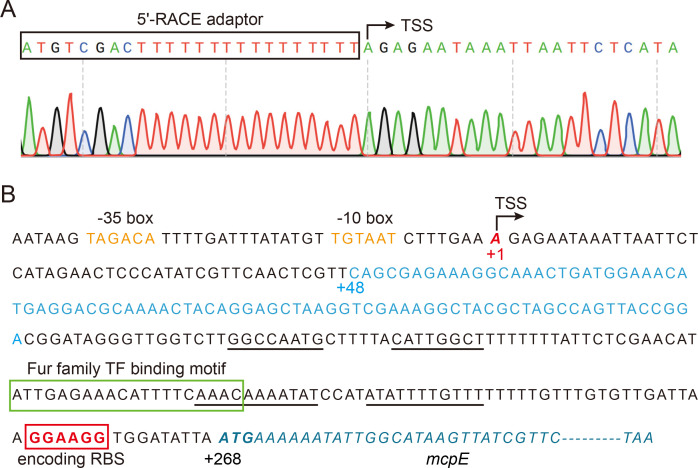
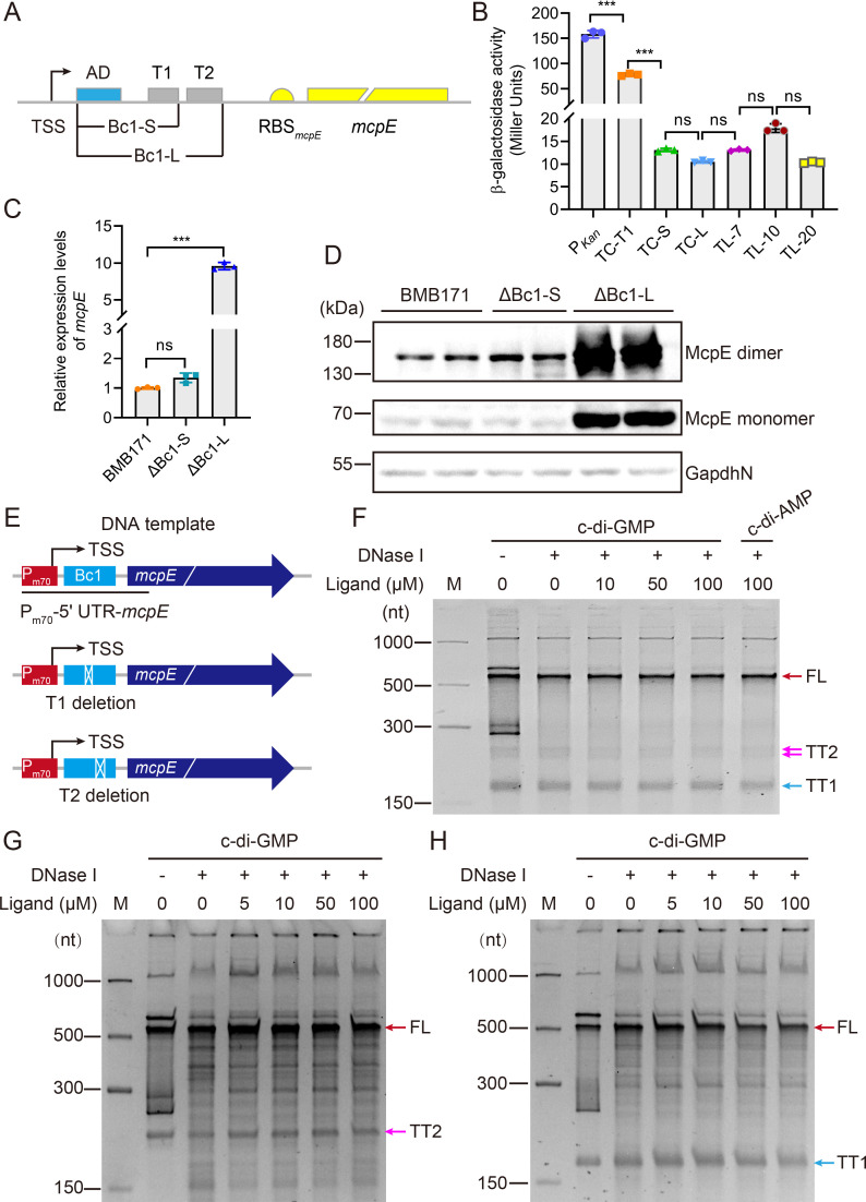
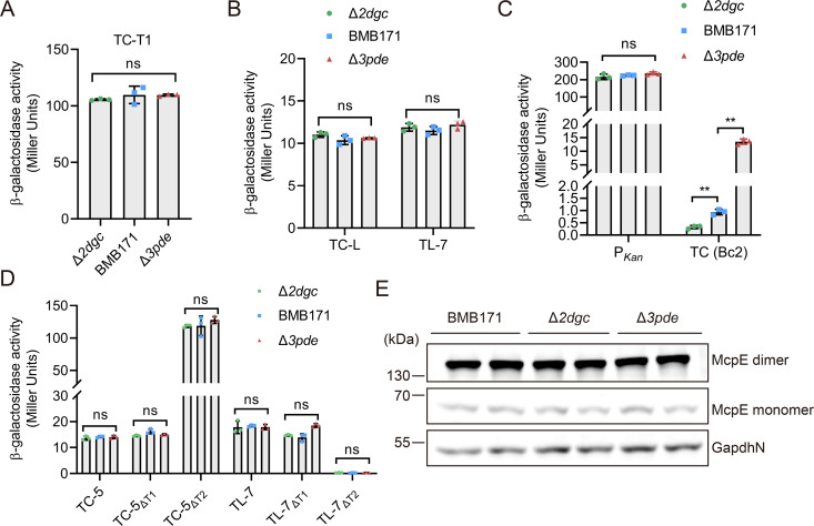
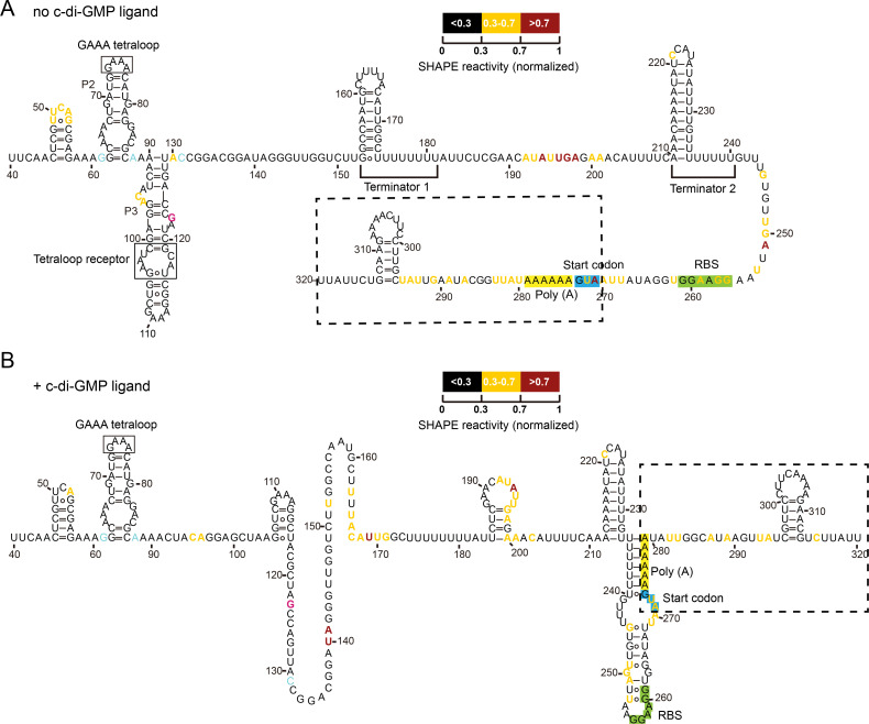
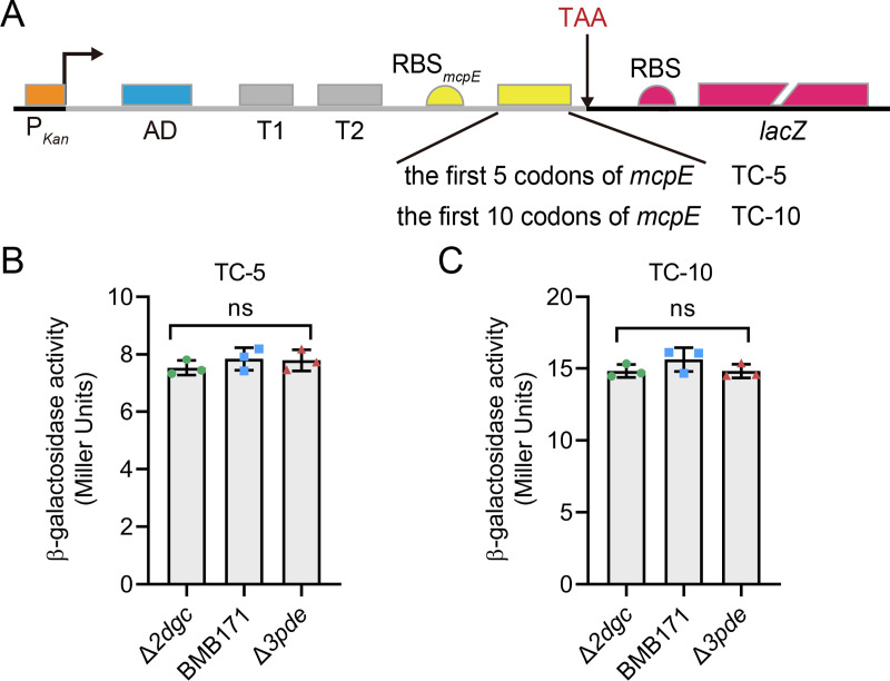
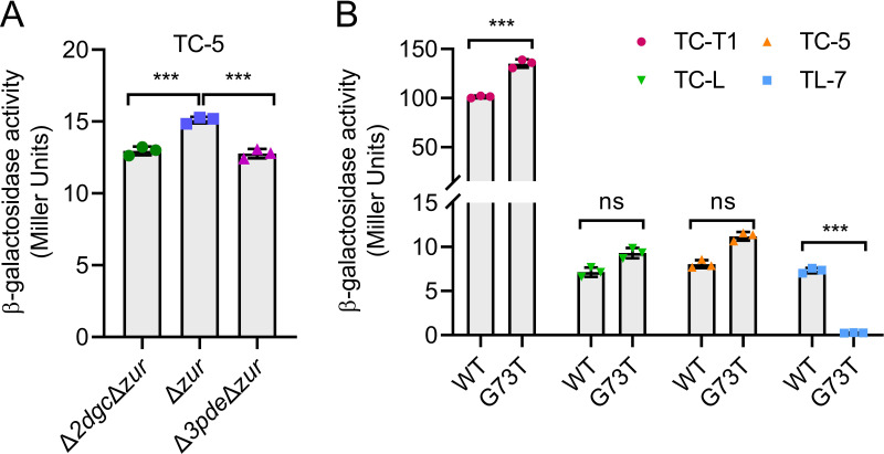
A riboswitch generally regulates the expression of its downstream genes through conformational change in its expression platform (EP) upon ligand binding. The cyclic diguanosine monophosphate (c-di-GMP) class I riboswitch Bc1 is widespread and conserved among Bacillus cereus group species. In this study, we revealed that Bc1 has a long EP with two typical ρ-independent terminator sequences 28 bp apart. The upstream terminator T1 is dominant in vitro, while downstream terminator T2 is more efficient in vivo. Through mutation analysis, we elucidated that Bc1 exerts a rare and incoherent "transcription-translation" dual regulation with T2 playing a crucial role. However, we found that Bc1 did not respond to c-di-GMP under in vitro transcription conditions, and the expressions of downstream genes did not change with fluctuation in intracellular c-di-GMP concentration. To explore this puzzle, we conducted SHAPE-MaP and confirmed the interaction of Bc1 with c-di-GMP. This shows that as c-di-GMP concentration increases, T1 unfolds but T2 remains almost intact and functional. The presence of T2 masks the effect of T1 unwinding, resulting in no response of Bc1 to c-di-GMP. The high Shannon entropy values of EP region imply the potential alternative structures of Bc1. We also found that zinc uptake regulator can specifically bind to the dual terminator coding sequence and slightly trigger the response of Bc1 to c-di-GMP. This work will shed light on the dual-regulation riboswitch and enrich our understanding of the RNA world.IMPORTANCEIn nature, riboswitches are involved in a variety of metabolic regulation, most of which preferentially regulate transcription termination or translation initiation of downstream genes in specific ways. Alternatively, the same or different riboswitches can exist in tandem to enhance regulatory effects or respond to multiple ligands. However, many putative conserved riboswitches have not yet been experimentally validated. Here, we found that the c-di-GMP riboswitch Bc1 with a long EP could form a dual terminator and exhibit non-canonical and incoherent "transcription-translation" dual regulation. Besides, zinc uptake regulator specifically bound to the coding sequence of the Bc1 EP and slightly mediated the action of Bc1. The application of SHAPE-MaP to the dual regulation mechanism of Bc1 may establish the foundation for future studies of such complex untranslated regions in other bacterial genomes.

Keywords: Bacillus cereus group; SHAPE-MaP; Zur (zinc uptake regulator); cyclic di-GMP riboswitch; dual regulation; dual terminator.


Conflict of interest statement

The authors declare no conflict of interest.

Figures

FIG 1
FIG 1
Analysis of the 5' UTR coding sequence of mcpE. (A) TSS mapping of mcpE through 5′-RACE assay. Black box indicates the 5′-RACE linker sequence, which contains poly(T)16, and black arrow refers to the +1 base A of the TSS. (B) Coding sequence of the 5′ UTR of the mcpE transcript. Red bold italic letter A indicates TSS, also highlighted with +1, TSS and black arrow; Orange indicates the −10 box and −35 box of the mcpE promoter; Light blue indicates the Bc1 AD coding sequence; Black underline represents the predicted hairpin sequence of both terminators; Green box represents the predicted Ferric uptake regulator (Fur) family TFs binding motif; Red box and red bold fonts emphasize the coding sequence of the RBS; and blue italics represent the mcpE gene sequence. See also Fig. S1 and S2.
FIG 2
FIG 2
Bc1 forms an intrinsic dual terminator and inhibits downstream gene expressions. (A) Schematic diagram of the Bc1-mcpE gene structure. The coding sequence of Bc1 AD is shown in blue, and its dual terminator is shown in grey, with AD, T1 and T2 labeled next to it. The short Bc1-S (containing the coding sequence from Bc1 AD to T1) and long Bc1-L (containing the coding sequence from Bc1 AD to T2) regions are annotated separately. Sequences encoding the RBS and mcpE gene are shown in yellow. (B) β-Galactosidase activity assays of BMB171 strain containing different transcriptional and translational fusion reporter constructs. To assist the reader, we abbreviate transcriptional fusion as TC and translational fusion as TL. PKan indicates a reporter construct, in which lacZ is transcriptionally fused to the promoter PKan. TC-T1, TC-S, TC-L denote constructs, in which lacZ is transcriptionally fused to PKan and varying lengths of 5′ UTR coding sequences up to terminator T1, two terminators, or the mcpE RBS, respectively. TL-7, TL-10 and TL-20 denote constructs in which lacZ is translationally fused to PKan, the entire 5′ UTR coding sequence, and the first 7, 10 or 20 codons of the mcpE gene, respectively. (C) RT-qPCR assay of mcpE transcript levels after deleting the single terminator T1 or the Bc1 dual terminator. Values in B and C are mean ± standard deviation of three biological replicates, and the significance of the difference was analyzed by one-way ANOVA in GraphPad Prism 8.0.6 with Bonferroni correction, ns means no significance, P > 0.05; *P < 0.05; **P < 0.01; ***P < 0.001. (D) Western blot assay of McpE protein levels after deleting the single terminator T1 or the Bc1 dual terminator. The target 6× His-McpE-6× His monomer is approximately 68.6 kDa, and its dimer is approximately 137.2 kDa. GapdhN (BMB171_RS04385, NADP-dependent glyceraldehyde-3-phosphate dehydrogenase) with a C terminal 6× His tag was used as a control loading protein with a total size of nearly 53.1 kDa. (E) Schematic diagram of the DNA template used in in vitro transcription termination assays in (F–H). The wild-type template is shown at the top. The native promoter Pm70 of mcpE is shown in red, the riboswitch Bc1 in light blue, and the mcpE gene in dark blue and italic. Deletion of terminator T1 (medium) or T2 (bottom) is indicated by white cross boxes. (F), (G) and (H) In vitro transcription termination assays using wild-type template, or mutant template with T1 or T2 deletion, respectively. M denotes the ssRNA marker, whose bands are indicated on the left as 1000, 500, 300, and 150 nt. FL transcripts are indicated by red arrows, and truncated transcripts terminated by T1 (TT1) or T2 (TT2) are indicated in blue or carmine arrows, respectively. Working concentrations of c-di-GMP are 0, 5, 10, 50 and 100 µM. 100 µM c-di-AMP represents the control group. See also Fig. S3.
FIG 3
FIG 3
Bc1 has little effect on the expressions of downstream genes under different intracellular c-di-GMP concentrations. (A) β-Galactosidase activity assay of Δ2dgc, BMB171, and Δ3pde strains containing the transcriptional fusion construct TC-T1. (B) β-Galactosidase activity assay of Δ2dgc, BMB171, and Δ3pde strains containing transcriptional fusion TC-L and translational fusion TL-7 constructs. (C) β-Galactosidase activity assay of Δ2dgc, BMB171, and Δ3pde strains harboring transcriptional fusion constructs PKan or TC (Bc2) (the coding sequence of 5′ UTR of the gene cap downstream of Bc2 is transcriptionally fused to PKan and lacZ). (D) β-Galactosidase activity assays of Δ2dgc, BMB171, and Δ3pde strains harboring transcriptional fusion constructs TC-5, TC-5ΔT1, TC-5ΔT2, and the translational fusion constructs TL-7, TL-7ΔT1, TL-7ΔT2. ΔT1, ΔT2 indicate that the test sequence is fused with T1 or T2 deletion alone. (E) Western blot assay of McpE protein levels in Δ2dgc, BMB171, and Δ3pde strains. GapdhN was served as a control loading protein. Values in A, B, C and D are mean ± standard deviation of three biological replicates, and the significance of the difference was analyzed by two-way ANOVA in GraphPad Prism 8.0.6 with Bonferroni correction. ns means no significance, P > 0.05; *P < 0.05; **P < 0.01; ***P < 0.001.
FIG 4
FIG 4
Bc1-mcpE transcript undergoes significant conformational changes in response to c-di-GMP identified by SHAPE-MaP. (A) In the absence of c-di-GMP, Bc1 forms a dual terminator structure, and the RBS and start codon AUG of the mcpE transcript are fully exposed. (B) In the presence of c-di-GMP, the dual terminator structure of Bc1 unfolds, and the mcpE RBS and start codon AUG are now trapped in the newly formed structure of Bc1. According to the normalized SHAPE reactivity value, each nucleotide in transcript region 40–320 is shown in black (<0.3), rich yellow (0.3–0.7), and rich red (>0.7), respectively. Both the Bc1 tetraloop and tetraloop receptor are indicated by solid black boxes. The P2, P3 stems of Bc1 AD, and terminators T1, T2 are also labeled. The mcpE transcript is indicated by a dashed box, with its RBS, start codon AUG, and preceding poly(A) sequence highlighted in green, blue, and yellow, respectively. See also Fig. S4–S6.
FIG 5
FIG 5
The poly(A) string within preceding mcpE coding sequence does not relieve the transcriptional inhibition of Bc1. (A) Schematic diagram of transcriptional fusions containing the 5′ UTR coding sequence up to the first 5 or 10 codons of the mcpE gene, denoted TC-5 and TC-10, respectively. It is worth noting that a “TAA” (shown in red) is inserted between the coding sequence of the mcpE gene and the RBS of the lacZ gene in the reporter plasmid. The respective elements are shown as Fig. S3A. (B) β-Galactosidase activity assay of Δ2dgc, BMB171 and Δ3pde strains containing TC-5 transcriptional fusion construct. (C) β-Galactosidase activity assay of Δ2dgc, BMB171 and Δ3pde strains containing TC-10 transcriptional fusion construct. Values in panels B and C are mean ± standard deviation of three biological replicates, and the significance of the difference was analyzed by one-way ANOVA in GraphPad Prism 8.0.6 with Bonferroni correction. ns means no significance, P > 0.05; *P < 0.05; **P < 0.01; ***P < 0.001.
FIG 6
FIG 6
Bc1 has a “transcription-translation” dual regulation mechanism. (A) β-Galactosidase activity assay of Δ2dgcΔzur, Δzur and Δ3pdeΔzur strains containing the TC-5 transcriptional fusion construct. (B) β-Galactosidase activity assay of BMB171 containing various reporter constructs TC-T1, TC-L, TC-5 and TL-7 upon G73T mutation of Bc1 AD. Values in A and B are mean ± standard deviation of three biological replicates, and the significance of the difference was analyzed by one-way ANOVA in GraphPad Prism 8.0.6 with Bonferroni correction, ns means no significance, P > 0.05; *P < 0.05; **P < 0.01; ***P < 0.001. See also Fig. S7.

Similar articles

References

    1. Ross P, Weinhouse H, Aloni Y, Michaeli D, Weinberger-Ohana P, Mayer R, Braun S, de Vroom E, van der Marel GA, van Boom JH, Benziman M. 1987. Regulation of cellulose synthesis in Acetobacter xylinum by cyclic diguanylic acid. Nature 325:279–281. doi:10.1038/325279a0 - DOI - PubMed
    1. Yu Z, Zhang W, Yang H, Chou SH, Galperin MY, He J. 2023. Gas and light: triggers of c-di-GMP-mediated regulation. FEMS Microbiol Rev 47:fuad034. doi:10.1093/femsre/fuad034 - DOI - PMC - PubMed
    1. Krasteva PV, Sondermann H. 2017. Versatile modes of cellular regulation via cyclic dinucleotides. Nat Chem Biol 13:350–359. doi:10.1038/nchembio.2337 - DOI - PMC - PubMed
    1. Kumar B, Sorensen JL, Cardona ST. 2018. A c-di-GMP-modulating protein regulates swimming motility of Burkholderia cenocepacia in response to arginine and glutamate. Front Cell Infect Microbiol 8:56. doi:10.3389/fcimb.2018.00056 - DOI - PMC - PubMed
    1. Liu X, Zhang C, Zhang R, Yuan J. 2022. The effect of the second messenger c-di-GMP on bacterial chemotaxis in Escherichia coli. Appl Environ Microbiol 88:e0037322. doi:10.1128/aem.00373-22 - DOI - PMC - PubMed

MeSH terms

LinkOut - more resources