. 2023 Nov 29:14:1237966.
doi: 10.3389/fpls.2023.1237966. eCollection 2023.

Microbiome and plant cell transformation trigger insect gall induction in cassava

Affiliations

Microbiome and plant cell transformation trigger insect gall induction in cassava

Omar Gätjens-Boniche et al. Front Plant Sci. .

Abstract

Several specialised insects can manipulate normal plant development to induce a highly organised structure known as a gall, which represents one of the most complex interactions between insects and plants. Thus far, the mechanism for insect-induced plant galls has remained elusive. To study the induction mechanism of insect galls, we selected the gall induced by Iatrophobia brasiliensis (Diptera: Cecidomyiidae) in cassava (Euphorbiaceae: Manihot esculenta Crantz) as our model. PCR-based molecular markers and deep metagenomic sequencing data were employed to analyse the gall microbiome and to test the hypothesis that gall cells are genetically transformed by insect vectored bacteria. A shotgun sequencing discrimination approach was implemented to selectively discriminate between foreign DNA and the reference host plant genome. Several known candidate insertion sequences were identified, the most significant being DNA sequences found in bacterial genes related to the transcription regulatory factor CadR, cadmium-transporting ATPase encoded by the cadA gene, nitrate transport permease protein (nrtB gene), and arsenical pump ATPase (arsA gene). In addition, a DNA fragment associated with ubiquitin-like gene E2 was identified as a potential accessory genetic element involved in gall induction mechanism. Furthermore, our results suggest that the increased quality and rapid development of gall tissue are mostly driven by microbiome enrichment and the acquisition of critical endophytes. An initial gall-like structure was experimentally obtained in M. esculenta cultured tissues through inoculation assays using a Rhodococcus bacterial strain that originated from the inducing insect, which we related to the gall induction process. We provide evidence that the modification of the endophytic microbiome and the genetic transformation of plant cells in M. esculenta are two essential requirements for insect-induced gall formation. Based on these findings and having observed the same potential DNA marker in galls from other plant species (ubiquitin-like gene E2), we speculate that bacterially mediated genetic transformation of plant cells may represent a more widespread gall induction mechanism found in nature.

Keywords: Iatrophobia brasiliensis; Manihot esculenta; endophytes; genetic transformation; induction mechanism; metagenomics; plant galls.


Conflict of interest statement

The authors declare that the research was conducted in the absence of any commercial or financial relationships that could be construed as a potential conflict of interest.

Figures
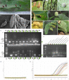
Figure 1
Figure 1
Identification of a potential universal marker in different gall systems. (A) Gall induced by the Cecidomyiidae Iatrophobia brasiliensis in Manihot esculenta (Montaldo, 1977). (B) Scanning electron micrograph of I. brasiliensis larva. (C) Scanning electron micrograph of the ventral area near the head of I. brasiliensis larva. (D1) Gall induced in Hirtella racemosa Lam. (Chrysobalanaceae), morphotype Hi_ra_1, by an unidentified Cecidomyiidae. (D2) Gall induced in Cydista diversifolia (Kunth) Miers (Bignoniaceae), morphotype Cy_di_1 by an unidentified Cecidomyiidae. (D3) Gall induced in Malvaviscus arboreus Dill. ex Cav. (Malvaceae), morphotype Ma_ar_1, by an unidentified Cecidomyiidae. (D4) Gall induced in Miconia oerstediana (Melastomataceae) by an unidentified Cecidomyiidae. (D5) Gall in Pisonia macranthocarpa (Donn. Sm.) Donn. Sm. (Nyctaginaceae), morphotype Pi_ma_4, induced by an unknown insect species. (D6) Gall in Coussarea hondensis (Standl.) C.M. Taylor & W.C. Burger (Rubiaceae) induced by an unknown insect. (D7) Gall induced in Lonchocarpus phlebophyllus Standl & Steyerm. (Fabaceae), morphotype Lo_phl_1, induced by a Psyllidae. (D8) Gall induced in Randia monantha Benth. (Rubiaceae), morphotype Ra_mo_1, induced by an unknown insect species. (D2–D8) Gätjens-Boniche et al. (2021). (E) Agarose gel electrophoresis of DNA fragments (specific gall fragment marker, SGF) amplified by PCR, comparing healthy leaf tissue DNA samples (H) and gall tissue DNA samples (G). Lane M, molecular weight marker (1 kb ladder); line NC, negative control (reagents only); lines S1–S16, samples of healthy leaf and gall tissues growing in the same plant organ (pair-compared). (F) Gel electrophoresis of PCR products using primers for the specific gall fragment marker (SGF) in gall morphotypes of different host plant species. Lane M, molecular weight marker (Gene Ruler 1 KB Plus); line NC, negative control (reagents only); lines 3–20, samples of healthy leaf and gall tissues of different plants growing in the same plant organ (pair-compared). The green circle indicates a positive sample for the specific gall fragment amplification (E, F). (G, H) Real-time PCR by Taq Man Probe showing the detection of the specific gall fragment from gall DNA samples of Manihot esculenta (amplification plot H) and from healthy leaf samples (amplification plot G). Each trace shows the ΔRn (normalised net fluorescence signal of the PCR product) plotted against the number of PCR cycles.
Figure 2
Figure 2
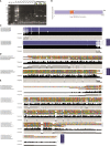
Genetic characterisation of the specific gall fragment marker. (A) Gel electrophoresis of PCR products using primers for the specific gall fragment marker (SGF) from purified wild-type plasmids of two putative endosymbiotic bacteria, Pseudomonas and Rhodococcus, isolated from the larval head of the inducing insect Iatrophobia brasiliensis (isolates ISB 1 and ISB 2), as well as from purified wild-type plasmids of seven possible endophytic bacteria isolates selected from the cassava plant gall tissue (IEB). PCR amplicons are also shown for the inducing insect salivary gland sample (SG). Samples of DNA purified from healthy leaf and gall cassava tissues were used as positive reaction controls (lines S1–S3). Lane M, molecular weight marker (Gene Ruler 1 KB Plus); line NC, negative control (reagents only). Green circles indicate positive samples for the specific gall fragment amplification (S1–S3). (B) Dot plot representation of the aligned and annotated specific gall fragment sequence showing overlapping regions with the UBE2Q2 gene of Fulvia fulva. The overlapping region between the sequences is shown as coloured triangles in each of the represented axes. (C) Alignments of specific gall fragments amplified and sequenced from six gall samples compared to the 479 consensus bp DNA reference sequence. (D) Alignments among the 479 consensus DNA sequences of the specific gall fragment from cassava and five sequenced gall morphotypes of different host plant species. (E) Alignment of sequenced PCR amplicons performed using specific gall fragment primers over purified wild-type plasmids of two alleged endosymbiotic bacteria isolated from the larval head of the inducing insect I. brasiliensis (colony-forming units ISB 1 and ISB 2) and from purified wild-type plasmids of seven possible endophytic bacteria isolations selected from the cassava plant gall tissue (colony-forming units IEB), as well as from PCR sequenced fragments from the inducing insect salivary gland (SG). The consensus sequence of the specific gall fragments (SGF) of cassava was used as a template sequence. Grey bar plots show the occupancy within each sequence position, and the black bar shows the base consensus within each sequence position in the resulting alignment.
Figure 3
Figure 3
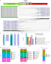
Bioinformatic analysis expands the catalogue of gall-specific sequences. (A) Theoretical diagram of the insertion regions according to the methodological approach applied. Position of the insertion sites into each host chromosome in the Manihot esculenta reference genome is shown on the left in a selected subgroup of hybrid contigs, mostly in forward orientation. Only the alignment framework of unmatched sequences to the cassava reference genome from hybrid/fusion contigs is shown on the right. Grey bar plots show the occupancy within each sequence position, and the black bar shows the base consensus within each sequence position in the resulting alignment scheme. (B) Dot plot representation of the aligned and annotated hybrid contigs region of unmatched sequences to the cassava reference genome, showing candidate insertion sequences in several of the hybrid contigs assembled, harbouring known DNA sequences revealing partial significant identity matches and covering with reported genes. Overlapping regions between the contigs and the annotated genes are shown as coloured triangles in each of the represented axes. Purple colour in the referenced gene represents forward strain orientation, and the blue-green colour (viridian) represents reverse strain orientation. (C) COG function classification histogram. Count of genes belonging to the COG categories related to exclusive gall reads involved in essential metabolic pathways and biological functions. (D, E) Microbiome profile of healthy plant tissue and gall tissue samples. Relative abundance of microorganism taxa identified in the microbiome of healthy leaves and gall samples of cassava. Taxonomic profiles were carried out to a 10K filter using raw reads generated by the shotgun sequencing approach. Each bar represents the organism taxon detected in one sample. The profile showed a similar abundance between both healthy samples (D), but a different relative abundance of microorganisms between gall samples. (E) Asterisks indicate enriched or exclusive microorganism species present only in gall tissue according to the taxonomic profile carried out, comparing sequenced heathy leaf samples with gall samples. The most common core endophyte taxa between leaf and gall tissues are also shown. (F) Taxonomic identity profile (10K) associated with some of the selected gall-specific reads. Gall-specific reads were bioinformatically filtered from the sequenced gall samples, which mismatched with the cassava reference genome and filtered against shared reads from healthy tissue samples.
Figure 4
Figure 4
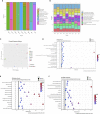
Microbiome taxonomic profile and functional analysis of gall endophytes and putative endosymbionts of the inducing insect. (A) Relative abundance of bacterial taxa identified in each of the samples of colony-forming units (CFUs) obtained from gall tissue of Manihot esculenta and from the larva head of the inducing insect Iatrophobia brasiliensis. The figure displays the most abundant taxa individually, with the remainder grouped together. Each bar represents the bacterial taxa detected in one sample. Each CFU from the insect head was called an isolated symbiotic bacteria (ISB). Each of the CFUs isolated and grown from internal sections of sterilised gall epidermis tissue was called an isolated endophytic bacteria (IEB). (B) Distribution of COG functional categories for CFU isolates from gall tissue and from the larval head of the inducing insect. (C) Genetic identity comparison of sequenced bacterial isolates classified up to genus by principal component analysis. (D–F) Functional analysis showing the top Gene Ontology enriched pathways of endophytic and alleged endosymbiont bacteria genomes. A high score indicates a high degree of enrichment.
Figure 5
Figure 5
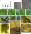
Bioassays show that potential insect endosymbiotic bacteria of the genus Rhodococcus induce gall-like structures in Manihot esculenta plants. (A) Graph of primary gall induction on leaves and micro-stakes with apical buds. Data for bacterial inoculation control are not shown due to the tissue damage caused since the first week of data collection. (B) Control inoculation, with only slight mechanical abrasion (without bacteria inoculation) in solid medium. (C) Leaf and apical buds control culture, inoculated with Pantoea ananatis (IEB 3-1) control bacterium. (D–F, H) Initial gall formation induced under natural field conditions on medium-mature young leaves. (G) Gall induced under natural field conditions on young leaf primordia. (I–L) Gall-like structure induced on leaves by inoculation with the isolated Rhodococcus strain (1–2 weeks of culture). The solid medium was supplemented with 3 mg L−1 Kathon, a bacteriostatic reagent used to inhibit bacterial growth in the culture medium. (M–P) Gall-like structure induced on apical buds by inoculation with the isolated Rhodococcus strain (M, 4 weeks of culture; N–P, 3 weeks of culture). Green or yellowish arrows show the formation of gall-like structures. Data are shown as the mean (± SE) of tissue response due to inoculation with the bacteria.

Similar articles

References

    1. Adams E., Spoel S. H. (2018). The ubiquitin-proteasome system as a transcriptional regulator of plant immunity. J. Exp. Bot. 69 (19), 4529–4537. doi: 10.1093/jxb/ery216 - DOI - PubMed
    1. Ahmad A., Wallau G. L., Ren Z. (2021). Characterization of Mariner transposons in seven species of Rhus gall aphids. Sci. Rep. 11, 16349. doi: 10.1038/s41598-021-95843-5 - DOI - PMC - PubMed
    1. Alexa A. (2022). Rahnenfuhrer J. topGO: Enrichment analysis for gene ontology (2.46. 0) [Computer software]. Bioconductor version: release (3.14).
    1. Ananthakrishnan T. (1998). Insect gall systems: patterns, processes and adaptive diversity. Curr. Sci. 75 (7), 672–676.
    1. Anders S., Pyl P. T., Huber W. (2015). HTSeq-a Python framework to work with high-throughput sequencing data. Bioinformatics 31 (2), 166–169. doi: 10.1093/bioinformatics/btu638 - DOI - PMC - PubMed

Grants and funding

The author(s) declare that no financial support was received for the research, authorship, and/or publication of this article. This research received no external funding. The postgraduate system at University of Costa Rica covered the Article Processing Fee as a Ph.D. student support.

LinkOut - more resources