

Distribution of ubiquilin 2 and TDP-43 aggregates throughout the CNS in UBQLN2 p.T487I-linked amyotrophic lateral sclerosis and frontotemporal dementia
- PMID: 38115557
- PMCID: PMC11007053
- DOI: 10.1111/bpa.13230
Distribution of ubiquilin 2 and TDP-43 aggregates throughout the CNS in UBQLN2 p.T487I-linked amyotrophic lateral sclerosis and frontotemporal dementia
Abstract
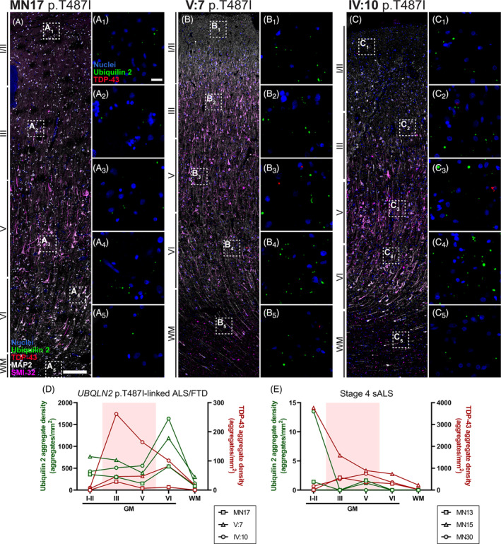
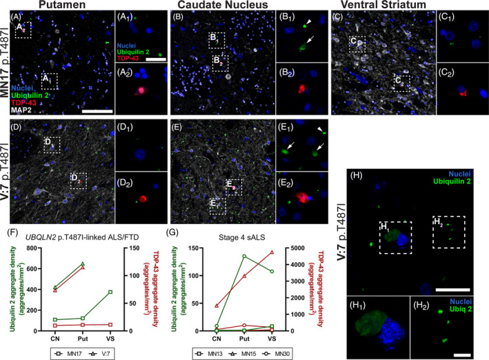
Mutations in the UBQLN2 gene cause amyotrophic lateral sclerosis (ALS) and frontotemporal dementia (FTD). The neuropathology of such UBQLN2-linked cases of ALS/FTD is characterised by aggregates of the ubiquilin 2 protein in addition to aggregates of the transactive response DNA-binding protein of 43 kDa (TDP-43). ALS and FTD without UBQLN2 mutations are also characterised by TDP-43 aggregates, that may or may not colocalise with wildtype ubiquilin 2. Despite this, the relative contributions of TDP-43 and ubiquilin 2 to disease pathogenesis remain largely under-characterised, as does their relative deposition as aggregates across the central nervous system (CNS). Here we conducted multiplex immunohistochemistry of three UBQLN2 p.T487I-linked ALS/FTD cases, three non-UBQLN2-linked (sporadic) ALS cases, and 8 non-neurodegenerative disease controls, covering 40 CNS regions. We then quantified ubiquilin 2 aggregates, TDP-43 aggregates and aggregates containing both proteins in regions of interest to determine how UBQLN2-linked and non-UBQLN2-linked proteinopathy differ. We find that ubiquilin 2 aggregates that are negative for TDP-43 are predominantly small and punctate and are abundant in the hippocampal formation, spinal cord, all tested regions of neocortex, medulla and substantia nigra in UBQLN2-linked ALS/FTD but not sporadic ALS. Curiously, the striatum harboured small punctate ubiquilin 2 aggregates in all cases examined, while large diffuse striatal ubiquilin 2 aggregates were specific to UBQLN2-linked ALS/FTD. Overall, ubiquilin 2 is mainly deposited in clinically unaffected regions throughout the CNS such that symptomology in UBQLN2-linked cases maps best to the aggregation of TDP-43.
Keywords: TDP‐43; UBQLN2; amyotrophic lateral sclerosis (ALS); frontotemporal dementia (FTD); neuropathology; ubiquilin 2.
© 2023 The Authors. Brain Pathology published by John Wiley & Sons Ltd on behalf of International Society of Neuropathology.
Conflict of interest statement
The authors declare that they have no competing interests.
Figures
Similar articles
-
Motor neuron disease, TDP-43 pathology, and memory deficits in mice expressing ALS-FTD-linked UBQLN2 mutations.Proc Natl Acad Sci U S A. 2016 Nov 22;113(47):E7580-E7589. doi: 10.1073/pnas.1608432113. Epub 2016 Nov 9. Proc Natl Acad Sci U S A. 2016. PMID: 27834214 Free PMC article.
-
Depressing time: Waiting, melancholia, and the psychoanalytic practice of care.In: Kirtsoglou E, Simpson B, editors. The Time of Anthropology: Studies of Contemporary Chronopolitics. Abingdon: Routledge; 2020. Chapter 5. In: Kirtsoglou E, Simpson B, editors. The Time of Anthropology: Studies of Contemporary Chronopolitics. Abingdon: Routledge; 2020. Chapter 5. PMID: 36137063 Free Books & Documents. Review.
-
Potential role of solid lipid curcumin particle (SLCP) as estrogen replacement therapy in mitigating TDP-43-related neuropathy in the mouse model of ALS disease.Exp Neurol. 2025 Jan;383:114999. doi: 10.1016/j.expneurol.2024.114999. Epub 2024 Oct 16. Exp Neurol. 2025. PMID: 39419433
-
Tar DNA-binding protein-43 (TDP-43) regulates axon growth in vitro and in vivo.Neurobiol Dis. 2014 May;65(100):25-34. doi: 10.1016/j.nbd.2014.01.004. Epub 2014 Jan 11. Neurobiol Dis. 2014. PMID: 24423647 Free PMC article.
-
Pathological mechanisms underlying TDP-43 driven neurodegeneration in FTLD-ALS spectrum disorders.Hum Mol Genet. 2013 Oct 15;22(R1):R77-87. doi: 10.1093/hmg/ddt349. Epub 2013 Jul 29. Hum Mol Genet. 2013. PMID: 23900071 Free PMC article. Review.
Cited by
-
Hippocampal aggregation signatures of pathogenic UBQLN2 in amyotrophic lateral sclerosis and frontotemporal dementia.Brain. 2024 Oct 3;147(10):3547-3561. doi: 10.1093/brain/awae140. Brain. 2024. PMID: 38703371 Free PMC article.
References
-
- Arai T, Hasegawa M, Akiyama H, Ikeda K, Nonaka T, Mori H, et al. TDP‐43 is a component of ubiquitin‐positive tau‐negative inclusions in frontotemporal lobar degeneration and amyotrophic lateral sclerosis. Biochem Biophys Res Commun. 2006;351(3):602–611. - PubMed
-
- Neumann M, Sampathu DM, Kwong LK, Truax AC, Micsenyi MC, Chou TT, et al. Ubiquitinated TDP‐43 in frontotemporal lobar degeneration and amyotrophic lateral sclerosis. Science. 2006;314(5796):130–133. - PubMed
-
- Mackenzie IR, Bigio EH, Ince PG, Geser F, Neumann M, Cairns NJ, et al. Pathological TDP‐43 distinguishes sporadic amyotrophic lateral sclerosis from amyotrophic lateral sclerosis with SOD1 mutations. Ann Neurol. 2007;61(5):427–434. - PubMed
-
- Mori K, Arzberger T, Grässer FA, Gijselinck I, May S, Rentzsch K, et al. Bidirectional transcripts of the expanded C9orf72 hexanucleotide repeat are translated into aggregating dipeptide repeat proteins. Acta Neuropathol. 2013;126(6):881–893. - PubMed
MeSH terms
Substances
Supplementary concepts
Grants and funding
LinkOut - more resources
Full Text Sources
Medical
Miscellaneous
