Review
. 2021 Jul 25;10(8):1885.
doi: 10.3390/cells10081885.

Ciliary Dyneins and Dynein Related Ciliopathies

Affiliations
Review

Ciliary Dyneins and Dynein Related Ciliopathies

Dinu Antony et al. Cells. .

Abstract

Although ubiquitously present, the relevance of cilia for vertebrate development and health has long been underrated. However, the aberration or dysfunction of ciliary structures or components results in a large heterogeneous group of disorders in mammals, termed ciliopathies. The majority of human ciliopathy cases are caused by malfunction of the ciliary dynein motor activity, powering retrograde intraflagellar transport (enabled by the cytoplasmic dynein-2 complex) or axonemal movement (axonemal dynein complexes). Despite a partially shared evolutionary developmental path and shared ciliary localization, the cytoplasmic dynein-2 and axonemal dynein functions are markedly different: while cytoplasmic dynein-2 complex dysfunction results in an ultra-rare syndromal skeleto-renal phenotype with a high lethality, axonemal dynein dysfunction is associated with a motile cilia dysfunction disorder, primary ciliary dyskinesia (PCD) or Kartagener syndrome, causing recurrent airway infection, degenerative lung disease, laterality defects, and infertility. In this review, we provide an overview of ciliary dynein complex compositions, their functions, clinical disease hallmarks of ciliary dynein disorders, presumed underlying pathomechanisms, and novel developments in the field.

Keywords: cilium; dynein; intraflagellar transport; primary ciliary dyskinesia; short rib polydactyly syndrome.


Conflict of interest statement

The authors declare no conflict of interest.

Figures
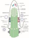
Figure 1
Figure 1
Structure of the cilia. Primary cilia with a 9 + 0 microtubule arrangement lacking dynein arms are usually immotile; respiratory epithelium cilia with a 9 + 2 microtubule arrangement with dynein arms, radial spokes, and a nexin dynein regulatory structure generate organized wave form movement while nodal cilia with a 9 + 0 microtubule arrangement with dynein arms generate a propelling rotational movement. Basal bodies show a triplet microtubule pattern.
Figure 2
Figure 2
Simplified structure of the dynein heavy chain, containing a heavy chain head with an AAA+ motor domain, a stalk with a microtubule binding domain, a linker, and a variable tail domain where other dynein subunits bind.
Figure 3
Figure 3
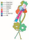
Sche matic of dynein-2, adapted from Toropova et al., 2019 [39]. Two copies of the dynein heavy chain form a homodimer, with one heavy chain adopting a zig-zag conformation at the tail region. One copy of the intermediate chain (DYNC2LI1 (LIC3)) is attached to each heavy chain. Two heterodimeric intermediate chains (WDR34 and WDR60) bind to DYNC2H1 via their C-terminal ends, and light chains are attached to the N-terminal ends of the intermediate chains.
Figure 4
Figure 4
Schematic representation of motile cilia. (A) Motile cilia structure showing a 9 + 2 arrangement of microtubules, inner and outer dynein arms attached to the A microtubule, the nexin dynein regulatory complex (N-DRC) that links the A microtubule of one peripheral doublet to the B microtubule of the adjacent doublet, and a central pair with associated projections and radial spokes. (B) Four ODAs, one double headed IDA and six single-headed IDAs, accompanied by three radial spokes and one N-DRC protein complex, form a 96 nm ruler distributed along the axoneme of the motile cilia. (C) Immunofluorescence of motile respiratory cilia (green: DNAI1, red: acetylated tubulin, and blue: DAPI). (D) Outer dynein arm subtypes in human respiratory cilia. Two ODA subtypes defined by the localization of dynein heavy chains can be distinguished: DNAH11 localizes to the proximal half, while DNAH9 is found in the distal half of human respiratory cilia.
Figure 5
Figure 5
IFT dynein dysfunction results in a complex developmental phenotype in mice. (A) Dync2h1 loss of function mouse embryo showing a turning defect and CNS defects compared to a control embryo, reprinted with permission from Ocbina et al. Nat Genet 2011 Jun;43(6):547-53 [46]. (B,C) Wdr34 mutant mouse embryos compared to controls, reprinted with permission from Wu et al. Hum Mol Genet. 2017 Jul 1;26(13):2386–2397 [128]. White arrows in (B) indicate microphthalmia Wdr34 mutant embryo compared to control; the arrow head indicates encephalocele in the Wdr34 mutant (D) Shortened stumpy primary cilia in Dync2h1 mutant mice compared to controls, visualized using electron microscopy. (E) Accumulation of IFT 88 within the stumpy Dync2h1 mutant cilia compared to controls. (F) Ciliary accumulation of Smo in Dynch1 dysfunctional cells in the absence of Shh, as well as after Shh stimulation, compared with control cells, reprinted with permission from Ocbina et al. Nat Genet 2011 Jun;43(6):547-53 [46].
Figure 6
Figure 6
Skeletal features observed in short rib polydactyly syndrome patients. The main hallmark is thoracic narrowing present, observed in utero or from birth (AC), becoming less pronounced with increasing age (D), (E) short horizontal ribs in a JATD case SRPS case with shortened long bones, long narrow thorax, pelvis configuration with acetabular spurs (F, close up in G, arrows indicate spurs). JATD case with a narrow thorax and shortened ribs, as well as handlebar clavicles, shown in the thorax X-ray, (A) reprinted with permission from Schmidts et al. J Med Genet 2013 May;50(5):309-23 [135], (B) reprinted with permission from Halbritter et al. Am J Hum Genet 2013 93, 915–925 [136], (CE) reprinted with permission from Schmidts et al. Am J Hum Genet. 2013 Nov 7;93(5):932–944 [131]; (F,G) reprinted with permission from Mc Inerney-Leo et al. Am J Hum Genet 2013 Sep 5;93(3):515-23 [133]. Polydactyly (H) is more frequently observed in SRPS compared with JATD, reprinted with permission from Schmidts et al. Nat Commun. 2015 Jun 5;6: 7074 [133].
Figure 7
Figure 7
Ciliary defects observed in IFT dynein patients. (A) Bulged ciliary tips and accumulation of IFT particles at the ciliary tips in fibroblast cilia from an individual carrying DYNC2H1 mutations; enlarged images of the cilia marked in the white squares are shown on the right side with pictures numbered accordingly and (B,C) reduced tubulin velocity compared with the control, indicating impaired IFT, reprinted with permission from Vig et al. Genet Med. 2020 Dec;22(12):2041–2051 [137]. (D) Slower flagella extension, but similar flagella end length in Tctex2b mutant Chlamydomonas compared with controls suggests partial functional redundancy for Tctex2b. (E) Reduced but not absent retrograde IFT in Tctex2b mutants compared with controls. (F) Normal anterograde IFT velocity and unchanged number of anterograde IFT trains, but a reduction of the retrograde velocity and a strong reduction in the number of retrograde IFT trains in Tctex2b mutant Chlamydomonas compared with controls, reprinted with permission from Schmidts et al. Nat Commun. 2015 Jun 5;6: 7074 [133].
Figure 8
Figure 8
The clinical phenotype of PCD and the representative EM defects observed. (A) Chest X ray of patient showing situs inversus, reprinted with permission from Loges et al. Am J Hum Genet. 2018 Dec 6; 103 (6):995–1008 [158]. (B) PCD patient with bronchiectasis of the right and left lower lobes, reprinted with permission from Onoufriadis et al. Am J Hum Genet. 2013 Jan 10; 92(1):88–98 [150]. (CI) Cross section of the cilia with ultra-structural defects in comparison with the control. (C) Control with outer and inner dynein arms (red arrows); (D) DNAAF3 patient’s cilia lacking both outer and inner dynein arms (arrows), reprinted with permission from Mitchison et al. Nat Genet. 2012 Mar 4;44(4):381–389 [159]. (E) Control with central pair appendage (arrow) and (F) Hydin patient cilia with missing central pair appendage (arrow) visualized by image averaging, reprinted with permission from Olbrich et al. Am J Hum Genet. 2012 Oct 5; 91(4):672–684 [160] (G) disorganized peripheral microtubules and (H) acentric central pair, as well as (I) supernumerary central pairs seen in the cilia of CCDC40 patients, reprinted with permission from Antony et al.Hum Mutat. 2013 Mar; 34(3):462–472 [161]. (J) Graphical demonstration of a normal motile cilia movement pattern compared with the pattern observed in DNAH9 dysfunction (immotile distal half), DNAH11 dysfunction (immotile proximal half), and DNAH5 dysfunction (complete immobility), with recovery strokes shown in black.

Similar articles

Cited by

References

    1. Satir P. Landmarks in cilia research from leeuwenhoek to US. Cell Motil. Cytoskelet. 1995;32:90–94. doi: 10.1002/cm.970320203. - DOI - PubMed
    1. Elliott K.H., Brugmann S.A. Sending mixed signals: Cilia-dependent signaling during development and disease. Dev. Biol. 2019;447:28–41. doi: 10.1016/j.ydbio.2018.03.007. - DOI - PMC - PubMed
    1. Choksi S.P., Lauter G., Swoboda P., Roy S. Switching on cilia: Transcriptional networks regulating ciliogene-sis. Development. 2014;141:1427–1441. doi: 10.1242/dev.074666. - DOI - PubMed
    1. Mitchison H.M., Valente E.M. Motile and non-motile cilia in human pathology: From function to phenotypes. J. Pathol. 2016;241:294–309. doi: 10.1002/path.4843. - DOI - PubMed
    1. Sironen A., Shoemark A., Patel M., Loebinger M.R., Mitchison H.M. Sperm defects in primary ciliary dyskinesia and related causes of male infertility. Cell Mol. Life Sci. 2020;77:2029–2048. doi: 10.1007/s00018-019-03389-7. - DOI - PMC - PubMed

Publication types

MeSH terms

LinkOut - more resources