

Epilepsy in Tubulinopathy: Personal Series and Literature Review
- PMID: 31269740
- PMCID: PMC6678821
- DOI: 10.3390/cells8070669
Epilepsy in Tubulinopathy: Personal Series and Literature Review
Abstract
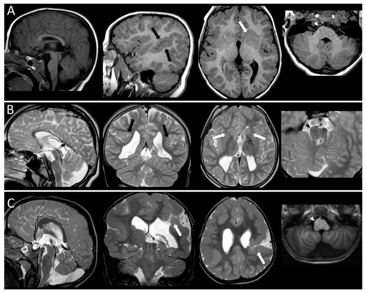
Mutations in tubulin genes are responsible for a large spectrum of brain malformations secondary to abnormal neuronal migration, organization, differentiation and axon guidance and maintenance. Motor impairment, intellectual disability and epilepsy are the main clinical symptoms. In the present study 15 patients from a personal cohort and 75 from 21 published studies carrying mutations in TUBA1A, TUBB2B and TUBB3 tubulin genes were evaluated with the aim to define a clinical and electrophysiological associated pattern. Epilepsy shows a wide range of severity without a specific pattern. Mutations in TUBA1A (60%) and TUBB2B (74%) and TUBB3 (25%) genes are associated with epilepsy. The accurate analysis of the Electroencephalogram (EEG) pattern in wakefulness and sleep in our series allows us to detect significant abnormalities of the background activity in 100% of patients. The involvement of white matter and of the inter-hemispheric connection structures typically observed in tubulinopathies is evidenced by the high percentage of asynchronisms in the organization of sleep activity recorded. In addition to asymmetries of the background activity, excess of slowing, low amplitude and Magnetic Resonance (MR) imaging confirm the presence of extensive brain malformations involving subcortical and midline structures. In conclusion, epilepsy in tubulinopathies when present has a favorable evolution over time suggesting a not particularly aggressive therapeutic approach.
Keywords: EEG; TUBA1A; TUBB2B; TUBB3; epilepsy; malformations cortical development; tubulin genes.
Conflict of interest statement
The authors declare no conflict of interest.
Figures


Similar articles
-
Brain malformations and mutations in α- and β-tubulin genes: a review of the literature and description of two new cases.Dev Med Child Neurol. 2014 Apr;56(4):354-60. doi: 10.1111/dmcn.12370. Epub 2014 Jan 7. Dev Med Child Neurol. 2014. PMID: 24392928 Review.
-
The wide spectrum of tubulinopathies: what are the key features for the diagnosis?Brain. 2014 Jun;137(Pt 6):1676-700. doi: 10.1093/brain/awu082. Brain. 2014. PMID: 24860126
-
Investigation of the most common clinical and imaging findings and the role of tubulin genes in the etiology of malformations of cortical development.Turk J Med Sci. 2020 Oct 22;50(6):1573-1579. doi: 10.3906/sag-1901-170. Turk J Med Sci. 2020. PMID: 32718119 Free PMC article.
-
Tubulin genes and malformations of cortical development.Eur J Med Genet. 2018 Dec;61(12):744-754. doi: 10.1016/j.ejmg.2018.07.012. Epub 2018 Jul 17. Eur J Med Genet. 2018. PMID: 30016746 Review.
-
A novel mutation in the β-tubulin gene TUBB2B associated with complex malformation of cortical development and deficits in axonal guidance.Dev Med Child Neurol. 2012 Aug;54(8):765-9. doi: 10.1111/j.1469-8749.2012.04316.x. Epub 2012 May 16. Dev Med Child Neurol. 2012. PMID: 22591407
Cited by
-
Gamma-Tubulin 1 (TUBG1) Mutation-Associated Lissencephaly and Microcephaly in an Indian Child: A Rare Case.Cureus. 2024 Jun 20;16(6):e62749. doi: 10.7759/cureus.62749. eCollection 2024 Jun. Cureus. 2024. PMID: 38912084 Free PMC article.
-
Forward Genetics-Based Approaches to Understanding the Systems Biology and Molecular Mechanisms of Epilepsy.Int J Mol Sci. 2023 Mar 9;24(6):5280. doi: 10.3390/ijms24065280. Int J Mol Sci. 2023. PMID: 36982355 Free PMC article. Review.
-
Complementing the phenotypical spectrum of TUBA1A tubulinopathy and its role in early-onset epilepsies.Eur J Hum Genet. 2022 Mar;30(3):298-306. doi: 10.1038/s41431-021-01027-0. Epub 2022 Jan 11. Eur J Hum Genet. 2022. PMID: 35017693 Free PMC article.
-
Epilepsy and Cognitive Impairment in Childhood and Adolescence: A Mini-Review.Curr Neuropharmacol. 2023;21(8):1646-1665. doi: 10.2174/1570159X20666220706102708. Curr Neuropharmacol. 2023. PMID: 35794776 Free PMC article. Review.
-
RELN gene-related drug-resistant epilepsy with periventricular nodular heterotopia treated with radiofrequency thermocoagulation: a case report.Front Neurol. 2024 Mar 27;15:1366776. doi: 10.3389/fneur.2024.1366776. eCollection 2024. Front Neurol. 2024. PMID: 38601336 Free PMC article.
References
-
- Nabbout R., Scheffer I.E. Genetics of idiopathic epilepsies. Handb. Clin. Neurol. 2013;111:567–578. - PubMed
Publication types
MeSH terms
Substances
LinkOut - more resources
Full Text Sources
Medical
