

ER complex proteins are required for rhodopsin biosynthesis and photoreceptor survival in Drosophila and mice
- PMID: 31263175
- PMCID: PMC7206144
- DOI: 10.1038/s41418-019-0378-6
ER complex proteins are required for rhodopsin biosynthesis and photoreceptor survival in Drosophila and mice
Abstract
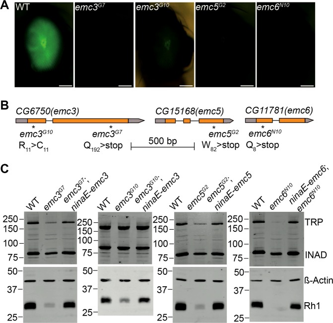
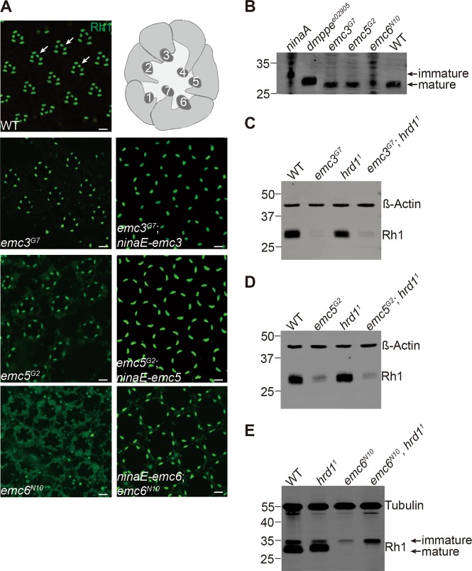
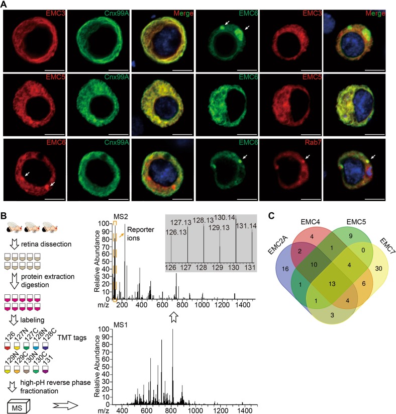
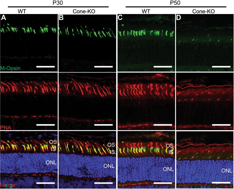
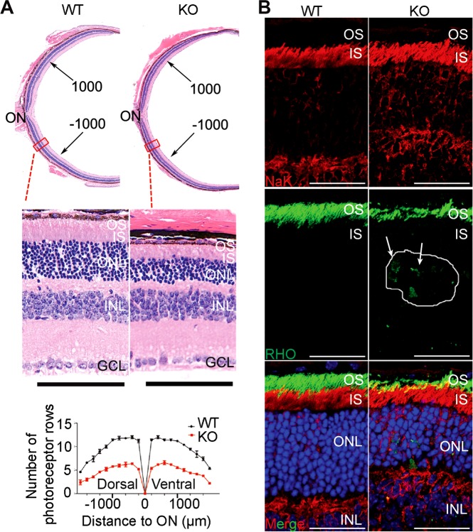
Defective rhodopsin homeostasis is one of the major causes of retinal degeneration, including the disease Retinitis pigmentosa. To identify cellular factors required for the biosynthesis of rhodopsin, we performed a genome-wide genetic screen in Drosophila for mutants with reduced levels of rhodopsin. We isolated loss-of-function alleles in endoplasmic reticulum membrane protein complex 3 (emc3), emc5, and emc6, each of which exhibited defective phototransduction and photoreceptor cell degeneration. EMC3, EMC5, and EMC6 were essential for rhodopsin synthesis independent of the ER associated degradation (ERAD) pathway, which eliminates misfolded proteins. We generated null mutations for all EMC subunits, and further demonstrated that different EMC subunits play roles in different cellular functions. Conditional knockout of the Emc3 gene in mice led to mislocalization of rhodopsin protein and death of cone and rod photoreceptor cells. These data indicate conserved roles for EMC subunits in maintaining rhodopsin homeostasis and photoreceptor function, and suggest that retinal degeneration may also be caused by defects in early biosynthesis of rhodopsin.
Conflict of interest statement
The authors declare that they have no conflict of interest.
Figures


Similar articles
-
dPob/EMC is essential for biosynthesis of rhodopsin and other multi-pass membrane proteins in Drosophila photoreceptors.Elife. 2015 Feb 26;4:e06306. doi: 10.7554/eLife.06306. Elife. 2015. PMID: 25715730 Free PMC article.
-
The endoplasmic reticulum membrane protein complex subunit Emc6 is essential for rhodopsin localization and photoreceptor cell survival.Genes Dis. 2023 May 23;11(2):1035-1049. doi: 10.1016/j.gendis.2023.03.033. eCollection 2024 Mar. Genes Dis. 2023. PMID: 37692493 Free PMC article.
-
Loss of the ER membrane protein complex subunit Emc3 leads to retinal bipolar cell degeneration in aged mice.PLoS One. 2020 Sep 4;15(9):e0238435. doi: 10.1371/journal.pone.0238435. eCollection 2020. PLoS One. 2020. PMID: 32886670 Free PMC article.
-
A model for transport of membrane-associated phototransduction polypeptides in rod and cone photoreceptor inner segments.Vision Res. 2008 Feb;48(3):442-52. doi: 10.1016/j.visres.2007.08.020. Epub 2007 Oct 18. Vision Res. 2008. PMID: 17949773 Free PMC article. Review.
-
Membrane protein trafficking in Drosophila photoreceptor cells.Eur J Cell Biol. 2017 Aug;96(5):391-401. doi: 10.1016/j.ejcb.2016.11.002. Epub 2016 Dec 7. Eur J Cell Biol. 2017. PMID: 27964885 Review.
Cited by
-
A novel and efficient murine model of Bietti crystalline dystrophy.Dis Model Mech. 2022 Mar 1;15(3):dmm049222. doi: 10.1242/dmm.049222. Epub 2022 Mar 1. Dis Model Mech. 2022. PMID: 35230417 Free PMC article.
-
EMC3 Is Essential for Retinal Organization and Neurogenesis During Mouse Retinal Development.Invest Ophthalmol Vis Sci. 2021 Feb 1;62(2):31. doi: 10.1167/iovs.62.2.31. Invest Ophthalmol Vis Sci. 2021. PMID: 33605987 Free PMC article.
-
De novo variants in EMC1 lead to neurodevelopmental delay and cerebellar degeneration and affect glial function in Drosophila.Hum Mol Genet. 2022 Sep 29;31(19):3231-3244. doi: 10.1093/hmg/ddac053. Hum Mol Genet. 2022. PMID: 35234901 Free PMC article.
-
Ribosome-associated quality control of membrane proteins at the endoplasmic reticulum.J Cell Sci. 2020 Nov 27;133(22):jcs251983. doi: 10.1242/jcs.251983. J Cell Sci. 2020. PMID: 33247003 Free PMC article. Review.
-
EMC10 homozygous variant identified in a family with global developmental delay, mild intellectual disability, and speech delay.Clin Genet. 2020 Dec;98(6):555-561. doi: 10.1111/cge.13842. Epub 2020 Sep 15. Clin Genet. 2020. PMID: 32869858 Free PMC article.
References
-
- Hartong DT, Berson EL, Dryja TP. Retinitis pigmentosa. Lancet. 2006;368:1795–809. - PubMed
-
- Nemet I, Ropelewski P, Imanishi Y. Rhodopsin trafficking and mistrafficking: signals, molecular components, and mechanisms. Prog Mol Biol Transl Sci. 2015;132:39–71. - PubMed
-
- Galy A, Roux MJ, Sahel JA, Leveillard T, Giangrande A. Rhodopsin maturation defects induce photoreceptor death by apoptosis: a fly model for RhodopsinPro23His human retinitis pigmentosa. Hum Mol Genet. 2005;14:2547–57. - PubMed
Publication types
MeSH terms
Substances
LinkOut - more resources
Full Text Sources
Molecular Biology Databases
