. 2019 Apr 8;9(1):5791.
doi: 10.1038/s41598-019-42113-0.

Angiotensin Receptor Neprilysin Inhibitor Attenuates Myocardial Remodeling and Improves Infarct Perfusion in Experimental Heart Failure

Affiliations

Angiotensin Receptor Neprilysin Inhibitor Attenuates Myocardial Remodeling and Improves Infarct Perfusion in Experimental Heart Failure

Daniel Pfau et al. Sci Rep. .

Abstract

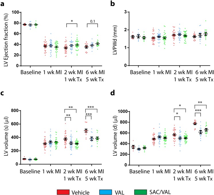
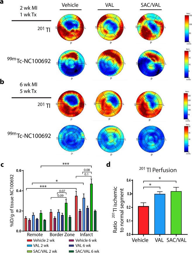
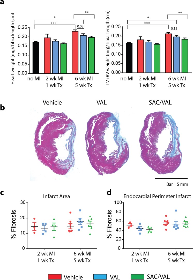
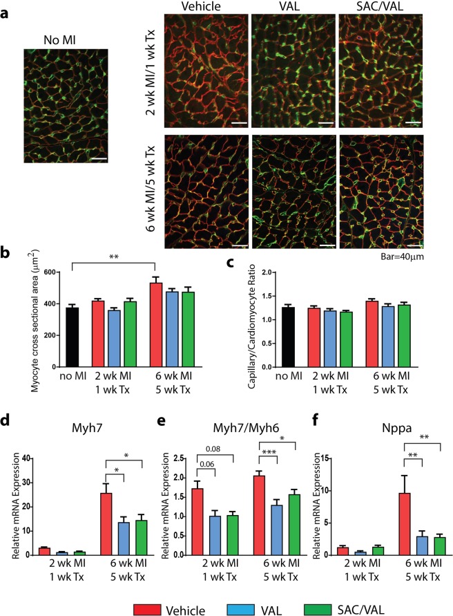
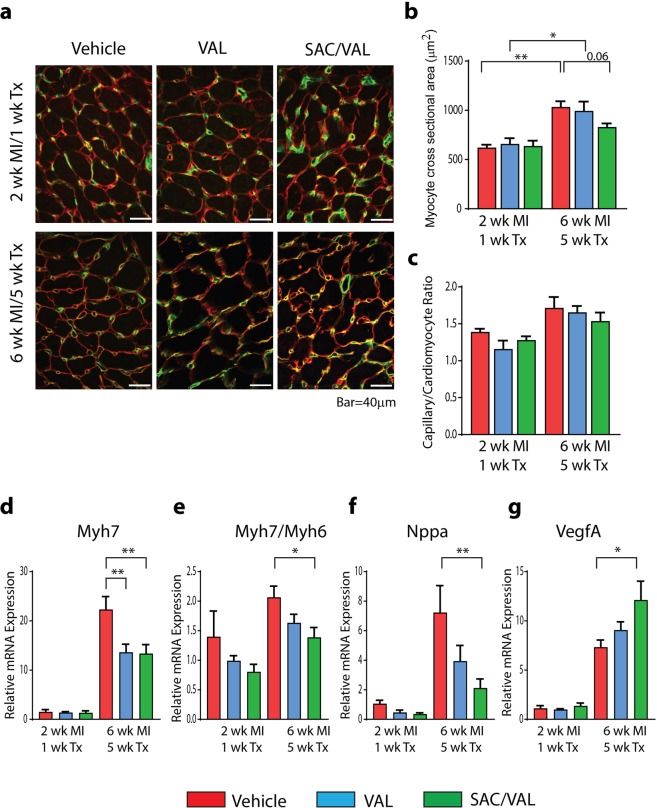
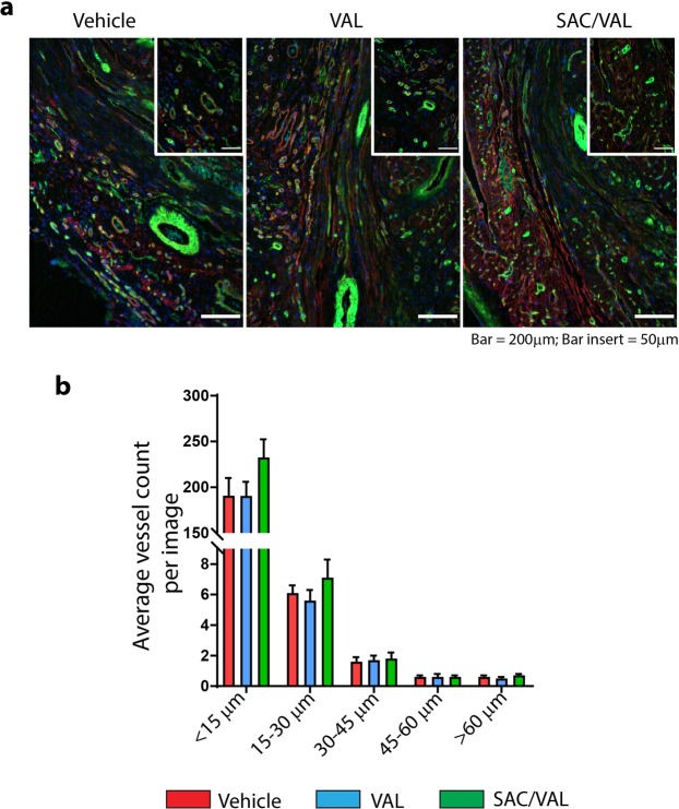
Angiotensin receptor blocker-neprilysin inhibitor (ARNi) therapy improves the prognosis of heart failure patients. However, the mechanisms remain unclear. This study investigated the biological effects of ARNi with neprilysin inhibitor sacubitril and angiotensin receptor blocker valsartan on myocardial remodeling and cardiac perfusion in experimental heart failure (HF) after myocardial infarction (MI). Male Lewis rats (10-weeks old) with confirmed HF were randomized one-week post-MI to treatment with vehicle (water), sacubitril/valsartan or valsartan, as comparator group, for either 1 or 5 weeks. Sacubitril/valsartan for 1-week limited LV contractile dysfunction vs. vehicle and both sacubitril/valsartan and valsartan attenuated progressive LV dilation after 1 and 5 weeks treatment. After 5 weeks, both sacubitril/valsartan and valsartan reduced CTGF expression in the remote myocardium, although only sacubitril/valsartan prevented interstitial fibrosis. In the border zone, sacubitril/valsartan and valsartan reduced hypertrophic markers, but only sacubitril/valsartan reduced cardiomyocyte size and increased VEGFA expression. In the infarct, sacubitril/valsartan induced an early uptake of 99mTc-NC100692 (a radiotracer of angiogenesis) and improved perfusion, as determined by 201Tl microSPECT/CT imaging. In conclusion, ARNi improved global LV function, limited remodeling in the remote and border zones, and increased perfusion to the infarct. Sacubitril/valsartan had more consistent effects than valsartan on LV remodeling in experimental HF.


Conflict of interest statement

The authors declare no competing interests.

Figures

Figure 1
Figure 1
Cardiac function and remodeling. (a) SAC/VAL treatment increased LV Ejection fraction post MI as compared with vehicle treated rats. (b) No significant changes in LV posterior wall thickness in diastole (LVPWd) post MI between treatment groups. (c) SAC/VAL and VAL treatment reduced LV volume in systole post MI compared to vehicle treated group. (d) SAC/VAL and VAL treatment reduced LV volume in diastole post MI compared to vehicle treated group. Changes in LVEF, LV volume (d) and LV volume (s) were statistically significant vs. baseline in all groups. 1 and 2 wk. MI/1 wk. Tx: Vehicle n = 29, VAL n = 27, SAC/VAL n = 30. 1, 2 and 6 wk. MI/5 wk. Tx: Vehicle n = 13, VAL n = 12, SAC/VAL n = 13. Statistical significance determined by repeated measures two-way ANOVA followed by Tukey’s post-hoc for multiple comparisons. *P < 0.05, **P < 0.01, ***P < 0.001 as indicated.
Figure 2
Figure 2
Myocardial 99mTc-NC100692 uptake and 201Tl perfusion. (a,b) Representative in vivo polar-maps displayed for the left ventricle with an uptake color scale (red high, blue low) in a two dimensional image with the apex in the center and the anterior, septal, posterior, and lateral wall in a counter-clockwise formation for the perfusion tracer 201Tl and concurrent uptake by the angiogenesis αvβ3 tracer 99mTc-NC100692 at 2 wk. MI/1 wk. Tx in panel (a) and 6 wk. MI/5 wk. Tx in panel (b) for each treatment group. (c) Post mortem gamma well counting of 99mTc-NC100692 in remote (non-infarcted myocardium), border zone, and infarct. 2 wk. MI/1 wk. Tx: Vehicle n = 6, VAL n = 5, SAC/VAL n = 6; 6 wk. MI/5 wk. Tx: Vehicle n = 6, VAL n = 6, SAC/VAL n = 6. (d) Dobutamine stress 201Tl perfusion. In vivo 201Tl ischemic to 201Tl non-ischemic under dobutamine-induced stress 6 weeks after MI. Both SAC/VAL and VAL increased dobutamine-stimulated flow to the infarct area 6 weeks after MI compared with vehicle treated rats. Vehicle n = 6, VAL n = 6, SAC/VAL n = 6. Statistical significance determined by one-way ANOVA followed by Tukey’s post-hoc for multiple comparisons. *P < 0.05, ***P < 0.001 as indicated.
Figure 3
Figure 3
LV structural remodeling. (a) Total heart weights and combined left and right ventricle weights of freshly excised hearts normalized to tibia length. SAC/VAL significantly reduced the heart remodeling 6 weeks after MI compared with vehicle treated rats. (b) Representative Masson’s trichrome stained heart sections from vehicle, VAL and SAC/VAL treated rats at 6 wk. MI/5 wk. Tx. (c) Infarct size assessed on cross sections of the mid-ventricle stained with Masson’s trichrome expressed as a percent of total tissue. (d) Endocardial perimeter infarct expressed as a percent of total endocardial perimeter. No MI (n = 4); 2 wk. MI/1 wk. Tx: Vehicle n = 3, VAL n = 4, SAC/VAL n = 4; 6 wk. MI/5 wk. Tx: Vehicle n = 7; VAL n = 6, SAC/VAL n = 6–7. Statistical significance determined by one-way ANOVA followed by Tukey’s post-hoc for multiple comparisons. *P < 0.05, **P < 0.01, ***P < 0.001 as indicated.
Figure 4
Figure 4
Interstitial fibrosis in the remote, non-infarcted myocardium 6 weeks after MI. (a) Representative images of Sirius Red staining in the remote myocardium in no MI control rats and vehicle, VAL or SAC/VAL treated rats at 6 wk. MI/5 wk. Tx. (b) Quantification of interstitial fibrosis. SAC/VAL significantly reduced interstitial fibrosis 6 weeks after MI, as compared with vehicle treated rats. (c) Both SAC/VAL and VAL significantly reduced Ctgf gene expression in remote myocardium 6 weeks after MI, as compared with vehicle treated group. No MI n = 4, Vehicle n = 7, VAL n = 6, SAC/VAL n = 7. Statistical significance determined by one-way ANOVA followed by Tukey’s post-hoc for multiple comparisons. *P < 0.05, **P < 0.01 as indicated.
Figure 5
Figure 5
Cardiomyocyte hypertrophy and capillary/myocyte ratio in the remote, non-infarcted myocardium. (a) Representative images of LV sections co-stained with laminin (in red) and isolectin B4 (in green) in no MI control rats and vehicle, VAL and SAC/VAL treated rats at 2 wk. MI/1 wk. Tx and 6 wk. MI/5 wk. Tx. (b) Quantification of myocyte cross sectional area. VAL and SAC/VAL reduced the hypertrophic response at 6 wk. MI/5 wk. Tx. (c) Capillary to myocyte ratio in no MI control rats and vehicle, VAL and SAC/VAL treated rats at 2 wk. MI/1 wk. Tx and 6 wk. MI/5 wk. Tx. (d) SAC/VAL and VAL significantly reduced Myh7 mRNA transcript levels at 6 wk. MI/5 wk. Tx compared to vehicle treated rats. (e) SAC/VAL and VAL significantly reduced Myh7/Myh6 ratio at 6 wk. MI/5 wk. Tx. (f) SAC/VAL and VAL significantly reduced Nppa mRNA transcript levels at 6 wk. MI/5 wk. Tx. No MI n = 4; 2 wk. MI/1 wk. Tx: Vehicle n = 3, VAL n = 4, SAC/VAL n = 4; 6 wk. MI/5 wk. Tx: Vehicle n = 7; VAL n = 6, SAC/VAL n = 7. Statistical significance determined by one-way ANOVA followed by Tukey’s post-hoc for multiple comparisons. *P < 0.05, **P < 0.01, ***P < 0.001 as indicated.
Figure 6
Figure 6
Cardiomyocyte hypertrophy and angiogenesis in the border zone. (a) Representative images of LV sections co-stained with laminin (in red) and isolectin B4 (in green) in vehicle, VAL or SAC/VAL treated rats at 2 wk. MI/1 wk. Tx and 6 wk. MI/5 wk. Tx. (b) Quantification of myocyte cross sectional area. SAC/VAL attenuated the cardiomyocyte hypertrophic response in the border zone at 6 wk. MI/5 wk. Tx, as compared with vehicle treated rats. (c) Capillary to myocyte ratio in vehicle, VAL or SAC/VAL treated rats at 2 wk. MI/1 wk. Tx and 6 wk. MI/5 wk. Tx. (d) SAC/VAL and VAL significantly reduced Myh7 mRNA transcript levels at 6 wk. MI/5 wk. Tx in the border zone. (e) SAC/VAL significantly reduced Myh7/Myh6 ratio at 6 wk. MI/5 wk. Tx compared to vehicle treated rats. (f) SAC/VAL significantly reduced Nppa mRNA transcript levels at 6 wk. MI/5 wk. Tx compared to vehicle treated rats. (g) Increased VegfA expression at 6 wk. MI with 5 wk. SAC/VAL treatment. 2 wk. MI/1 wk. Tx: Vehicle n = 3, VAL n = 4, SAC/VAL n = 4; 6 wk MI/5 wk. Tx: Vehicle n = 7; VAL n = 6, SAC/VAL n = 7. Statistical significance determined by one-way ANOVA followed by Tukey’s post-hoc for multiple comparisons. *P < 0.05, **P < 0.01 as indicated.
Figure 7
Figure 7
Vessel remodeling in the infarct region at 6 weeks post MI. (a) Representative images of infarct region co-stained with laminin (in red), isolectin B4 (in green), and DAPI (in blue) in vehicle, VAL and SAC/VAL treated rats at 6 wk. MI/5 wk. Tx. (b) Quantification of vessel density by vessel diameter. Values expressed as average vessel count per image. Vehicle n = 7, VAL n = 6, SAC/VAL n = 7.

Similar articles

Cited by

References

    1. von Lueder TG, et al. Renin-angiotensin blockade combined with natriuretic peptide system augmentation: novel therapeutic concepts to combat heart failure. Circ Heart Fail. 2013;6:594–605. doi: 10.1161/CIRCHEARTFAILURE.112.000289. - DOI - PMC - PubMed
    1. Yancy CW, et al. ACC/AHA/HFSA Focused Update on New Pharmacological Therapy for Heart Failure: An Update of the 2013 ACCF/AHA Guideline for the Management of Heart Failure: A Report of the American College of Cardiology/American Heart Association Task Force on Clinical Practice Guidelines and the Heart Failure Society of America. J Am Coll Cardiol. 2016;68:1476–1488. doi: 10.1016/j.jacc.2016.05.011. - DOI - PubMed
    1. McMurray JJ, et al. Angiotensin-neprilysin inhibition versus enalapril in heart failure. N Engl J Med. 2014;371:993–1004. doi: 10.1056/NEJMoa1409077. - DOI - PubMed
    1. Gu J, et al. Pharmacokinetics and pharmacodynamics of LCZ696, a novel dual-acting angiotensin receptor-neprilysin inhibitor (ARNi) J Clin Pharmacol. 2010;50:401–414. doi: 10.1177/0091270009343932. - DOI - PubMed
    1. Schiering N, et al. Structure of neprilysin in complex with the active metabolite of sacubitril. Sci Rep. 2016;6:27909. doi: 10.1038/srep27909. - DOI - PMC - PubMed

Publication types

MeSH terms