. 2016 Oct;12(10):876-84.
doi: 10.1038/nchembio.2166. Epub 2016 Aug 29.

Covalent targeting of remote cysteine residues to develop CDK12 and CDK13 inhibitors

Affiliations

Covalent targeting of remote cysteine residues to develop CDK12 and CDK13 inhibitors

Tinghu Zhang et al. Nat Chem Biol. 2016 Oct.

Abstract

Cyclin-dependent kinases 12 and 13 (CDK12 and CDK13) play critical roles in the regulation of gene transcription. However, the absence of CDK12 and CDK13 inhibitors has hindered the ability to investigate the consequences of their inhibition in healthy cells and cancer cells. Here we describe the rational design of a first-in-class CDK12 and CDK13 covalent inhibitor, THZ531. Co-crystallization of THZ531 with CDK12-cyclin K indicates that THZ531 irreversibly targets a cysteine located outside the kinase domain. THZ531 causes a loss of gene expression with concurrent loss of elongating and hyperphosphorylated RNA polymerase II. In particular, THZ531 substantially decreases the expression of DNA damage response genes and key super-enhancer-associated transcription factor genes. Coincident with transcriptional perturbation, THZ531 dramatically induced apoptotic cell death. Small molecules capable of specifically targeting CDK12 and CDK13 may thus help identify cancer subtypes that are particularly dependent on their kinase activities.


Figures
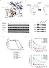
Figure 1
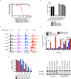
Figure 1. THZ531 covalently inhibits CDK12 and 13
a, Overlay of CDK7 and CDK12 crystal structures reveals similarly positioned cysteines in the C-terminal extension segments of the two kinase domains. Cys-312 was modeled into the crystal structure of CDK7 as previously described. PDB codes: 1UA2 (CDK7) and 4NST (CDK12). b, Structures of THZ1 and THZ531 and rational design strategy to synthesize THZ531 from THZ1. c, THZ531 binds intracellular CDK12-cyclin K complexes with higher affinity than CDK7-cyclin H complexes out competing lysate –introduced bioTHZ1 in target engagement assay. d, Biotinylated THZ531 (bioTHZ531, 1 µM) pulls down CDK12-cyclin K and CDK13-cyclin K complexes, but not CDK7-cyclin H complexes, whereas bioTHZ1 (1 µM) pulls down CDK7, CDK12, and 13 complexes. e, THZ531 potently inhibits CDK12 and CDK13. IC50 values for CDK12 and 13 were 158 and 69 nM, respectively, compared to 10.5 µM for CDK9 and 8.5 µM for CDK7. Measurements were performed in triplicate and data represent mean values ± S.D. f, THZ531 inhibition of CDK12 and 13 is time –dependent. In vitro kinase activity assay of CDK12-cyclin K (top) and CDK13-cyclin K (bottom) with different concentrations of THZ531 and varying preincubation times. For all incubation time series, the counts per minute of the kinase activity measurements were normalized to the relative [32P] transfer. Measurements were performed in triplicate and data represent the mean values ± S.D. Uncut western blots are in Supplementary Fig. 10.
Figure 2
Figure 2. THZ531 co-crystal structure with CDK12-cyclin K
a, THZ531 binds to M816 in the kinase hinge region and connects to Cys-1039 in two conformations via the compound’s flexible linker. Solvent-exposed regions of THZ531 with poor electron density are represented by thin sticks. b. Omit map contoured at 2.5σ for THZ531 bound to CDK12 chain C. c. Omit map contoured at 2.5σ for THZ531 bound to CDK12 chain D.
Figure 3
Figure 3. THZ531 induces apoptosis in Jurkat cells
a. THZ531, but not THZ531R or THZ532, exhibits strong antiproliferative effect in Jurkat T-cell acute lymphoblastic leukemia cells. Cells were treated with the indicated compounds for 72 hrs and assessed for antiproliferative effect using CellTiter Glo. b, THZ531 demonstrates irreversible suppression of Jurkat cell proliferation. Jurkat cells were treated with the indicated compounds for 6 hrs. Cell growth medium containing inhibitors was then removed (washout) and cells were allowed to grow for the remainder of the 72 hr proliferation assay without inhibitors. CellTiter Glo results were compared to cellular treatments whereby cells were treated with inhibitors for the full 72 hrs (no washout). c. THZ531 increases sub-G1 population. Jurkat T-ALL cells were treated with THZ531 for the indicated time periods. Cell cycle progression was assessed using FACS cell cycle analysis. d. Treatment with THZ531 induces apoptosis. Jurkat cells were treated with THZ531 for the indicated times. Cells were stained with Annexin V and propidium iodide. All proliferation and apoptosis assays were performed in biological triplicate and error bars are +/− SD. #p-value=3.33e-07, +p-value=1.04e-04, ++p-value=1.35e-04, +++p-value=9.94e-04, ++++p-value=1.53e-07. e, 72-hour antiproliferation assay using WT and C1039S HAP1 cells. Cells were analyzed using Cell Titer Glo. Experiment was performed in biological triplicate and error bars are +/− SD. *p-value=5.84e-04, **p-value=1.76e-04, ***p-value=9.09e-05, ****p-value=4.25e-04. f, Expression of C1039S CDK12 partially reduces PARP cleavage in HAP1 cells. Western blot analysis of indicated proteins in HAP1 CDK12 WT and C1039S cells. Uncut western blots are in Supplementary Fig. 10. P-values were determined with a two-tailed Student’s T test.
Figure 4
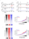
Figure 4. CDK12 binds regulatory and coding regions of active genes
a. Pol II, H3K27Ac, and CDK12 co-occupy active enhancers and promoters. Metagene representation of average Pol II, H3K27Ac, and CDK12 occupancy at H3K27ac-defined enhancers (left) and 37974 RefSeq promoters (right). b. Pol II, H3K27Ac, and CDK12 bind at promoters and across gene bodies. Metagene representation of global Pol II, H3K27Ac, and CDK12 occupancy at promoters and gene bodies. Average ChIP-seq signal in 13906 RefSeq genes expressed in 6h DMSO conditions. c. CDK12, Pol II, and H3K27Ac co-occupy active gene promoters. Left: Heatmaps show the density of Pol II- (left), H3K27Ac- (center), and CDK12- (right) ChIP-Seq reads relative to RefSeq promoters and comprising the region +/−2kb from RefSeq transcription start sites. Right: ChIP-seq read densities for CDK12 and H3K27ac at RefSeq promoters of 4kb centered on transcription start sites. d. CDK12, Pol II, and H3K27Ac co-occupy active enhancers. Left: Heatmaps show the density of Pol II- (left), H3K27Ac- (center), and CDK12- (right) ChIP-Seq reads relative to 32855 H3K27ac –enriched enhancer sites. For each H3K27ac-bound site (y axis), the densities of H3K27Ac (red) and CDK12 (blue) are displayed within a 2 kb window centered on the center of the H3K27ac-bound site. Right: ChIP-seq read densities for CDK12 and H3K27ac at enhancers of 4kb centered on the middle of H3K27ac peaks. For all heatmaps, promoters and enhancers were ranked by Pol II density (black) and a smoothing function was applied. All metagenes are presented in units of rpm/bp.
Figure 5
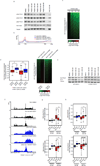
Figure 5. THZ531 inhibits transcription elongation and gene expression
a, THZ531 reduces Pol II CTD Ser2 phosphorylation (pSer2) in a concentration-dependent manner. b, THZ531 reduces 3’ pSer2 Pol II signal density. Metagene representation of global pSer2 Pol II occupancy at gene bodies. Average ChIP-seq signal (rpm/bp) across 13906 genes expressed in DMSO. c, THZ531 downregulates mRNA levels in a concentration-dependent manner. Heatmaps display the Log2 fold-change in gene expression vs. DMSO for 14,745 transcripts expressed in DMSO. Heatmap is ranked by 500 nM THZ531. d, C1039S CDK12 expression partially restores gene expression in HAP1 cells. Box plots (left) and heatmaps (right) displaying expression of 14,261 expressed transcripts. *p-value < 10−16 and **p-value < 10−16. Heatmaps are ranked by WT HAP1 fold-change. e, C1039S CDK12 expression partially restores pSer2 in HAP1 cells. f, THZ531 reduces elongating Pol II at THZ531-response genes. g and h, THZ531 reduces 3’ pSer2 Pol II (g) and Pol II (h) ChIP-seq signal at THZ531-responsive genes. Box plots of pSer2 Pol II and Pol II signal density at transcriptional start sites (TSS, top) and termination sites (TES, bottom) at THZ531–responsive genes (Resp.) and equal numbers of randomly selected non-responsive genes (Non-resp.) or genes exhibiting no change (no Δ). 7 and 3801 genes were sensitive to THZ531 at 50 and 500 nM THZ531, respectively. +p-value = 1.64e-252, ++p-value = 0, *p-value = 1.31e-136, **p-value = 9.00e-180. ^p-value = 1.17e-231, ^^p-value = 0, #p-value = 4.43e-101, ##p-value = 1.21e-155. Sensitive genes are defined by > 2 log2 fold-change in gene expression. Uncut western blots are in Supplementary Fig. 10. P-values were determined with a two-tailed Student’s T test.
Figure 6
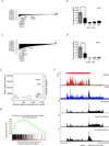
Figure 6. THZ531 inhibits DDR and transcription factor gene expression
a, DDR genes are sensitive to 50 nM THZ531. b, RT-qPCR of BRCA1 gene expression following THZ531 treatment. *p-value=9.42e-05, c, Transcription factor genes are sensitive to 200 nM THZ531. d, RT-qPCR of RUNX1 gene expression following THZ531 treatment. +p-value=5.33e-06. e, Total H3K27Ac ChIP-seq signal in enhancer regions for all stitched enhancers ranked by increasing H3K27Ac ChIP-seq signal. Gene names in gray either have no associated enhancer or associated typical enhancers. f, RUNX1 gene contains exceptional CDK12 and H3K27Ac signal and suffers profound loss of elongating Pol II following THZ531 treatment. CDK12 (blue), H3K27Ac (red) and Pol II (black) ChIP-seq occupancy at the promoter and across the gene body of RUNX1 locus following THZ531 treatment. The red bar indicates the genomic coordinates of a super –enhancer. g, Transcripts downregulated by 200 nM THZ531 are enriched for transcripts whose gene promoters contain high levels of CDK12. Gene set enrichment analysis of top 500 genes downregulated following THZ531 treatment in comparison to CDK12 promoter-bound ChIP-seq signal of these transcripts, GSEA-supplied p-value < 0.001. Heatmaps (a and c) display the log2 fold-change in gene expression vs. DMSO for the 14,745 transcripts expressed in Jurkat cells. RT qPCRs were performed in biological triplicate and error bars are +/− SD. P-values were determined with a two-tailed Student’s T test.
Scheme 1
Scheme 1
The synthesis of THZ531
Scheme 2
Scheme 2
The synthesis of BioTHZ531
Scheme 3
Scheme 3
The synthesis of THZ531R

Comment in

Similar articles

Cited by

References

    1. Young RA. RNA polymerase II. Annual review of biochemistry. 1991;60:689–715. - PubMed
    1. Buratowski S. Progression through the RNA polymerase II CTD cycle. Molecular cell. 2009;36:541–546. - PMC - PubMed
    1. Fuda NJ, Ardehali MB, Lis JT. Defining mechanisms that regulate RNA polymerase II transcription in vivo. Nature. 2009;461:186–192. - PMC - PubMed
    1. Munoz MJ, de la Mata M, Kornblihtt AR. The carboxy terminal domain of RNA polymerase II and alternative splicing. Trends in biochemical sciences. 2010;35:497–504. - PubMed
    1. Sanso M, Fisher RP. Pause, play, repeat: CDKs push RNAP II's buttons. Transcription. 2013;4:146–152. - PMC - PubMed

Methods-only References

    1. Kwiatkowski N, et al. Targeting transcription regulation in cancer with a covalent CDK7 inhibitor. Nature. 2014;511:616–620. - PMC - PubMed
    1. Bosken CA, et al. The structure and substrate specificity of human Cdk12/Cyclin K. Nature communications. 2014;5:3505. - PMC - PubMed
    1. Czudnochowski N, Bosken CA, Geyer M. Serine-7 but not serine-5 phosphorylation primes RNA polymerase II CTD for P-TEFb recognition. Nature communications. 2012;3:842. - PubMed
    1. Zhang T, et al. Discovery of potent and selective covalent inhibitors of JNK. Chemistry & biology. 2012;19:140–154. - PMC - PubMed
    1. Zhang Z, Marshall AGA. universal algorithm for fast and automated charge state deconvolution of electrospray mass-to-charge ratio spectra. Journal of the American Society for Mass Spectrometry. 1998;9:225–233. - PubMed

Publication types

MeSH terms