. 2016 May 23;12(5):e1006080.
doi: 10.1371/journal.pgen.1006080. eCollection 2016 May.

A Minimal Threshold of c-di-GMP Is Essential for Fruiting Body Formation and Sporulation in Myxococcus xanthus

Affiliations

A Minimal Threshold of c-di-GMP Is Essential for Fruiting Body Formation and Sporulation in Myxococcus xanthus

Dorota Skotnicka et al. PLoS Genet. .

Abstract

Generally, the second messenger bis-(3'-5')-cyclic dimeric GMP (c-di-GMP) regulates the switch between motile and sessile lifestyles in bacteria. Here, we show that c-di-GMP is an essential regulator of multicellular development in the social bacterium Myxococcus xanthus. In response to starvation, M. xanthus initiates a developmental program that culminates in formation of spore-filled fruiting bodies. We show that c-di-GMP accumulates at elevated levels during development and that this increase is essential for completion of development whereas excess c-di-GMP does not interfere with development. MXAN3735 (renamed DmxB) is identified as a diguanylate cyclase that only functions during development and is responsible for this increased c-di-GMP accumulation. DmxB synthesis is induced in response to starvation, thereby restricting DmxB activity to development. DmxB is essential for development and functions downstream of the Dif chemosensory system to stimulate exopolysaccharide accumulation by inducing transcription of a subset of the genes encoding proteins involved in exopolysaccharide synthesis. The developmental defects in the dmxB mutant are non-cell autonomous and rescued by co-development with a strain proficient in exopolysaccharide synthesis, suggesting reduced exopolysaccharide accumulation as the causative defect in this mutant. The NtrC-like transcriptional regulator EpsI/Nla24, which is required for exopolysaccharide accumulation, is identified as a c-di-GMP receptor, and thus a putative target for DmxB generated c-di-GMP. Because DmxB can be-at least partially-functionally replaced by a heterologous diguanylate cyclase, these results altogether suggest a model in which a minimum threshold level of c-di-GMP is essential for the successful completion of multicellular development in M. xanthus.


Conflict of interest statement

The authors have declared that no competing interests exist.

Figures

Fig 1
Fig 1. c-di-GMP accumulates at increased levels in developing and starving M. xanthus cells.
DK1622 WT cells were starved in MC7 buffer on a solid surface in submerged culture or in suspension. At the indicated time points the c-di-GMP levels were determined and correlated to protein concentration. The c-di-GMP level is shown as mean ± standard deviation (SD) calculated from three biological replicates. * p < 0.05, ** p < 0.001 in Student’s t-test in which samples from individual time points were compared to the relevant 0 hrs sample.
Fig 2
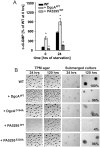
Fig 2. c-di-GMP level is important for development.
(A) c-di-GMP levels in cells expressing the indicated proteins during starvation in suspension. The c-di-GMP levels are shown as mean ± SD from three biological replicates relative to WT at 0 hrs. * p < 0.05 in Student’s t-test comparing different mutants to the WT at the respective time points. Note that the data for WT are the same as in Fig 1. (B) Fruiting body formation and sporulation under two different starvation conditions. Numbers after 120 hrs of starvation in submerged culture indicate heat- and sonication resistant spores formed after 120 hrs of starvation in submerged culture in percentage of WT (100%) from one representative experiment. Scale bars, TPM agar 500 μm, submerged culture 100 μm.
Fig 3
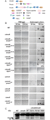
Fig 3. Complementation experiments with ΔdmxB, ΔpmxA and ΔtmoK mutants.
(A) Domain structure of DmxB, PmxA and TmoK. The primary sequences of the indicated proteins were analyzed for domain structure using [47]. (B) Fruiting body formation and sporulation under two different starvation conditions. Cells were treated and spores enumerated as described in Fig 2B. Scale bars: TPM agar 500 μm, submerged culture 100 μm. (C) Immunoblot detection of DmxB in total cell extracts. Total cell lysates from cells of the indicated genotypes were harvested from starvation agar at 24 hrs of development, separated by SDS-PAGE and probed with rabbit, polyclonal α-DmxB serum. Protein from the same calculated number of cells was loaded per lane. DmxB has a calculated molecular mass of 35.3 kDa. Molecular mass marker is indicated on the left. The non-specific band above the band corresponding to DmxB serves as an internal loading control.
Fig 4
Fig 4. In vitro assay for enzyme activity and c-di-GMP binding.
(A) DGC assay of DmxB variants. The indicated His6-tagged full-length DmxB variants were incubated with [α-32P]-GTP for the indicated periods of time followed by separation of nucleotides by TLC. Full-length DgcAWT was used as a positive control. GTP and c-di-GMP are indicated. The intermediate product indicated was described as a product formed during the DGC-dependent synthesis of c-di-GMP [49]. Numbers at the bottom indicate levels of c-di-GMP in % of the total signal in each lane. (B) PDE activity assay of PmxA. The indicated PmxA variants were incubated with [α-32P]-labeled c-di-GMP for the indicated periods of time followed by separation of nucleotides by TLC. pGpG and c-di-GMP are indicated. Numbers at the bottom indicate levels of pGpG in % of the total signal in each lane. (C) DRaCALA to detect c-di-GMP binding. The indicated full-length DmxB variants were incubated with [α-32P]-c-di-GMP with or without unlabeled c-di-GMP or GTP as competitors as indicated. 10 μl of the reaction mixtures were transferred to a nitrocellulose filter, dried and imaged.
Fig 5
Fig 5. c-di-GMP levels in cells of the indicated mutants during starvation.
(A, B) c-di-GMP levels in cells of the indicated genotypes were determined from three biological replicates as described in Fig 1. Note that the data for the WT are the same as in Fig 1 and in (B) the data for the WT and ΔdmxB strains are the same as in (A) and are shown again for the indicated time points for comparison. Note the different scale in (B) and for the ΔdmxB/dmxBR210A strain. * p < 0.05, ** p < 0.001 in Student’s t-test comparing the different mutants to the WT at the same time points.
Fig 6
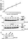
Fig 6. Determination of dmxB transcript and DmxB accumulation levels.
(A) qRT-PCR analysis of dmxB expression. Total RNA was isolated from WT developed in submerged culture at the indicated time points. dmxB transcript level is shown as the mean ± standard deviation from two biological replicates with each three technical replicates relative to WT at 0 hrs. (B) Immunoblot detection of DmxB in total cell extracts. Total cell lysates were prepared from cells of the indicated genotypes harvested from starvation agar plates at the indicated time points of development. Immunoblots were prepared as described in Fig 3C. Protein from the same calculated number of cells was loaded per lane. Molecular mass marker is indicated on the left. The non-specific band above the band corresponding to DmxB serves as an internal loading control. (C) Immunoblot detection of DmxB in total cell extract of WT, ΔdmxB and difE strains. Total cell lysates from cells harvested from starvation agar after 24 hrs of development were prepared. Immunoblots were prepared as described in Fig 3C. Protein from the same calculated number of cells was loaded per lane. Molecular mass marker is indicated on the left. The non-specific band above the band corresponding to DmxB serves as an internal loading control.
Fig 7
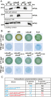
Fig 7. T4P formation and EPS accumulation in various mutants.
(A) Immunoblot detection of PilA in total cell extracts and in the sheared T4P fraction. In the upper and lower blots, total cell extracts were isolated from the indicated strains developed in submerged culture at the indicated time points. In the middle blot, T4P were sheared off from the same number of cells and concentrated by MgCl2 precipitation. In all three blots, protein from the same calculated number of cells was loaded per lane. The upper and middle blots were probed with α-PilA antibodies. The lower blot was probed against PilC, which is important for T4P assembly and served as a loading control. PilA and PilC have a calculated molecular mass 23.4 kDa and 45.2 kDa, respectively. Molecular mass marker is indicated on the left. (B) Quantification of EPS accumulation in dmxB mutants. 20 μl aliquots of exponentially growing cells of the indicated genotypes were spotted at a density of 7 × 10⁹ cells/ml on 1.5% agar supplemented with 0.5% CTT and 20 μg/ml trypan blue or on TPM starvation agar supplemented with 20 μg/ml trypan blue and incubated at 32°C for 24 hrs. (C) Extracellular complementation assay of ΔdmxB mutant. Cells of the indicated genotypes were either developed alone as described in Fig 2B in submerged culture or mixed at a 1:1 ratio and co-developed in submerged culture. Sporulation levels are enumerated after 120 hrs of starvation as the number of germinating heat- and sonication resistant spores relative to WT (100%).
Fig 8
Fig 8. c-di-GMP regulates epsABD transcription.
Total RNA was isolated at the indicated time points from cells of WT (closed circles) and the ΔdmxB mutant (open squares) developed in submerged culture. Transcript levels are shown as mean ± standard deviation from two biological replicates with each three technical replicates relative to WT at 0 hrs.
Fig 9
Fig 9. Transcriptional regulator EpsI/Nla24 binds c-di-GMP.
(A) Pull-down experiment with the soluble fraction of E. coli cell lysates before and after induction of EpsI/Nla24 synthesis using biotinylated c-di-GMP immobilized on streptavidin magnetic beads. Cell extract from the uninduced sample was used as a negative control. Pulled-down protein is indicated with an arrow. EpsI/Nla24-His6 has a calculated molecular mass of 50 kDa. (B) SPR sensorgrams and resulting affinity fit data for EpsI/Nla24 binding to biotinylated c-di-GMP. Upper panel, sensorgrams of EpsI/Nla24 binding to biotinylated c-di-GMP immobilized on sensor chip. The concentration of EpsI/Nla24 ranged from 62.5 nM (lowest curve) to 4 μM (highest curve) and concentration replicates were included as appropriate. The protein binding and dissociation phases for all sensorgrams are shown. Lower panel, affinity fit of EpsI/Nla24 binding to biotinylated c-di-GMP. For this fit, binding responses were measured 4 sec before the end of the injection.

Similar articles

Cited by

References

    1. Römling U, Galperin MY, Gomelsky M. Cyclic di-GMP: the first 25 years of a universal bacterial second messenger. Microbiol Mol Biol Rev. 2013; 77: 1–52. 10.1128/MMBR.00043-12 - DOI - PMC - PubMed
    1. Krasteva PV, Giglio KM, Sondermann H. Sensing the messenger: The diverse ways that bacteria signal through c-di-GMP. Protein Sci. 2012; 21: 929–948. 10.1002/pro.2093 - DOI - PMC - PubMed
    1. Boyd CD, O'Toole GA. Second messenger regulation of biofilm formation: Breakthroughs in understanding c-di-GMP effector systems. Annu Rev Cell Dev Biol 2012; 28: 439–462. 10.1146/annurev-cellbio-101011-155705 - DOI - PMC - PubMed
    1. Neunuebel MR, Golden JW. The Anabaena sp. strain PCC 7120 gene all2874 encodes a diguanylate cyclase and is required for normal heterocyst development under high-light growth conditions. J Bacteriol. 2008; 190: 6829–6836. 10.1128/JB.00701-08 - DOI - PMC - PubMed
    1. Tschowri N, Schumacher MA, Schlimpert S, Chinnam NB, Findlay KC, Brennan RG, et al. Tetrameric c-di-GMP mediates effective transcription factor dimerization to control Streptomyces development. Cell. 2014; 158: 1136–1147. 10.1016/j.cell.2014.07.022 - DOI - PMC - PubMed

Publication types

MeSH terms

Grants and funding

This work was funded by: German Research Council within the framework of the Collaborative Research Center SFB987 “Microbial Diversity in Environmental Signal Response” (www.dfg.de/en/): to LSA; Max Planck Society (http://www.mpg.de/en): to LSA; and National Science Foundation grant MCB-1024989 (http://www.nsf.gov/): to MS. The funders had no role in study design, data collection and analysis, decision to publish, or preparation of the manuscript.