Review
. 2016 Sep;22(6):463-93.
doi: 10.1111/odi.12446. Epub 2016 Apr 14.

Vitamin C: the known and the unknown and Goldilocks

Affiliations
Review

Vitamin C: the known and the unknown and Goldilocks

S J Padayatty et al. Oral Dis. 2016 Sep.

Abstract

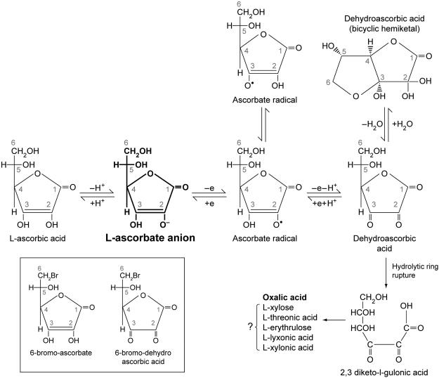
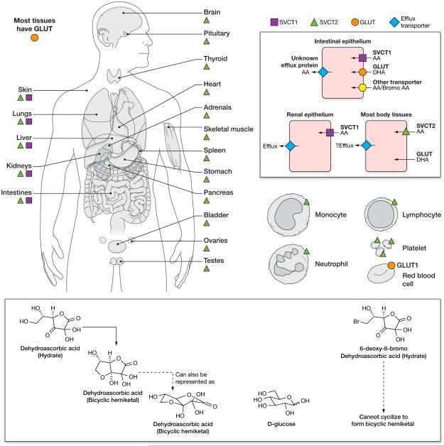
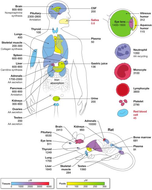
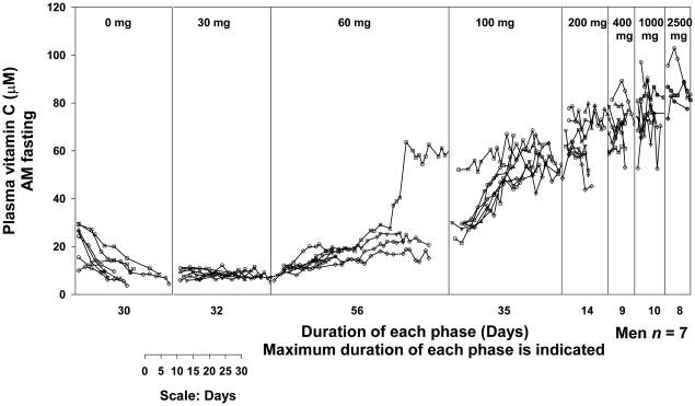
Vitamin C (Ascorbic Acid), the antiscorbutic vitamin, cannot be synthesized by humans and other primates, and has to be obtained from diet. Ascorbic acid is an electron donor and acts as a cofactor for fifteen mammalian enzymes. Two sodium-dependent transporters are specific for ascorbic acid, and its oxidation product dehydroascorbic acid is transported by glucose transporters. Ascorbic acid is differentially accumulated by most tissues and body fluids. Plasma and tissue vitamin C concentrations are dependent on amount consumed, bioavailability, renal excretion, and utilization. To be biologically meaningful or to be clinically relevant, in vitro and in vivo studies of vitamin C actions have to take into account physiologic concentrations of the vitamin. In this paper, we review vitamin C physiology; the many phenomena involving vitamin C where new knowledge has accrued or where understanding remains limited; raise questions about the vitamin that remain to be answered; and explore lines of investigations that are likely to be fruitful.

Keywords: dehydroascorbic acid; dose-concentration relationship; recommended dietary allowance; scurvy; vitamin C; vitamin C transport.


Figures

Figure 1
Figure 1. Ascorbic acid metabolism
Ascorbic acid metabolism and halogenated analogues of ascorbic acid. Vitamin C (ascorbic acid) under physiological conditions is >99% in the form of ascorbate anion (shown in bold)(Beuttner & Schafer, 2004). It can sequentially donate two electrons from the double bond between carbons two and three. Loss of the first electron (oxidation) produces the free radical ascorbate radical (semidehydroascorbic acid). Some reactive free radicals produced by biological processes can be harmful because of their highly reactive nature. These can be reduced by ascorbic acid but in the process ascorbic acid is itself is converted (oxidized) into ascorbate radical. Ascorbate radical has a half-life of 10−3 seconds to several minutes, depending on the presence of oxygen and metals. Under physiologic conditions, ascorbate radical is comparatively unreactive compared to other free radicals. Ascorbate radical can be reduced back to vitamin C. Alternately, it can lose a second electron (oxidized) to form dehydroascorbic acid. Thus, because vitamin C loses electrons, it acts as an antioxidant or free radical scavenger (Buettner, 1993). Dehydroascorbic acid is also unstable with a half-life of several minutes (Lewin, 1976). While dehydroascorbic acid exists in different forms, the dominant form in vivo is likely to be a hydrated hemiketal (Lewin, 1976, Corpe et al., 2005). Dehydroascorbic acid undergoes hydrolysis, with irreversible ring rupture to form 2, 3–diketogulonic acid, whose metabolic products include oxalate, threonate, and possibly xylose, xylonic acid, and lynxonic acid (Lewin, 1976). Oxalic acid is the clinically significant metabolic product in humans (bold). In some animals, products of vitamin C catabolism may enter the pentose phosphate pathway or other pathways of carbohydrate metabolism (Banhegyi & Loewus, 2004). Dehydroascorbic acid may be reduced back sequentially to ascorbate radical and ascorbic acid by glutathione or directly to ascorbic acid by enzyme dependent mechanisms (Linster & Van Schaftingen, 2007). Under suitable conditions (millimolar concentrations of ascorbic acid, presence of metal ions), hydrogen peroxide may be formed. Analogues of vitamin C have been synthesized by replacing the OH group at carbon 6 with bromine or iodine. These halogenated analogues of vitamin C (shown in box) can be oxidized to bromo dehydroascorbic acid and iodo dehydroascorbic acid respectively. However, oxidized halogen ascorbate analogues cannot form a cyclized hydrated hemiketal (Fig. 2). Note that many of the reactions above have been shown only in vitro or in animals and not under physiological conditions in vivo or in humans. From (Washko et al., 1992). Modified and reproduced with permission of Analytical Biochemistry.
Figure 2
Figure 2. Vitamin C transporters
Distribution of vitamin C transporters in human tissues. Sodium Vitamin C Transporters (SVCTs) are mainly responsible for vitamin C transport into cells in humans and other mammals. SVCT1 is primarily expressed in absorptive tissues, including the intestinal epithelium and the proximal convoluted tubules and the descending part of the loop of Henle in the kidney. SVCT 1 is also expressed in liver. SVCT2 is expressed in most body tissues, as are Glucose Transporters (GLUTs). SVCT 1 and 2 transport ascorbic acid but not dehydroascorbic acid into cells. GLUTs 1, 2, 3, 4 and 8 (but not other GLUTS) (Corpe et al., 2013, Rumsey et al., 2000a, Rumsey et al., 1998, Burzle & Hediger, 2012), transport dehydroascorbic acid but not ascorbic acid into cells. Some GLUTs have a higher affinity for DHA than for glucose. The transporter responsible for exporting ascorbic acid from cells into the extracellular fluid or plasma has not been identified. The only ascorbate containing cell known to lack an SVCT is the mature red blood cell. The red blood cell obtains its ascorbate by transporting dehydroascorbic acid and immediately reducing it internally. Dehydroascorbic acid is transported into human red blood cells by GLUT1, and into mouse red blood cells by GLUT 3 and/or 4 (Tu et al., 2015). D-Glucose closely resembles the ring form (hydrated hemiketal form) of dehydroascorbic acid, which is likely to account for its transport by some GLUTs. When bromo ascorbic acid is oxidized to form bromo dehydroascorbic acid, this compound is not transported by GLUTs. SVCT distribution was inferred by the presence of specific mRNA for SVCT1 and 2, and in some cases by antibodies. Figure based on data from: (Tsukaguchi et al., 1999, Savini et al., 2008) (Daruwala et al., 1999, Wang et al., 1999, Wang et al., 2000).
Figure 3
Figure 3. Vitamin C concentrations in human and rat tissues and fluids
Concentration of vitamin C in body tissues and body fluids are shown in µM. Ascorbic acid concentrations are much higher in tissues than in plasma. Tissues and body fluids are shown in separate concentration dependent color schemes. Organs that are uncolored (white) indicate that data on vitamin C concentrations are unavailable. For clarity, some organs shown are slightly displaced from their correct anatomical positions. Vitamin C concentrations shown are derived from published reports. Values for white blood cells and urinary vitamin C concentrations were obtained from depletion repletion studies in young healthy women, at the study phase when they were at steady state for vitamin C intake of 100 mg per day (Levine et al., 2001b), which is close to the recommended dietary allowance for vitamin C. Note that the red blood cell is the only cell with internal concentrations of vitamin C that are below those in plasma (values less than plasma are shown in red). Of body fluids, only saliva has a vitamin C concentration lower than that of plasma. Putative functions of vitamin C as a cofactor for enzymes are indicated, as are selected clinical or experimental observations of phenomenon involving vitamin C. Values shown should be considered approximate as they were obtained from tissues collected under non uniform conditions including clinical and post mortem samples. Some vitamin C concentrations shown are the mean values of results from two or more studies. Further, the dietary intake or plasma concentrations of vitamin C were not known in many cases and the methods used in sample collection and storage, and for vitamin C assays, varied widely. Values may also vary with age and possibly in disease states. Data obtained from (Hornig, 1975, Voigt et al., 2002)(Dostalova, 1982, Koliakos et al., 2002, Vobecky et al., 1982, Evans et al., 1982, Omaye et al., 1987, Berger et al., 1996, Taylor et al., 1997, Rathbone et al., 1989, Sobala et al., 1989, Levine et al., 2001b, Jacob et al., 1987). Reported salivary vitamin C concentrations varied widely. Salivary vitamin C concentration shown (median 0.6 µM, range 0.2-19 µM) in nonsmokers was measured by HPLC. Corresponding median plasma vitamin C concentration was 53 µM (range 16 µM-89 µM)(Schock et al., 2004). Note that tissue vitamin C concentrations shown for the rat are much higher than for humans. Explanations are that fresh tissue samples can be obtained from animals (while many human tissue samples were obtained post mortem), or that there are true species differences. Compared to other tissues, AA concentrations in the rat pituitary and adrenal glands are higher, and therefore they are shown in arbitrary colors (and not according to the concentration dependent color scale) (Hornig, 1975). Vitamin C concentration data for mice are similar to that of the rat (Sotiriou et al., 2002, Corpe et al., 2010).
Figure 4
Figure 4. Vitamin C dose concentration relationship
Vitamin C concentrations in plasma and circulating cells were studied in young healthy men (Levine et al., 1996b) and women (Levine et al., 2001b) each of whom were given six to seven different doses of the vitamin in two depletion-repletion studies. Seven healthy men (4A) and fifteen healthy women (4B), all nonsmokers, age 19-27 years were studied as inpatients. To decrease hospitalization time, outpatient subjects prior to admission were instructed to consume a diet containing < 60 mg of vitamin C. When inpatients, throughout hospitalization they consumed a defined diet containing less than 5 mg of vitamin C daily (King et al., 1997). Deficiencies of other nutrients were prevented by supplementation. When plasma vitamin C concentrations achieved nadir of <10 µM, vitamin C in solution was administered at 15 mg orally in the fasted state twice daily (30 mg total per day) until steady state for the dose was achieved. Vitamin C dose was increased to 30 mg twice daily (60 mg total per day) until steady state was achieved for this dose. In this way subjects received the following doses in mg/day: 30, 60, 100, 200, 400, 1000, and 2500. Vitamin C was measured by HPLC with coulometric electrochemical detection. Doses are indicated at the top of the figure. Each symbol represents a different subject. There is a one-day gap between all doses for bioavailability sampling. Each vertical line represents the start of a new dose. Some subjects reached steady state concentrations earlier than others; the duration shown is longest time taken by a subject to reach steady state. Two subjects were studied at one time and the graphs show data collated at the end of all studies. Modified and reproduced with permission from Biofactors (Levine et al., 2001a) and The Proceedings of the National Academy of Sciences (Levine et al., 2001b).
Figure 5
Figure 5. Ascorbate flux in humans
The relationship between the daily intake of known doses of vitamin C and its absorption, distribution and excretion are shown for four different daily intakes of vitamin C. Data were obtained from studies in seven healthy young men (Levine et al., 1996b) detailed in Fig 4. Ascorbate flux is shown in four panels. The upper part of each panel shows the relationship between an oral dose of vitamin C and: bioavailability; amount absorbed by the gastrointestinal tract; peak plasma concentrations reached; and the amount of the vitamin excreted in urine. Bioavailability studies were performed when patients were at steady-state for that dose (with some modifications, see below). Following each bioavailability study, subjects were given twice daily doses of the vitamin until they reached steady state for that dose. The lower part of each panel shows the steady-state condition for a specific dose. The panels for 50 and 100 mg doses can be taken as examples. When subjects were at steady-state for vitamin C intake of 50 mg twice daily (100mg total daily dose), fasting plasma vitamin C concentrations were 56µM, and neutrophil and lymphocyte vitamin C concentrations were 1250 µM and 3100 µM respectively. At this stage, bioavailability was studied in part by using a test dose of 100mg by mouth (labelled “test dose”). Urine excretion data (25 mg) are shown for this oral dose. Not shown are data for the same dose administered intravenously, to permit bioavailability to be calculated. Bioavailability for the 100 mg test dose was 80%: 80 mg of vitamin C was absorbed, and the resultant peak plasma vitamin C concentration attained was 78 µM. 25 mg of vitamin C was excreted in the urine in the following 24 hours. The upper part of each of the four panels shows bioavailability doses of 15, 50, 100 or 500 mg. The lower part of the four panels show steady state for vitamin C intake of 30 mg/day, 100 mg/day (close to the recommended dietary allowance), 200 mg/day (the approximate amount provided by five servings of fruits and vegetables per day), or 1000mg/day (a dose that is used as a dietary supplement). Data shown are mean values for all patients studied. Amount absorbed was calculated from bioavailability data (Graumlich et al., 1997, Levine et al., 1996b). Separate color schemes, for which the intensity of color is related to the amount or concentration of vitamin C, are used for test doses; amounts absorbed and excreted in the urine; and for fasting steady state intracellular vitamin C concentrations. Bioavailability studies were performed as follows: bioavailability for 15 mg dose at the end of depletion phase, for 50 mg when the subjects were at steady state for 60 mg, for 100 mg when the subjects were at steady state for 100 mg, and for 500 mg when the subjects were at steady state for 400 mg. Note that the end of the depletion phase was not a true steady-state. Mean fasting plasma vitamin C concentration at the end of depletion was 7.62 +/− 1.64 µM. Fasting steady state plasma vitamin C concentrations at the time of oral bioavailability tests were: for 50 mg dose, 24.8 µM +/− 14.1; for 100 mg dose, 56 µM +/− 4.5; and for 500 mg dose, 70 µM +/− 6.9. Each subject received a total of seven different doses of vitamin C but only data from four bioavailability studies and for four steady state conditions are shown. Details of methods used were previously published (King et al., 1997, Graumlich et al., 1997, Levine et al., 1996b, Levine et al., 2001b).
Figure 6
Figure 6. Ascorbic acid recycling in Human Neutrophils
Ascorbic acid (AA) and dehydroascorbic acid (DHA) transport and recycling in human neutrophils in vitro (Stankova et al., 1975) (Bigley & Stankova, 1974) (Hemila et al., 1984) (Anderson & Lukey, 1987, Wang et al., 1997). When normal human neutrophils in vitro were activated by pathogens (E coli, Enterococcus faecalis, Moraxella catarrhlis, Klebsiella oxytoca, Acinetobacter baumanii or C. albicans) (Wang et al., 1997), neutrophils accumulated ascorbic acid. Intracellular vitamin C concentrations increased from ~ 1.3mM to 8mM, and vitamin C in the culture media decreased to undetectable concentrations from 50µM. These findings did not occur when neutrophils were used from patients with Chronic Granulomatous Disease (Wang et al., 1997). Patients with this condition are unable to make superoxide, and have dysfunctional neutrophils that cannot kill certain pathogens. The observed in vitro phenomena can be accounted for by the proposed model described below. Normal human neutrophils maintain internal vitamin C concentrations of about ~ 1.3mM by uptake of AA by sodium-dependent vitamin C transporter 2 (SVCT2). Activated neutrophils secrete reactive oxygen species because membrane associated NADPH oxidase is able to transfer electrons across the cell membrane, using NADPH in the process. H2O2 is formed, which is a precursor for production of reactive oxygen species (ROS and RNOS). These oxidize extracellular AA to DHA. Neutrophil activation with oxidant formation and its consequent results are shown in red. DHA is rapidly transported into the neutrophil by glucose transporters, probably GLUT1 and GLUT3, and immediately reduced to AA by glutaredoxin, producing a 4 to10-fold increase in neutrophil internal AA concentration. When bacteria are engulfed by the neutrophil, a phagolysosome is formed. Myeloperoxidase containing vesicles fuse with the phagolysosome, such that the oxidant generating reactions occur in a sequestered space. Although superoxide cannot cross the plasma membrane, H2O2 can readily diffuse into the cell. Oxidants generated by H2O2 can be reduced by internal AA. Glutathione (GSH), used by glutaredoxin during DHA reduction, is regenerated from glutathione disulfide (GSSG) by glutathione reductase (GRD) and NADPH. NADPH essential for these reactions is derived predominantly from the pentose phosphate pathway. Two molecules of NADPH are produced by the oxidative phase of pentose phosphate pathway (shown in green) when glucose 6 phosphate is converted to ribulose 6 phosphate (shown in green). Ribulose 6 phosphate undergoes further metabolism in the non-oxidative phase of the pentose phosphate pathway. As NADPH is oxidized to NADP, electrons are transferred to GRD so it can reduce GSSG to GSH. Modified and reproduced from (Rumsey & Levine, 1998), with permission of the Journal of Nutritional Biochemistry.
Figure 7
Figure 7. Vitamin C secretion by human adrenal glands
Vitamin C concentrations were measured in the adrenal and peripheral veins of 26 patients with primary hyperaldosteronism. Under radiographic guidance, catheters were placed in both adrenal veins, and blood samples were taken after stimulation with adrenocorticotrophic hormone (ACTH). Vitamin C concentrations in each of the adrenal (n = 47) and peripheral (n = 26) veins sampled are shown. In 5 patients, blood samples were obtained from only one adrenal vein because of unusual venous anatomy or difficulties with adrenal vein catheterization. In the adrenal veins, peak vitamin C concentrations (Mean ± SD: 176 ± 71 µmol/L) were reached between 1 and 4 min, and were significantly (P < 0.0001, paired t test) higher than corresponding peripheral plasma vitamin C concentrations (35 ± 15 µmol/L). In patients in whom adrenal vein vitamin C concentration could be measured in only one adrenal gland, that single value was used in the calculation. In patients in whom both adrenals were successfully sampled, the mean of the two adrenal vein vitamin C concentrations was used for calculation, but all values are shown. Modified and reproduced from (Padayatty et al., 2007), with permission from American Journal of Clinical Nutrition.
Figure 8
Figure 8. Ascorbate radical formation in vivo
Concentrations of ascorbate, ascorbate radical (Asc• −) (radical is denoted by a superscript dot) and H2O2 in blood and extracellular space, and proposed mechanisms that result in measurable Asc• − and H2O2 in extracellular fluid but not in blood. Whether given by oral or parenteral routes, ascorbate rapidly equilibrates between blood and extracellular fluid, possibly through intercellular junctions. When pharmacological doses are administered parenterally, blood and extracellular fluid ascorbate concentrations reached 10 to 20 mM. Concentrations of Asc• − in blood reached 10-30 nM but H2O2 was not detectable in blood. Asc• − and H2O2 reached concentrations of 250-300 nM and 150 µM respectively in extracellular fluid. A proposed scheme that accounts for these observations are as follows: In extracellular fluid, ascorbate loses one electron to form Asc• − (solid lines). The electron reduces a protein-centered metal, for example Fe3+ to Fe2+. Candidate proteins may be either in extracellular fluid, or on cell membranes facing outward. Fe2+ donates an electron to oxygen, forming superoxide (O2 • −) which undergoes dismutation to form H2O2 (Qian & Buettner, 1999). In blood, these reactions are damped or inhibited (dashed lines), or the resultant products diffuse into red blood cells where they are rapidly extinguished/reduced. In blood, the formation of Asc• − may be inhibited by red blood cell membrane bound reducing proteins (May et al., 2001) and/or by large plasma proteins that do not distribute into the extracellular space. H2O2 that is formed in blood will be immediately destroyed by plasma catalase and red blood cell GSH peroxidase, so that no H2O2 is detectable (Chen et al., 2007, Gaetani et al., 1996, Johnson et al., 2005) (Chen et al., 2005). The identities of the metal-centered proteins are not known. H2O2 that diffuses into the blood or tissues from extracellular fluid is inactivated by reducing substances in these compartments. Ascorbate was measured by HPLC, Asc• − by Electron Paramagnetic Resonance (EPR) and H2O2 by fluorescence (Chen et al., 2007). The values shown are approximations from measurements in animals given ascorbate by the intra peritoneal or intravenous routes (Chen et al., 2007)(Chen et al., 2005, Chen et al., 2008). Modified and reproduced from (Chen et al., 2007), with permission from the National Academy of Sciences.

Similar articles

Cited by

References

    1. Aditi A, Graham DY. Vitamin C, gastritis, and gastric disease: a historical review and update. Dig Dis Sci. 2012;57:2504–15. - PMC - PubMed
    1. Agus DB, Gambhir SS, Pardridge WM, Spielholz C, Baselga J, Vera JC, Golde DW. Vitamin C crosses the blood-brain barrier in the oxidized form through the glucose transporters. J Clin Invest. 1997;100:2842–8. - PMC - PubMed
    1. Amir Shaghaghi M, Bernstein CN, Serrano Leon A, El-Gabalawy H, Eck P. Polymorphisms in the sodium-dependent ascorbate transporter gene SLC23A1 are associated with susceptibility to Crohn disease. Am J Clin Nutr. 2014;99:378–83. - PubMed
    1. Anderson R, Lukey PT. A biological role for ascorbate in the selective neutralization of extracellular phagocyte-derived oxidants. Ann N Y Acad Sci. 1987;498:229–47. - PubMed
    1. Anonymous Ascorbic acid intake and salivary ascorbate levels. Nutr Rev. 1986;44:328–30. - PubMed