Advances in diagnosis and management of celiac disease
- PMID: 25662623
- PMCID: PMC4409570
- DOI: 10.1053/j.gastro.2015.01.044
Advances in diagnosis and management of celiac disease
Abstract
Celiac disease is an autoimmune disorder that is induced by dietary gluten in genetically predisposed individuals. It has a prevalence of approximately 1% in many populations worldwide. New diagnoses have increased substantially, owing to increased awareness, better diagnostic tools, and probable real increases in incidence. The breadth of recognized clinical presentations continues to expand, making the disorder highly relevant to all physicians. Newer diagnostic tools, including serologic tests for antibodies against tissue transglutaminase and deamidated gliadin peptide, greatly facilitate diagnosis. Tests for celiac-permissive HLA-DQ2 and HLA-DQ8 molecules are useful in defined clinical situations. Celiac disease is diagnosed by histopathologic examination of duodenal biopsy specimens. However, according to recent controversial guidelines, a diagnosis can be made without a biopsy in certain circumstances, especially in children. Symptoms, mortality, and risk for malignancy each can be reduced by adherence to a gluten-free diet. This treatment is a challenge, however, because the diet is expensive, socially isolating, and not always effective in controlling symptoms or intestinal damage. Hence, there is increasing interest in developing nondietary therapies.
Keywords: Autoimmune; Cereal; Diet; Enteritis; Enteropathy; Gluten; Gluten-Free Diet; Lymphoma; Malabsorption; Nutritional Deficiency; Refractory; Serology; Villous Atrophy; Wheat.
Copyright © 2015 AGA Institute. Published by Elsevier Inc. All rights reserved.
Figures

Serologic markers of celiac disease: IgA against tTG, Endomysial antibody (IgA), IgG against DGD, IgA against deamidated gliadin peptide, IgG against tTG.
A small number of patients with celiac disease have negative results from serologic tests. Biopsies should therefore be performed if the clinical suspicion for celiac disease is high, regardless of these results.
Tests for HLA DQ2 and DQ8 can be performed. Negative results mean that celiac disease can be permanently excluded. However, many individuals without celiac disease are carriers of these alleles—especially those with a family history of celiac disease or related autoimmune disorder.
For symptomless patients, especially children, with mild increases in serologic markers of disease, biopsy analysis can be delayed, pending results from serologic tests performed at intervals of 3–6 months.
Potential celiac disease has been defined as a normal small intestinal mucosa with an increased risk for celiac disease based on results from serologic analysis.

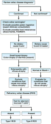
Confirm the diagnosis of celiac disease by reviewing findings from serologic tests (not anti-gliadin antibody tests) and small bowel histology findings. If patients tested negative for tTG and EMA antibodies, perform HLA DQ2 DQ8 typing.
Investigate other possible etiologies for clinical presentation and/or abnormal histology findings.
Increased serum levels of IgA against tTG indciate continued gluten ingestion as a cause
Non-celiac villous atrophy can be caused by intestinal infections (eg, giardiasis, small intestinal bacterial overgrowth, and viral enteritis, including HIV enteropathy), autoimmune enteropathy, hypogammaglobulinemia, as well as combined variable immunodeficiency, tropical sprue, Crohn' s disease, peptic duodenitis, or collagenous sprue.
Conditions that present as NRCD without villous atrophy include irritable bowel syndrome, microscopic colitis, food intolerances, small intestinal bacterial overgrowth, Crohn's disease, and microscopic colitis.
Aberrant small intestinal mucosal and intraepithelial lymphocytes in patients with RCD Type II can be identified by immunohistochemistry or flow cytometry (an excess of CD3+ cells without CD4 or CD8 surface proteins) or by T-cell receptor gene rearrangement analysis showing clonal expansion.
Similar articles
-
[Adult Celiac Disease].Praxis (Bern 1994). 2016 Jul 6;105(14):803-10; quiz 809-12. doi: 10.1024/1661-8157/a002413. Praxis (Bern 1994). 2016. PMID: 27381303 Review. German.
-
Biomarkers for diagnosis and monitoring of celiac disease.J Clin Gastroenterol. 2013 Apr;47(4):308-13. doi: 10.1097/MCG.0b013e31827874e3. J Clin Gastroenterol. 2013. PMID: 23388848 Review.
-
[CELIAC DISEASE].Rev Prat. 2015 Dec;65(10):1299-304. Rev Prat. 2015. PMID: 26979028 French.
-
Celiac disease in children.Clin Res Hepatol Gastroenterol. 2015 Oct;39(5):544-51. doi: 10.1016/j.clinre.2015.05.024. Epub 2015 Jul 15. Clin Res Hepatol Gastroenterol. 2015. PMID: 26186878 Review.
-
Celiac disease: from pathogenesis to novel therapies.Gastroenterology. 2009 Dec;137(6):1912-33. doi: 10.1053/j.gastro.2009.09.008. Epub 2009 Sep 18. Gastroenterology. 2009. PMID: 19766641 Review.
Cited by
-
Gluten Free Diet for the Management of Non Celiac Diseases: The Two Sides of the Coin.Healthcare (Basel). 2020 Oct 14;8(4):400. doi: 10.3390/healthcare8040400. Healthcare (Basel). 2020. PMID: 33066519 Free PMC article. Review.
-
Gluten Detection Methods and Their Critical Role in Assuring Safe Diets for Celiac Patients.Nutrients. 2019 Dec 2;11(12):2920. doi: 10.3390/nu11122920. Nutrients. 2019. PMID: 31810336 Free PMC article. Review.
-
Is duodenal biopsy always necessary for the diagnosis of coeliac disease in adult patients with high anti-tissue transglutaminase (TTG) antibody titres?Frontline Gastroenterol. 2021 Jun 25;13(4):287-294. doi: 10.1136/flgastro-2020-101728. eCollection 2022. Frontline Gastroenterol. 2021. PMID: 35722610 Free PMC article.
-
Effect of a Gluten-Free Diet on Whole Gut Transit Time in Celiac Disease (CD) and Non-Celiac Gluten Sensitivity (NCGS) Patients: A Study Using the Wireless Motility Capsule (WMC).J Clin Med. 2024 Mar 16;13(6):1716. doi: 10.3390/jcm13061716. J Clin Med. 2024. PMID: 38541941 Free PMC article.
-
Meta-Analysis on Associations of RGS1 and IL12A Polymorphisms with Celiac Disease Risk.Int J Mol Sci. 2016 Mar 30;17(4):457. doi: 10.3390/ijms17040457. Int J Mol Sci. 2016. PMID: 27043536 Free PMC article.
References
-
- Rostom A, Murray JA, Kagnoff MF. American Gastroenterological Association (AGA) Institute technical review on the diagnosis and management of celiac disease. Gastroenterology. 2006;131:1981–2002. - PubMed
-
- Green PH, Cellier C. Celiac disease. N Engl J Med. 2007;357:1731–43. - PubMed
-
- Lionetti E, Castellaneta S, Francavilla R, et al. Introduction of gluten, HLA status, and the risk of celiac disease in children. N Engl J Med. 2014;371:1295–303. - PubMed
-
- Vriezinga SL, Auricchio R, Bravi E, et al. Randomized feeding intervention in infants at high risk for celiac disease. N Engl J Med. 2014;371:1304–15. - PubMed
Publication types
MeSH terms
Substances
Grants and funding
LinkOut - more resources
Full Text Sources
Other Literature Sources
Medical
Research Materials

