Cerebrotendinous xanthomatosis: a comprehensive review of pathogenesis, clinical manifestations, diagnosis, and management
- PMID: 25424010
- PMCID: PMC4264335
- DOI: 10.1186/s13023-014-0179-4
Cerebrotendinous xanthomatosis: a comprehensive review of pathogenesis, clinical manifestations, diagnosis, and management
Abstract
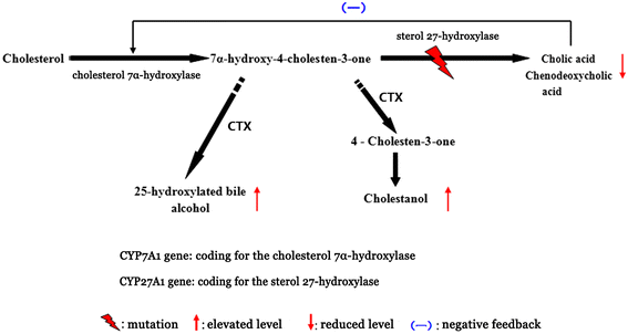
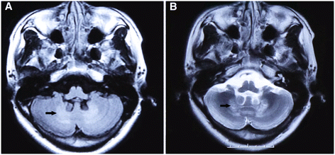
Cerebrotendinous xanthomatosis (CTX) OMIM#213700 is a rare autosomal-recessive lipid storage disease caused by mutations in the CYP27A1 gene; this gene codes for the mitochondrial enzyme sterol 27-hydroxylase, which is involved in bile acid synthesis. The CYP27A1 gene is located on chromosome 2q33-qter and contains nine exons. A CYP27A1 mutation leads to decreased synthesis of bile acid, excess production of cholestanol, and consequent accumulation of cholestanol in tissues. Currently there is no consensus on the prevalence of CTX, one estimate being <5/100,000 worldwide. The prevalence of CTX due to the CYP27A1 mutation R362C alone is approximately 1/50,000 in Caucasians. Patients with CTX have an average age of 35 years at the time of diagnosis and a diagnostic delay of 16 years. Clinical signs and symptoms include adult-onset progressive neurological dysfunction (i.e., ataxia, dystonia, dementia, epilepsy, psychiatric disorders,peripheral neuropathy, and myopathy) and premature non-neurologic manifestations (i.e., tendon xanthomas, childhood-onset cataracts, infantile-onset diarrhea, premature atherosclerosis, osteoporosis, and respiratory insufficiency). Juvenile cataracts, progressive neurologic dysfunction, and mild pulmonary insufficiency are unique symptoms that distinguish CTX from other lipid storage disorders including familial dysbetalipoproteinemia, homozygous familial hypercholesterolemia, and sitosterolemia, all of which might also present with xanthomas and cardiovascular diseases. Brain magnetic resonance imaging (MRI) shows bilateral lesions in the dentate nucleus of the cerebellum and mild white matter lesions. The classical symptoms and signs, namely elevated levels of cholestanol and bile alcohols in serum and urine, brain MRI, and the mutation in the CYP27A1 gene confirm the diagnosis of CTX. Early diagnosis and long-term treatment with chenodeoxycholic acid (750 mg/d) improve neurological symptoms and contribute to a better prognosis.
Figures
Similar articles
-
Cerebrotendinous Xanthomatosis: Molecular Pathogenesis, Clinical Spectrum, Diagnosis, and Disease-Modifying Treatments.J Atheroscler Thromb. 2021 Sep 1;28(9):905-925. doi: 10.5551/jat.RV17055. Epub 2021 May 8. J Atheroscler Thromb. 2021. PMID: 33967188 Free PMC article.
-
[Pathophysiology of cerebrotendinous xanthomatosis].Rinsho Shinkeigaku. 2016 Dec 28;56(12):821-826. doi: 10.5692/clinicalneurol.cn-000962. Epub 2016 Nov 12. Rinsho Shinkeigaku. 2016. PMID: 27840382 Review. Japanese.
-
[Cerebrotendinous xanthomatosis: physiopathology, clinical manifestations and genetics].Rev Med Chil. 2014 May;142(5):616-22. doi: 10.4067/S0034-98872014000500010. Rev Med Chil. 2014. PMID: 25427019 Review. Spanish.
-
Cerebrotendinous xanthomatosis (CTX): a treatable lipid storage disease.Pediatr Endocrinol Rev. 2009 Sep;7(1):6-11. Pediatr Endocrinol Rev. 2009. PMID: 19696711 Review.
-
A novel frameshift mutation in the sterol 27-hydroxylase gene in an Egyptian family with cerebrotendinous xanthomatosis without cataract.Metab Brain Dis. 2017 Apr;32(2):311-315. doi: 10.1007/s11011-017-9971-x. Epub 2017 Feb 22. Metab Brain Dis. 2017. PMID: 28229379
Cited by
-
Evaluation of the effect of chenodeoxycholic acid treatment on skeletal system findings in patients with cerebrotendinous xanthomatosis.Turk Pediatri Ars. 2019 Jul 11;54(2):113-118. doi: 10.14744/TurkPediatriArs.2019.23281. eCollection 2019. Turk Pediatri Ars. 2019. PMID: 31384146 Free PMC article.
-
Common genetic etiology between "multiple sclerosis-like" single-gene disorders and familial multiple sclerosis.Hum Genet. 2017 Jun;136(6):705-714. doi: 10.1007/s00439-017-1784-9. Epub 2017 Mar 23. Hum Genet. 2017. PMID: 28337550
-
c.1263+1G>A Is a Latent Hotspot for CYP27A1 Mutations in Chinese Patients With Cerebrotendinous Xanthomatosis.Front Genet. 2020 Jul 1;11:682. doi: 10.3389/fgene.2020.00682. eCollection 2020. Front Genet. 2020. PMID: 32714376 Free PMC article.
-
Patients with cerebrotendinous xanthomatosis diagnosed with diverse multisystem involvement.Metab Brain Dis. 2021 Aug;36(6):1201-1211. doi: 10.1007/s11011-021-00714-7. Epub 2021 Mar 11. Metab Brain Dis. 2021. PMID: 33704661
-
Practical approach to the diagnosis of adult-onset leukodystrophies: an updated guide in the genomic era.J Neurol Neurosurg Psychiatry. 2019 May;90(5):543-554. doi: 10.1136/jnnp-2018-319481. Epub 2018 Nov 22. J Neurol Neurosurg Psychiatry. 2019. PMID: 30467211 Free PMC article. Review.
References
Publication types
MeSH terms
Substances
LinkOut - more resources
Full Text Sources
Other Literature Sources
Medical
Molecular Biology Databases

