Case Reports
. 2014 Jul;124(7):3107-20.
doi: 10.1172/JCI73778. Epub 2014 Jun 9.

Missense dopamine transporter mutations associate with adult parkinsonism and ADHD

Case Reports

Missense dopamine transporter mutations associate with adult parkinsonism and ADHD

Freja H Hansen et al. J Clin Invest. 2014 Jul.

Abstract

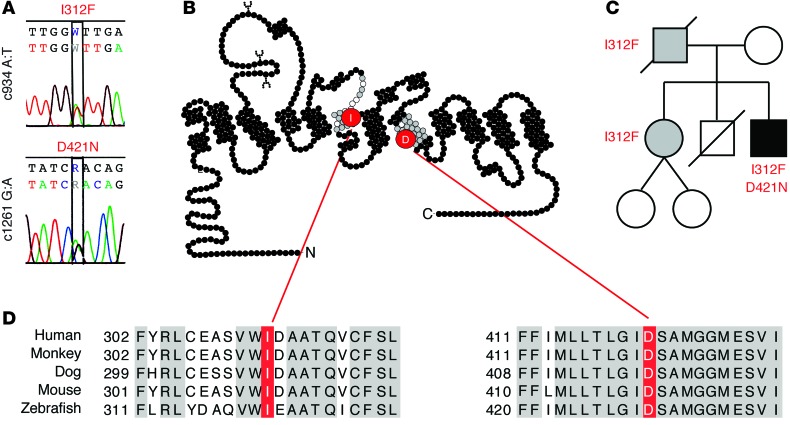
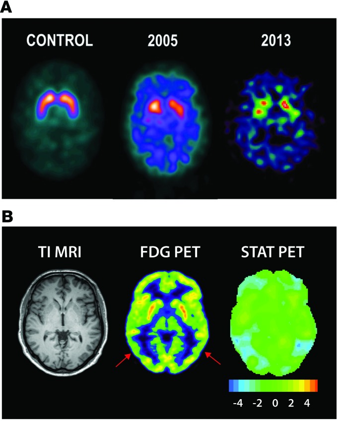
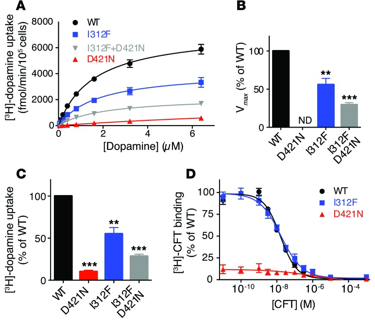
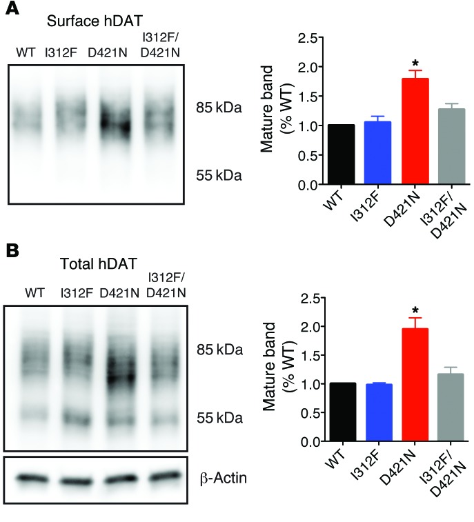
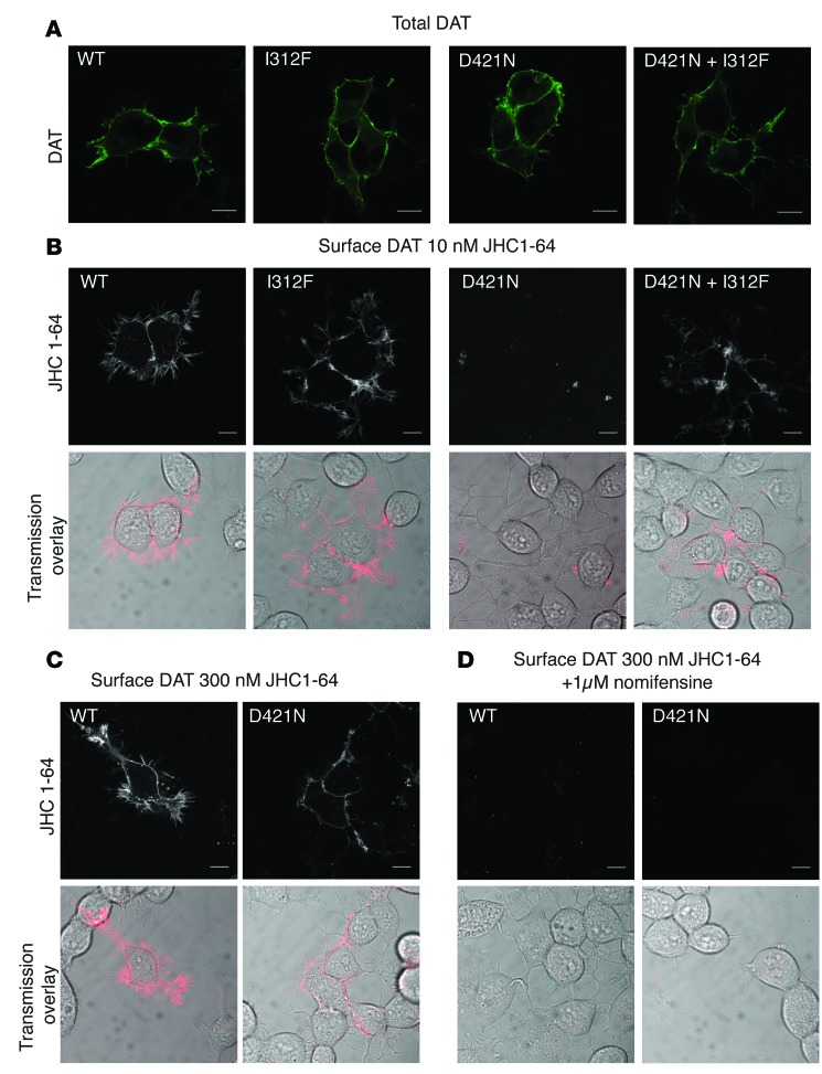
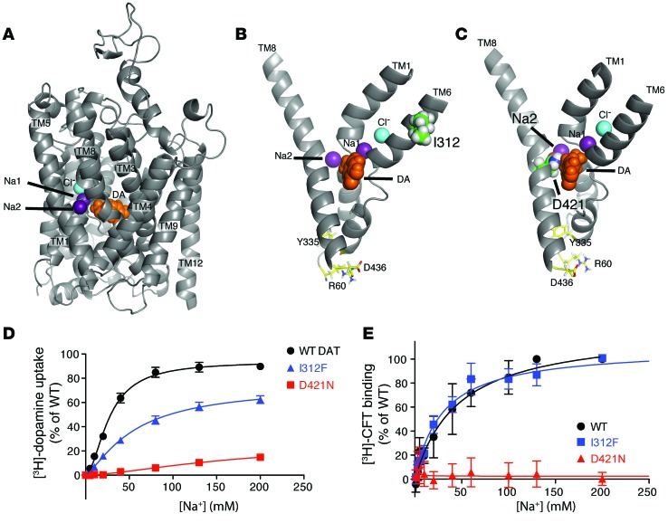
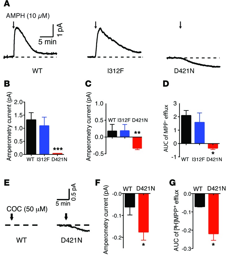
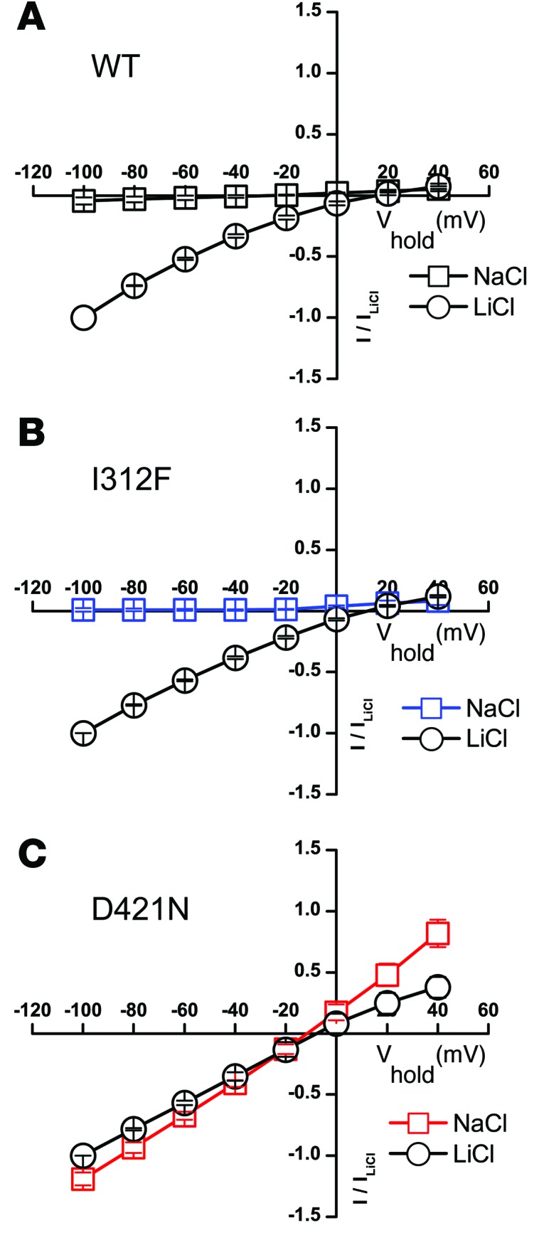
Parkinsonism and attention deficit hyperactivity disorder (ADHD) are widespread brain disorders that involve disturbances of dopaminergic signaling. The sodium-coupled dopamine transporter (DAT) controls dopamine homeostasis, but its contribution to disease remains poorly understood. Here, we analyzed a cohort of patients with atypical movement disorder and identified 2 DAT coding variants, DAT-Ile312Phe and a presumed de novo mutant DAT-Asp421Asn, in an adult male with early-onset parkinsonism and ADHD. According to DAT single-photon emission computed tomography (DAT-SPECT) scans and a fluoro-deoxy-glucose-PET/MRI (FDG-PET/MRI) scan, the patient suffered from progressive dopaminergic neurodegeneration. In heterologous cells, both DAT variants exhibited markedly reduced dopamine uptake capacity but preserved membrane targeting, consistent with impaired catalytic activity. Computational simulations and uptake experiments suggested that the disrupted function of the DAT-Asp421Asn mutant is the result of compromised sodium binding, in agreement with Asp421 coordinating sodium at the second sodium site. For DAT-Asp421Asn, substrate efflux experiments revealed a constitutive, anomalous efflux of dopamine, and electrophysiological analyses identified a large cation leak that might further perturb dopaminergic neurotransmission. Our results link specific DAT missense mutations to neurodegenerative early-onset parkinsonism. Moreover, the neuropsychiatric comorbidity provides additional support for the idea that DAT missense mutations are an ADHD risk factor and suggests that complex DAT genotype and phenotype correlations contribute to different dopaminergic pathologies.


Figures

Figure 1
Figure 1. Identification of missense mutations in SLC6A3.
(A) Identified mutations in SLC6A3. Upper panel: nucleotides c.930-938; the c934 A:T mutation, giving rise to DAT-I312F, is denoted as W. Lower panel: nucleotides c.1257-1265; the c1261 G:A mutation, giving rise to DAT-D421N, is denoted as R. (B) Snake diagram of DAT sequence illustrating the location of I312 and D421 in TMs 6 and 8, respectively. (C) Family tree of the patient. The affected patient is indicated by the black square. An unaffected sister also carries the I312F variant (indicated by the gray circle). The mother carries only WT DAT. The diseased father was not available for testing, but was presumably a carrier of the I312F allele (indicated by the gray square). Females are indicated by circles and males by squares. (D) Sequence alignment of 5 vertebrate species showing conservation of I312 as well as D421.
Figure 2
Figure 2. Nuclear brain imaging with SPECT and FDG PET.
(A) SPECT imaging of DAT. The figure shows images of [123I]FP-CIT binding to DAT in a healthy control (left) and in the proband at 34 years of age (middle) and 43 years of age (right). DAT availability in the striatum, i.e., the ratio of specifically bound radioligand to that of nondisplaceable radioligand, was severely reduced bilaterally and was approximately 35% in 2005 and 15% in 2013 of the expected value in a group of age-matched controls. (B) Transaxial sections through the striatum of the proband with coregistered T1-weighted MRI (left), [18F]-FDG PET (middle), and quantitative statistical comparisons of [18F]-FDG PET (right) with healthy controls showing preserved metabolic activity in the basal ganglia with a slight to moderate reduction (–4 SD; see color scale) in the occipitotemporal junction (red arrows).
Figure 3
Figure 3. Functional characterization of DAT-I312F and DAT-D421N by [3H]-dopamine uptake and [3H]-CFT–binding experiments.
(A) [3H]-dopamine uptake in transiently transfected HEK293 cells. The curves represent average curves from 6 independent experiments, each with triplicate uptake determinations (5 minutes) at the indicated dopamine concentrations. (B) Vmax values of [3H]-dopamine uptake compared with WT. In each experiment, the Vmax values of mutants were normalized to WT. Michaelis-Menten kinetics was applied to fit data. **P < 0.01 and ***P < 0.0001 by 1-sample t test; n = 6. Uptake by DAT-D421N could not be fitted reliably by Michaelis-Menten kinetics and is therefore denoted as not determined (ND). Km values were fitted to 1.7 ± 0.3 μM for WT; 1.8 ± 0.4 μM for DAT-I312F; and 2.1 ± 0.4 for DAT-I312F plus DAT-D421N. (C) Evaluation of [3H]-dopamine uptake at 6.4 μM of dopamine. The uptake was normalized to WT for each experiment. **P < 0.01 and ***P < 0.0001 by 1-sample t test; n = 6. (D) [3H]-CFT binding to transiently transfected COS-7 cells. Curves represent the averages of 4 experiments, each performed in triplicate. Binding data were fitted by nonlinear regression and normalized to WT in each experiment. While DAT-I312F shows preserved binding properties (Kd values [SE intervals] for DAT-I312F and WT were 16.9 [12.1–23.6] nM and 13.7 [12.7–14.8] nM, respectively), no specific binding to DAT-D421N was detected. All data are the means ± SEM. Bmax for WT was 149 ± 39 fmol/105 cells.
Figure 4
Figure 4. Surface expression of DAT mutants assessed by surface biotinylation.
Lysates were made from transiently transfected HEK293 cells. Representative immunoblot of DAT content in biotinylated surface fraction (A) and total protein (B). Right panels show percentage quantifications of the mature DAT glycoforms in the respective fractions compared with WT. DAT-D421N had significantly increased levels of the mature glycoforms compared with WT, whereas mature DAT-I312F levels were similar to those of WT. Data are the means ± SEM; *P < 0.05 by 1-sample t test; n = 5. Full, uncut gels are shown in the Supplementary Material.
Figure 5
Figure 5. Visualization of DAT mutants by confocal microscopy.
(A) Cellular distribution of WT and DAT mutants in transiently transfected CAD cells. DAT was visualized by immunostaining with an N-terminal antibody. No differences were observed in the localization of DAT-I312F or DAT-D421N compared with WT. (B) Confocal live imaging of transfected CAD cells. Cells were stained with the fluorescent cocaine analog JHC 1-64 (10 nM) to obtain specific labeling of DAT present in the plasma membrane. While cells expressing WT, DAT-I312F, or both DAT-I312F and DAT-D421N achieved clear membrane labeling with 10 nM of JHC 1-64, no membrane labeling was observed in cells expressing the DAT-D421N mutant alone. (C) Upon labeling of transfected CAD cells with 300 nM JHC1-64, both WT and DAT-D421N–transfected cells showed clear membrane labeling, which was not observed when cells were preincubated with 1 μM nomifensine, a selective blocker of DAT (D). Images shown are representative of 3 independent experiments. Images of WT and mutants were taken with identical settings.
Figure 6
Figure 6. Molecular modeling of hDAT.
(A) Homology model of hDAT based on the outward-open crystal structure of LeuT, a bacterial homolog. (B) I312, indicated in green in TM 6, was seen to have no direct contact with dopamine or ion binding sites. (C) D421, indicated in green in TM 8, was involved in coordinating binding of sodium at the second sodium binding site (Na2). Sodium and chloride ions are indicated in purple and turquoise, respectively. (D) Sodium dependence of [3H]-dopamine uptake. Dopamine uptake was evaluated at increasing concentrations of sodium. The sodium dependence of DAT-I312F–mediated uptake was comparable to that of WT (Kd = 70 ± 20 mM for DAT-I312F vs. Kd = 30 ± 4 mM for WT). DAT-D421N, on the other hand, showed impaired binding of sodium, seen as a linear sodium dependence curve with no tendency to saturate at 200 mM of NaCl. (E) Sodium dependence of [3H]-CFT binding. Sodium dependence of [3H]-CFT binding to DAT-I312F was similar to that of WT (Kd = 29 ± 3.6 mM for DAT-I312F vs. Kd = 46 ± 10 mM for WT), while specific binding of [3H]-CFT to DAT-D421N could not be detected. The curves of D and E are average curves from 3 and 4 experiments, respectively, each performed in triplicate and normalized to WT Bmax. ChoCl was used for equimolar cation substitution of sodium to obtain indicated sodium concentrations. Bmax and Kd values were derived using a one-site–specific binding model.
Figure 7
Figure 7. Amphetamine- and cocaine-induced amperometric currents and MPP+ efflux.
HEK293 cells were transiently transfected with WT, DAT-I312F, or DAT-D421N. (A) Representative amperometric currents; arrows indicate the application of amphetamine (10 μM). (B) Quantification of the amphetamine-induced peak amperometric currents. (C) Amperometric data are reported as the amphetamine-induced amperometric current recorded 10 minutes after amphetamine application. Data are the means ± SEM; n = 7–9. **P < 0.01 and ***P < 0.001 by Mann-Whitney U test. (D) Quantification of amphetamine-induced [3H]MPP+ efflux presented as the AUC of MPP+ efflux. [3H]MPP+ release was calculated by subtracting the basal release from total release during the first 8 minutes following amphetamine exposure. Data are the means ± SEM; n = 4–5. *P < 0.025 by Mann-Whitney U test. (E) Representative amperometric currents following cocaine exposure (50 μM) of HEK293 cells transiently expressing WT DAT or DAT-D421N. (F) Amperometric data are reported as the cocaine-induced amperometric current 10 minutes after cocaine application. Data are the means ± SEM; n = 3. *P < 0.05 by 1-tailed t test. (G) Quantification of cocaine-induced [3H]MPP+ efflux presented as the AUC of MPP+ efflux. Cocaine treatments led to a significantly higher reduction in MPP+ efflux from cells expressing DAT-D421N compared with WT. *P < 0.05 by Mann-Whitney U test, consistent with a constitutive leak of dopamine.
Figure 8
Figure 8. DAT-D421N possesses a cocaine-sensitive cation leak in sodium.
(AC) I/V diagrams of the cocaine-sensitive steady-state currents (control - cocaine) in sodium chloride (NaCl; squares) and lithium chloride (LiCi, circles) for WT (A), DAT-I312F (B), and DAT-D421N (C). WT and mutant constructs were expressed in Xenopus oocytes and analyzed by the 2-electrode voltage clamp technique as described in Methods. The I/V diagrams were generated in 20-mV steps from –100 to +40 mV. The currents are normalized to the maximum current in lithium at a membrane potential of –100 mV (WT, –402 ± 59 nA; DAT-I312F, –175 ± 27 nA; DAT-D421N 219 ± 31 nA; means ± SEM; n = 4–6).

Similar articles

Cited by

References

    1. Iversen SD, Iversen LL. Dopamine: 50 years in perspective. Trends Neurosci. 2007;30(5):188–193. doi: 10.1016/j.tins.2007.03.002. - DOI - PubMed
    1. Gainetdinov RR, Caron MG. Monoamine transporters: from genes to behavior. Annu Rev Pharmacol Toxicol. 2003;43:261–284. doi: 10.1146/annurev.pharmtox.43.050802.112309. - DOI - PubMed
    1. Torres GE, Gainetdinov RR, Caron MG. Plasma membrane monoamine transporters: structure, regulation and function. Nat Rev Neurosci. 2003;4(1):13–25. doi: 10.1038/nrn1008. - DOI - PubMed
    1. Sotnikova TD, Beaulieu JM, Gainetdinov RR, Caron MG. Molecular biology, pharmacology and functional role of the plasma membrane dopamine transporter. CNS Neurol Disord Drug Targets. 2006;5(1):45–56. doi: 10.2174/187152706784111579. - DOI - PubMed
    1. Kristensen AS, et al. SLC6 neurotransmitter transporters: structure, function, and regulation. Pharmacol Rev. 2011;63(3):585–640. doi: 10.1124/pr.108.000869. - DOI - PubMed

Publication types

MeSH terms